Contents lists available atScienceDirect
Thrombosis Research
journal homepage:www.elsevier.com/locate/thromres
Full Length Article
Involvement of the ADAMTS13-VWF axis in acute Kawasaki disease and effects of intravenous immunoglobulin
☆Nobuyuki Tsujiia, Keiji Nogamia,⁎, Masanori Matsumotob, Hiroyuki Yoshizawaa, Toshio Takasec, Ichiro Tanakac, Toshiyuki Sakaid, Kazuyoshi Fukudae, Masaki Hayakawab, Kazuya Sakaib, Ayami Isonishib, Kayoko Matsuurab, Yoshihiro Fujimuraf, Midori Shimaa
aDepartment of Pediatrics, Nara Medical University, Kashihara, Nara 634-8522, Japan
bDepartment of Blood Transfusion Medicine, Nara Medical University, Nara, Japan
cPediatrics, Yao Municipal Hospital, Osaka, Japan
dPediatrics, Kokuho Central Hospital, Nara, Japan
ePediatrics, Saiseikai Chuwa Hospital, Nara, Japan
fJapanese Red Cross Kinki Block Blood Center, Osaka, Japan
A R T I C L E I N F O Keywords:
Kawasaki disease ADAMTS13 von Willebrand factor Intravenous immunoglobulin Isoelectro-focusing analysis
A B S T R A C T
Introduction:ADAMTS13 modulates shear-dependent platelet thrombus formation (PTF) by limited proteolysis of von Willebrand factor (VWF). A high-plasma-ratio of VWF antigen to ADAMTS13 activity (VWF:Ag/
ADAMTS13:AC) promotes PTF and aggravates shear-induced inflammation mediated by VWF. A role of ADAMTS13 in Kawasaki disease (KD) remains unknown, however. We investigated the involvement of ADAMTS13-VWF axis in the acute-phase of KD (acute-KD).
Methods:VWF:Ag and ADAMTS13:AC in 77 KD infants were measured at three time-points; immediately before (Pre), one-week (1 W) and one-month (1 M) after intravenous-immunoglobulin (IVIG) treatment. VWF multimer (VWFM) distribution and ADAMTS13-isoelectrofocusing (IEF) patterns were compared between the responders and non-responders to IVIG.
Results:A high VWF:Ag (195.7 ± 85.6%,p< 0.05), low ADAMTS13:AC (60.3 ± 23.8%,p< 0.05) and high VWF:Ag/ADAMTS13:AC ratio (3.70 ± 2.12,p< 0.05) at Pre were seen compared to control plasmas. These parameters returned to normal levels time-dependently after IVIG treatment. Non-responders to IVIG demon- strated high VWF:Ag and low ADAMTS13:AC at Pre, and high VWF:Ag/ADAMTS13:AC ratio at 1 W compared to responders, but there were no significant differences in VWFM distribution between both groups. IEF analyses revealed the decreased free form of ADAMTS13 and increased complex form with ADAMTS13 and high-mole- cular-weight-VWFM at Pre in non-responders. A high VWF:Ag/ADAMTS13:AC ratio was associated with in- creased white blood cell counts, together with decreased serum albumin and sodium at Pre and 1 W.
Conclusions:A high VWF:Ag/ADAMTS13:AC ratio in acute-KD persisted after primary treatment in non-re- sponders, and unbalanced substrate-to-enzyme ratio appeared to associate with vascular endothelial damage.
Analysis of existing mode of ADAMTS13 may help to clarify pathogenesis of IVIG resistance in acute-KD.
1. Introduction
Kawasaki disease (KD) is an acute systemic vasculitis of unknown etiology that occurs mainly in infants and young children, typically manifested by fever, and affecting the skin, mucous membranes, lymph nodes, and blood vessels [1]. Damage to vascular endothelial cells (VEC) develops in KD in small- and medium-sized arteries, most
commonly in coronary arteries, and may lead to coronary thrombosis, myocardial infarction and the development of life-threatening aneur- ysms [2]. Intravenous immunoglobulin (IVIG) therapy is the first-line choice for treatment of this disease with a high rate of responsiveness, although the risk of cardiovascular damage appears to be increased in patients that fail to respond to IVIG [3]. In the acute phase of KD (acute- KD), perivasculitis and vasculitis of the micro-vessels and small arteries,
https://doi.org/10.1016/j.thromres.2019.04.026
Received 22 February 2019; Received in revised form 17 April 2019; Accepted 23 April 2019
☆Statement of prior presentation: An account of this work was presented at the 26th Congress of the International Society on Thrombosis and Haemostasis, 2017, Berlin, Germany.
⁎Corresponding author at: Department of Pediatrics, Nara Medical University, 840 Shijo-cho, Kashihara, Nara 634-8522, Japan.
E-mail address:roc-noga@naramed-u.ac.jp(K. Nogami).
Available online 24 April 2019
0049-3848/ © 2019 Elsevier Ltd. All rights reserved.
T
together with perivascular endarteritis in the coronary arteries are evident [4]. The inflammatory cells that invade coronary artery lesions are mainly monocytes and/or macrophages. Neutrophils also appear to contribute to coronary vascular damage [5].
Von Willebrand factor (VWF) is an acute-phase reactant protein that increases in circulating plasma in response to systemic inflammatory mediators. Inflammatory cytokines and VEC damage further stimulate the release of unusually large VWF multimers (UL-VWFM) [6].
ADAMTS13 is a metalloprotease that specifically cleaves the Try1605- Met1606 scissile bond in the VWF molecule [7,8]. Accumulation of UL- VWFM in the blood leads to an imbalance of substrate and enzyme, and a markedly high ratio of substrate-to-enzyme, when the plasma level of UL-VWFM is substantially higher relative to ADAMTS13. These dis- ordered mechanisms promote the formation of platelet thrombi in the pathologic disorder termed thrombotic thrombocytopenic purpura (TTP) [9]. ADAMTS13 also modulates VWF-mediated, shear-induced inflammation [10]. Metabolic disturbances of this nature are observed in patients with severe sepsis or septic shock in the presence of excess UL-VWFM in plasma [11]. The extent of altered substrate-to-enzyme ratio appears to be negatively correlated with platelet counts, and po- sitively associated with the severity of inflammation and the degree of organ failure [11,12]. In the present context, ADAMTS13 and VWF levels have been shown to be associated with the risk of cardiovascular disease in young patients [13].
Several diverse studies have indicated, therefore, that systemic in- flammation mediates increased concentrations of UL-VWFM together with decreased ADAMTS13 activity (ADAMTS13:AC) in plasma. The mechanisms by which ADAMTS13:AC is reduced under pro-in- flammatory conditions are poorly understood, but may include; (i) down-regulation during transcription, (ii) proteolytic degradation and (iii) consumption due to high substrate levels [11]. VWF antigen (VWF:Ag) in children with KD has been reported previously [14,15], but little information is available on ADAMTS13 in these patients. We have recently described the presence of at least 2 types of ADAMTS13:Ag in plasma using isoelectro-focusing (IEF); a free form or a complex with high-molecular weight (HMW)-VWFM. Our data sug- gested that ADAMTS13 could differentially modulate VWF-dependent platelet aggregation under high shear [16]. In the present study, we investigated the involvement of ADAMTS13-VWF axis in children with acute-KD, especially comparing patients that responded to IVIG (re- sponders) and those that failed to respond (non-responders).
2. Materials and methods 2.1. Patients
Pediatric patients with KD were investigated after written informed parental consent. Patients were admitted to Nara Medical University Hospital, Kokuho Central Hospital, Yao Municipal Hospital and Saiseikai Chuwa Hospital in Japan between January 2011 and June 2017, and were diagnosed with KD according to criteria published by the Japanese Circulation Society [17]. Therapeutic management during the acute phase was standardized in keeping with these guidelines. All patients received anti-platelet agents and IVIG treatment as the first- line therapy. IVIG was administered as a single infusion of 2 g/kg or two infusions of 1 g/kg daily. Aspirin was given as an anti-platelet agent at a dose of 30–50 mg/kg/day during the acute phase, and subsequently at 3–5 mg/kg/day during convalescence. In patients with elevated levels of liver enzymes or in the presence of influenza virus, flurbiprofen was given at 3–5 mg/kg/day. Individuals that required additional treatment for fever lasting > 24 h after the end of IVIG infusion or recrudescent fever associated with KD symptoms after an afebrile period were de- fined as non-responders [17].
Coronary artery lesions (CAL), including aneurysms, were defined using Japanese diagnostic guidelines [17]. Coronary artery anomalies were assessed by cardio-specialists using two-dimensional
echocardiography. Coronary arteries were defined as abnormal if the luminal diameter was > 3 mm in a child aged younger than 5 years or > 4 mm in those aged 5 years and older. Cardiovascular lesions were graded if the internal diameter of an arterial segment was at least 1.5- fold as large as that of the adjacent segment, or if the lumen was irre- gular.
2.2. Blood samples
This study was approved by the Medical Research Ethics Committees in the participating institutions. Blood samples were ob- tained after parental written informed consent. Blood was obtained by venipuncture at three time-points; immediately before treatment (‘Pre’), one week (‘1 W’) and one month (‘1 M’) after starting treatment.
Samples were collected into plastic tubes containing 3.2% sodium ci- trate at a 9:1 ratio. Pooled normal plasma (PNP) was prepared from twenty normal healthy individuals aged from 20 to 40 years old and used as the standard reference. Platelet poor plasma was recovered after centrifugation of citrated whole blood for 15 min at 1500 ×g. All plasmas were stored at −80 °C, and thawed at 37 °C immediately prior to assays. Blood samples that were considered unsuitable (e.g.hemo- lysis) or not stored appropriately were excluded.
2.3. VWF:Ag and ADAMTS13:AC measurements
VWF:Ag value was measured by an enzyme-linked immune-sorbent assay (ELISA) using rabbit anti-human VWF polyclonal antiserum (DAKO, Glostrup, Denmark) [18]. VWF:Ag values in normal individuals (n= 20) were 102 ± 33% of control (mean ± standard deviation (SD)). ADAMTS13:AC value was measured using a chromogenic assay (ADAMTS13 act-ELISA, Kainos Laboratories, INC, Tokyo, Japan). The assay is an ELISA using VWF73 substrate (GST-VWF73-His) coupled to anti-GST monoclonal antibody (mAb) on a solid phase microplate with horseradish-peroxidase conjugated anti-N10 mAb recognizing the C- terminal edge of VWF A2 domain cleaved by ADAMTS13 [19].
ADAMTS13:AC values obtained in normal individuals (n= 55) were 99 ± 22% of control. VWF:Ag, ADAMTS13:AC and VWF:Ag/
ADAMTS13:AC ratio were assessed in the samples ‘Pre’, at ‘1 W’, and at
‘1 M’. The 100% reference values were defined using PNP. Control ex- periments confirmed that neither VWF:Ag nor ADAMTS13:AC was present in high-dose pooled human immunoglobulin (Venoglobulin IH®, Japan Blood Products Organization, Tokyo, Japan) used for the IVIG therapy (data not shown).
2.4. Quantitative VWF multimer (VWFM) analysis
VWFM analyses were performed with samples from IVIG responders (n= 10) and non-responders (n = 10) at the three-time points, together with normal control plasmas (n= 15), using sodium dodecyl sulphate- 1.2% agarose gel electrophoresis followed by Western blotting and lu- minographic detection [20,21]. The blots were scanned and subjected to densitometric analysis using Image J (National Institutes of Health, Bethesda, MD). Multimers were classified as low-molecular weight (LMW; bands 1–5 from base), intermediate-molecular weight (IMW;
bands 6–10), and high-molecular weight (HMW; bands ≥11) [22]. For quantitative assessment, the ratio of band density of each group relative to total multimers was calculated. The band densities of total VWFM were regarded as 100%.
2.5. Isoelectro-focusing (IEF) analysis on ADAMTS13:Ag
The identical samples used for VWFM analysis were analyzed by IEF using large-pore agarose-acrylamide composite gel [16,23]. Briefly, sucrose (4 g) and agarose (0.3 g) (GE Healthcare BioScience, Uppsala, Sweden) were mixed with distilled water (34.2mL) and left at 56 °C.
Thirty % acrylamide/bis-acrylamide (1.67 mL, final 1.25%), distilled
water (1.67 mL), 40% carrier ampholyte (2.5 mL, Pharmalyte 3–10, GE Healthcare), ammonium peroxodisulfate (0.27 mL, 22.8mg/mL) and N,N,N′,N′-tetramethylethylenediamine (0.01 mL) were added, and the mixture poured into the IEF gel plate and left for 2 h at 22 °C. The IEF gel was placed in the Multiphor apparatus (GE Healthcare) equilibrated at 10 °C, and sample electrophoresis performed at 100V-5mA-15W for an initial 30 min; 200V-10mA-6W for a further 60 min; and 1500V- 15mA-6W for at final 90 min. After IEF, the isolated proteins were electrophoretically transferred to nitrocellulose membranes. Three bands of ADAMTS13:Ag were detected; a major band (band I) re- presenting a free form with pI of 4.9–5.6, a second band (band II) at pI 5.8–6.7 and a third band (band III) at pI 7.0–7.5, comprising a complex with HMW-VWFM. The composition of band II remains to be de- termined. For a quantitative assessment, the ratio of band density of individual band relative to sum of total band (I + II + III) was calcu- lated by band densitometry using Image J. Total band densities of ADAMTS13:Ag were regarded as 100%. Data obtained from fifteen measurement of normal plasma from healthy individuals was regarded as normal control.
2.6. Clinical and laboratory data
Clinical records in all children with KD included age, sex, blood counts, serum biochemistry, hemostasis tests, IVIG-resistance, treat- ment with/without prednisolone and with/without CAL.
2.7. Statistical analysis
Data were analyzed were using JMP10 (SAS Institute Inc., Cary, NC). Results were reported as median [interquartile range (IQR)] or mean ± SD. The Shapiro-Wilk test was used to evaluate normality.
Differences between groups were compared by the Wilcoxon rank-sum test. Multiple comparisons among different patient groups were de- termined by Kruskal-Wallis one way analysis of variance. Groups were compared with the t-test or the Mann-Whitney U test, and the re- lationships between two variables were evaluated using Spearman rank correlations.P< 0.05 was considered to be statistically significant.
3. Results
3.1. Patients' characteristics
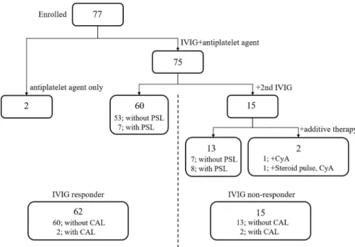
A total of 77 pediatric patients with KD (47 male, 30 female) were enrolled. The clinical characteristics are summarized in Table 1. The age of onset ranged from 3 to 153 months (median 22 months old). Two patients received only anti-platelet agents (one received aspirin, the other received flurbiprofen) and required no additional treatment.
Among all children, 49 patients (69%) received oral aspirin and 28
patients (31%) received oral flurbiprofen. Sixty patients (80%) that received the IVIG responded well. Seven of them received IVIG together with oral prednisolone. Fifteen patients (20%) that received IVIG failed to respond (non-responders), and subsequently received additional IVIG without and with prednisolone. Thirteen of these non-responders re- sponded to additional IVIG treatment. One of the non-responders re- ceived cyclosporine alone. The other received steroid-pulse therapy, cyclosporine and anticoagulant drugs to prevent coronary artery com- plications. CAL developed in 4 patients; in 3 patients, transient dilation returned to normal within one month of onset. One patient developed a coronary aneurysm and required anticoagulant therapy (Fig. 1).
3.2. Changes in VWF:Ag and ADAMTS13:AC in acute-KD
Immediately prior to treatment (Pre), high levels of VWF:Ag (195.7 ± 85.6%) and low levels of ADAMTS13:AC (60.3 ± 23.8%) were detected in children with KD compared to normal controls.
Consequently, high ratios of VWF:Ag/ADAMTS13:AC (3.70 ± 2.12) were demonstrated, indicating a marked imbalance between substrate and enzyme (Fig. 2). At one week (1 W) and 1 month (1 M) after starting treatment, however, levels of VWF:Ag (174.8 ± 86.6%;
p< 0.001 and 134.0 ± 69.1%; p< 0.05, respectively), ADAMTS13:AC (98.1 ± 43.1%; p< 0.001, and 105.8 ± 35.1%;
p< 0.001, respectively), and the ratios of VWF:Ag/ADAMTS13:AC (2.04 ± 1.16;p< 0.001 and 1.38 ± 0.79;p< 0.001, respectively) were significantly different compared to Pre (Fig. 2). The data indicated that the parameters gradually returned close to normal, suggesting a re- adjustment of the unbalanced VWF and ADAMTS13 levels in circulating blood.
3.3. VWF:Ag and ADAMTS13:AC in IVIG responders and non-responders The association between the ADAMTS13-VWF mechanism and clinical response to IVIG in patients with acute-KD is illustrated in Fig. 3. VWF:Ag (panel A) and ADAMTS13:AC (panel B) levels and VWF:Ag/ADAMTS13:AC ratio (panelC) were compared between IVIG responders and non-responders at the three time points. At Pre-treat- ment, high levels of VWF:Ag (185.7 ± 76.2% and 236.7 ± 110.6%, respectively), low levels of ADAMTS13:AC (61.6 ± 24.0% and 54.9 ± 22.5%, respectively) and high ratios (3.48 ± 2.06 and 4.58 ± 2.20, respectively), relative to normal controls, were observed in both groups. VWF:Ag levels in non-responders were significantly greater than those in responders (p= 0.045), but there were no sig- nificant differences between both groups in either ADAMTS13:AC or VWF:Ag/ADAMTS13:AC ratio.
At 1 W, VWF:Ag was not significantly different between both groups (171.9 ± 88.8% and 186.1 ± 79.0%, respectively). Whilst, the ADAMTS13:AC and VWF:Ag/ADAMTS13:AC ratio in responders were significant higher and lower compared to non-responders (104.2 ± 42.9% and 73.6 ± 35.3%, p= 0.013; 1.85 ± 1.08 and 2.81 ± 1.19, p= 0.003), respectively. At 1 M, no significant differ- ences were observed.
3.4. Relationship between VWF multimers (VWFM) and IVIG responsiveness
Quantitative VWFM distribution of circulating VWF was analyzed using each of ten plasma samples from the IVIG responders and non- responders. A representative VWFM pattern in normal plasma, IVIG- responder and non-responder at Pre-treatment visualized by Western blotting are shown inFig. 4A (lower panel). In addition, a representative band density of normal plasma classified by densitometry as LMW, IMW and HMW are illustrated inFig. 4A (upper panel). The ratio of each band density of LMW, IMW, and HMW relative to total band density was compared between both groups (Fig. 4B). At Pre-treatment, the median [IQR] ratios of HMW in responders (22.9% [6.2:29.1],p= 0.013) and Table 1
Characteristics of enrolled patients with KD.
Patients (n) 77 (100%)
Sex (M/F) (n) 47/30 (61/39%)
Mean age at onset (month) 32.9 ± 31.4 [3–153]
Treatment
With IVIG (n) 75 (97.4%)
Without IVIG (n) 2 (2.6%)
Non-responder for IVIG (n) 15 (19.5%)
Additional therapy
Anti-platelet drug (n) 77 (100%)
Aspirin 49 (63.6%)
Flurbiprofen 28 (36.4%)
Steroid (n) 15 (19.5%)
Coronary artery lesion (n) 4 (5.2%)
Dilation/aneurysm 3/1
Values are mean ± SD. [min-max], IVIG; intravenous immunoglobulin.
non-responders (19.2% [5.6:23.5],p< 0.001) were lower compared to controls (27.3% [24.9:29.7]). In addition, the ratios of LMW in re- sponders (45.5% [39.2:70.1], p= 0.003) and non-responders (51.0%
[48.6:70.9], p< 0.0001) were higher compared to controls (36.4%
[34.8:39.3]). These trends were also observed in 1 W and 1 M (data not shown). There were no significant differences between the groups at any time point, however (data not shown).
3.5. Existing form of ADAMTS13:Ag in IVIG responders and non- responders
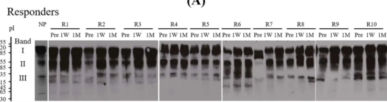
The existing form of ADAMTS13:Ag in acute-KD patients was fur- ther analyzed using an IEF assay. The IEF bands of ADAMTS13:Ag at the three time-points in each of the ten responders (upper panels) and non-responders (lower panels), using the identical samples analyzed by Fig. 1.IVIG treatment, responsiveness, and complications in children with acute KD - IVIG; intravenous immunoglobulin, PSL; prednisolone, CyA; cyclosporine, CAL;
coronary artery lesion.
Fig. 2.Dynamic changes in VWF:Ag, ADAMTS13:AC, and VWF:Ag/ADAMTS13:AC ratios during acute-KD - VWF:Ag (panel A) and ADAMTS13:AC (panelB) were measured Pre, at 1 W and at 1 M obtained in all patients as described in Methods. The calculated VWF:Ag/ADAMTS13:AC ratios are illustrated inpanel C. Boxplots of para- meter changes in the Pre, 1 W and 1 M groups are shown. The median values of parameters are de- picted within the boxes. The boxes end at the 25th and 75th percentiles, and the whiskers extend to the furthest points that are not outliers. *P< 0.05;
significant difference. Pre; immediately prior to treatment, 1 W and 1 M; one week and one month after starting IVIG treatment.
VWFM, are shown in Fig. 5A. As noted above, the origin of band II remains to be determined, and quantitative calculations of band den- sities were restricted, therefore, to band I (free form of ADAMTS13), and band III (ADAMTS13 bound to HMW-VWFM). The respective median [IQR] percentage of band I and band III in normal plasmas were 53.1% [50.1:57.2] and 11.9% [9.1:15.0] (Fig. 5B). In the non-re- sponders, the percentages of band I were lower than normal at all times (Pre 37.6% [33.9:40.5], 1 W 39.6% [36.6:43.2], 1 M 43.0%
[38.5:50.2]) (panela), and those of band III were higher than normal Pre (20.1% [14.9:21.2]) and at 1 W (15.7% [12.5:22.2]) (panelb). By contrast, the percentage of band I in the responders was decreased at only 1 W (43.4% [37.8:47.7]) (panela). The findings suggested that compared to responders, free form of ADAMTS13 in non-responders was significantly decreased Pre (p= 0.028) and at 1 M (p= 0.026) and bound form of ADAMTS13 and HMW-VWFM was significantly in- creased Pre (p= 0.013).
3.6. Relationships between clinical and biochemical variables at each phase of treatment
We studied the additional biochemical data to assess rate and grade of disease pathogenesis and analyzed how these correlated with our primary variables of interest. At Pre-treatment (Table 2), high levels of VWF:Ag moderately correlated with prothrombin times (PT), and weakly correlated with the failure to respond to IVIG and steroid treatment. High ratios of VWF:Ag/ADAMTS13:AC weakly correlated with increased total white blood cell counts (WBC) and percentage of
neutrophils, and decreased serum albumin and sodium. None of the Pre-clinical variables correlated with ADAMTS13:AC, however.
At 1 W (Table 3), high levels of VWF:Ag mildly correlated with in- creased WBC and serum creatinine. Low levels of ADAMTS13:AC moderately correlated with IVIG non-responding status, increased C- reactive protein (CRP) and decreased serum albumin. ADAMTS13:AC weakly correlated with increased brain natriuretic peptide and fibrin degradation products (FDP), decreased serum total protein and sodium, and steroid treatment. High ratios of VWF:Ag/ADAMTS13:AC moder- ately correlated with increased WBC, decreased serum albumin, and steroid treatment, but weakly correlated with non-responding status, increased CRP, FDP and D-dimer, and decreased serum sodium. At 1 M, little significant correlation was evident between clinical variables, VWF:Ag, ADAMTS13:AC and VWF:Ag/ADAMTS13:AC ratio (data not shown). The prevalence of CAL did not correlate with either VWF:Ag or ADAMTS13:AC at any time point.
4. Discussion
The present study has, for the first time, identified low levels of ADAMTS13:AC and high ratios of VWF:Ag/ADAMTS13:AC prior to IVIG treatment in acute-KD. These parameters improved gradually to normal levels after treatment. In addition, the circulating form of ADAMTS13 had some bearing on the effects of IVIG in acute-KD pa- tients.
High VWF:Ag levels in acute-KD have been previously reported [14,15]. Our findings demonstrated that the increased levels of Fig. 3.Comparisons of VWF:Ag and ADAMTS13:AC between IVIG responders and non-responders-VWF:Ag (panelA), ADAMTS13:AC (panelB) and VWF:Ag/
ADAMTS13:AC ratio (panelC) were compared in IVIG responders and non-responders at the indicated times. The median values of parameters are depicted within the boxes. The boxes end at the 25th and 75th percentiles, and the whiskers extend to the furthest points that are not outliers. *p< 0.05; significant difference, n.s.; not significant, R; responder, non-R; non-responder, Pre; immediately prior to treatment, 1 W and 1 M; one week and one month after starting IVIG treatment.
circulating VWF were associated with the decrease in HMWM and the increase in LMWM, together with lower ADAMTS13:AC. We attribute this finding to proteolysis of large amounts of VWF by ADAMTS13 led to this distribution of VWFM, resulting in excessive consumption of this enzyme, while other contributory mechanisms may also be possible.
These findings were in keeping with previous studies in patients with influenza-associated thrombotic microangiopathy [24,25]. In those cases, cytokine-induced HMWM and UL-VWFM was found in the pre- sence of reduced ADAMTS13:AC, suggesting the consumption of ADAMTS13. The accumulation of UL-VWFM in the presence of the high VWF:Ag/ADAMTS13:AC ratio could have promoted the formation of platelet thrombi. There was no evidence of thrombotic microangio- pathy in our KD children, however, and the precise reason for the un- balance of VWF:Ag and ADAMTS13:AC levels in acute-KD remains to be fully clarified.
Coronary artery aneurysms caused by KD develop mainly at branching sites of the major coronary artery, where restricted flow velocity, reduced shear stress, and disturbed flow patterns are evident [26]. The VWF-cleavage potential of ADAMTS13 is less under low shear conditions rather than at high shear [27], and leukocytes appear to tether to and roll on platelets adhered to UL-VWF at high shear, at least in vitro, without ADAMTS13 [28]. Under these conditions, leukocyte interactions with VWF strings could contribute to inflammatory me- chanisms independent of ADAMTS13 [29]. Several studies have shown that various integrins, together with glycoprotein Ib-IX-V complex and P selectin, supported neutrophil-rolling and tethering on stimulated endothelial cells at low shear [30]. The evidence indicates, therefore, that in response to inflammatory stimuli, circulating leukocytes adhere, roll and then arrest on VWF from activated endothelial cells prior to migrating to surrounding tissues. Aspirin or flurbiprofen given for acute KD function as anti-platelet agent mainly by inactivation of cycloox- ygenase 1, leading the decreased production of prostaglandin E2 and thromboxane A2 [31]. Therefore, either aspirin or flurbiprofen does not directly suppress neutrophil-rolling and -tethering on stimulated
endothelial cells (but suppress the following discharge of thromboxane A2). In addition, the vasa vasorum, which supplies nutrition within the adventitia of blood vessels, may be proliferated by inflammatory mediators at bifurcation sites, and could contribute to the progression of vasculitis in KD [32,33]. In arterioles, sufficient levels of ADAMTS13:AC appear to be required to prevent the formation of pla- telet thrombi induced by shear-related VWF conformation. Low levels of ADAMTS13:AC may influence, therefore, the development of CAL.
There were no significant differences in ADAMTS13:AC between pa- tients with and without CAL in our study, perhaps because of a small number of individuals with CAL available for analysis. Nevertheless, it seemed reasonable to conclude that shear-induced inflammatory me- chanisms involving VWF was associated with the development of CAL in KD.
We investigated the association between VWF and ADAMTS13 in responders and non-responders to IVIG treatment. In summary these data indicated; i) IVIG non-responders had higher VWF:Ag Pre-treat- ment, and lower ADAMTS13:AC with higher VWF:Ag/ADAMTS13:AC ratios at 1 W, relative to responders, ii) The VWFM patterns in both groups were not significantly different, and iii) At Pre-treatment, the free form of ADAMTS13 was decreased and bound form to ADAMTS13 and HMWM was increased in non-responders compared to responders.
These results suggest that the VWF-ADAMTS13 axis, involving the circulating form of ADAMTS13 and unbalanced VWF and ADAMTS13 levels have some bearing on the effects of IVIG in acute-KD. To clarify the association with the responsiveness to IVIG, however, additional supporting findings such as the role of free form or complex with HMW- VWFM is further required. On the other hand, previous studies provided evidence that higher MMP-8, S100A8, S100A9, carcinoembryonic an- tigen-related cell adhesion molecule 1 and IL-1 transcript levels were associated with IVIG resistance [34]. Since we did not evaluate these factors, analyses of relationship between the VWF-ADAMT13 axis and these factors in IVIG resistance would be required.
The relatively high VWF:Ag levels pre-treatment in non-responders Fig. 4.Ratios of HMWM or LMWM relative to total of VWFM in IVIG responders and non-responders - (PanelA) VWFM was analyzed in normal plasma, Pre-treatment plasma with IVIG responder (R1-Pre) and Pre-treatment plasma with non-responder (NR1- Pre) as described in Methods (lower panels). A re- presentative band pattern of VWFM and the intensity of band density of LMWM, IMWM and HMWM of normal plasma are shown inupper panel. (PanelB) The ratios of band density of HMWM (left panel) and LMWM (right panel) relative to total of VWFM in samples from IVIG responders (n= 10) and non-re- sponders (n = 10) each together with normal control plasma (n= 15) are shown. The densities of total VWFM were expressed as 100%. Boxplots of ratio in both groups and normal plasmas are shown. The median values of HMW or LMW ratio are depicted within the boxes. The boxes end at the 25th and 75th percentiles, and the whiskers extend to the furthest points that are not outliers. *p< 0.05; significant difference. R; responder, non-R; non-responder, NP;
normal plasma, LMW; low molecular weight, HMW;
high molecular weight, VWFM; VWF multimers.
Fig. 5.IEF analysis of ADAMTS13:Ag in IVIG responders and non-responders - (PanelA)IEF pattern of ADAMTS13:Ag- Plasmas Pre, at 1 W, and at 1 M in IVIG responders (R1–10;upper panel) and non-responders (NR1–10;lower panel) were analyzed by IEF as described in Methods. Plasma levels of ADAMTS13:AC and VWF:Ag in the identical samples are shown below the scans. The IEF pattern of ADAMTS13:Ag in normal plasma is illustrated at the left of each group. (PanelB) Comparison between IVIG responders and non-responders-The median values of parameters are depicted within the boxes. The percentages of band I (panela) and band III (panelb) densities relative to total densities (band I + II + III) in samples from IVIG responders (n = 10) and non-responders (n = 10) depicted inFig. 4together with control plasmas are shown. The boxes end at the 25th and 75th percentiles, and the whiskers extend to the furthest points that are not outliers. *p< 0.05;
significant difference, n.s.; not significant, Pre; immediately prior to treatment, 1 W and 1 M; one week and one month after starting IVIG treatment, NP; normal plasma, R; responder, non-R; IVIG non-responder.
compared to responders, together with similarly low ADAMTS13 in each group, suggested that an advanced inflammatory status and/or more VEC damage was present in non-responders. Increased VWF:Ag in KD children has been previously reported [14,15], and this is the first report to demonstrate quantitatively that VWF:Ag in non-responders was significantly higher than that in responders. In addition, the re- storation of ADAMTS13:AC at 1 W was delayed in non-responders, re- sulting in a persistently unbalanced VWF:Ag/ADAMTS13:AC ratio. The
increased ADAMTS13-VWF bound form and the reduced free ADAMTS13 at 1 W in non-responders may be due to the reduced en- zyme capacity following the proteolysis of excessive VWF by ADAMTS13, but a further research is required to conclude.
We have previously described the usefulness of IEF to analyze the existing form of ADAMTS13:Ag in circulating plasma [16], and de- monstrated that band I represented the free form of ADAMTS13:Ag whilst band III consisted of ADAMTS13:Ag bound to HWM-VWFM. The Table 2
Relationships between clinical variables, biochemical data, VWF:Ag and ADAMTS13:AC in KD patients Pre-treatment.
Variables Patients Control VWF:Ag ADAMTS13:AC VWF:Ag/ADMTS13:AC
Mean ± SD N Reference R P R P R P
Age (month) 32.9 ± 31.4 (77) − 0.041 0.73 −0.011 0.92 −0.015 0.90
Sex (M/F) (n) 47/30 (77) − 0.42 0.66 0.99
IVIGR/non-R (n) 62/15 (77) − 0.048 0.32 0.091
Steroid (−/+) (n) 63/14 (77) − 0.026 0.097 0.005
CAL (−/+) (n) 73/4 (77) − 0.60 0.21 0.55
Laboratory results
WBC (×103μl) 13.9 ± 5.0 (77) [3.3–8.6] 0.189 0.11 −0.178 0.13 0.278 0.020
Neut (%) 67.6 ± 15.6 (77) [38.0–74.0] 0.209 0.080 −0.130 0.28 0.269 0.024
RBC (×104/μl) 436 ± 40 (77) [386–492] −0.076 0.53 0.152 0.20 −0.179 0.14
Plt (×104/μl) 33.8 ± 11.0 (76) [15.8–34.8] 0.045 0.71 −0.108 0.37 0.026 0.83
TP (g/dl) 6.6 ± 0.5 (76) [6.6–8.1] −0.017 0.88 0.189 0.11 −0.128 0.29
Alb (g/dl) 3.9 ± 0.4 (76) [4.1–5.1] −0.106 0.38 0.230 0.053 −0.300 0.012
GOT (IU/l) 98 ± 152 (77) [13–30] 0.212 0.075 −0.019 0.88 −0.003 0.98
GPT (IU/l) 89 ± 145 (77) [7–23] 0.166 0.17 0.182 0.13 0.047 0.70
Cre (mg/dl) 0.26 ± 0.09 (72) [0.46–0.79] 0.140 0.26 −0.080 0.52 0.135 0.28
Na (mEq/l) 134 ± 3 (77) [138–145] −0.182 0.13 0.129 0.28 −0.239 0.046
PT (sec) 13.3 ± 1.4 (39) [10.0–15.0] 0.476 0.003 0.021 0.91 0.213 0.21
aPTT (sec) 34.6 ± 4.7 (38) [22.0–32.0] 0.130 0.45 −0.028 0.87 0.126 0.47
FDP (μg/ml) 4.8 ± 1.4 (20) [ < 5.0] −0.058 0.81 −0.303 0.12 0.318 0.17
D-D (μg/ml) 1.3 ± 0.4 (26) [≤1.0] −0.167 0.41 −0.205 0.33 −0.086 0.68
CRP (mg/dl) 7.9 ± 5.0 (76) [≤0.14] 0.069 0.57 −0.082 0.49 0.140 0.25
BNP (mol/ml) 61.8 ± 80.1 (29) [0.0–18.4] −0.075 0.71 −0.303 0.12 0.255 0.19
WBC/RBC; white/red blood cell count, Neut; neutrophil, Plt; platelet count, TP; total protein, Alb; albumin, GOT; glutamic oxaloacetic transaminase, GPT; glutamic pyruvic transaminase, Cre; creatinine, PT; prothrombin time, aPTT; activated partial thromboplastin time, FDP; fibrin degradation product, D-D; D dimer, CRP; C- reactive protein, BNP; brain natriuretic peptide, IVIGR; intravenous Immunoglobulin responder, non-R; IVIG non-responder, CAL, coronary artery lesion. R;
Coefficient correlation,P; Significant difference asP< 0.05. The bold emphases indicate the significance differences.
Table 3
Relationship between clinical and biochemical variables, VWF:Ag and ADAMTS13:AC in KD patients at 1 W.
Variables Patient Control VWF:Ag ADAMTS13:AC VWF:Ag/ADMTS13:AC
Mean ± SD N Reference R P R P R P
Age (month) 32.9 ± 31.4 (77) − 0.174 0.14 0.012 0.92 0.133 0.25
Sex (M/F) (n) 47/30 (77) − 0.078 0.58 0.17
Steroid (−/+) (n) 63/14 (77) − 0.088 0.027 0.0005
CAL (−/+) (n) 73/4 (77) − 0.42 0.41 0.37
Laboratory results
WBC (×103μl) 10.1 ± 4.0 (77) [3.3–8.6] 0.261 0.024 −0.121 0.30 0.371 0.001
Neut (%) − (77) [38.0–74.0]
RBC (×104/μl) 431 ± 43 (77) [386–492] −0.018 0.88 0.217 0.059 −0.129 0.27
Plt (×104/μl) 53.5 ± 15.6 (77) [15.8–34.8] 0.025 0.83 0.084 0.47 −0.050 0.67
TP (g/dl) 8.0 ± 0.7 (75) [6.6–8.1] 0.065 0.59 0.256 0.027 −0.143 0.23
Alb (g/dl) 3.7 ± 0.4 (74) [4.1–5.1] −0.165 0.17 0.354 0.002 −0.413 0.0003
GOT (IU/l) 43.5 ± 28.9 (77) [13–30] −0.170 0.14 −0.111 0.34 0.020 0.87
GPT (IU/l) 33.1 ± 27.3 (77) [7–23] −0.058 0.62 −0.067 0.57 0.041 0.72
Cre (mg/dl) 0.26 ± 0.07 (73) [0.46–0.79] 0.307 0.009 0.060 0.62 0.154 0.20
Na (mEq/l) 137 ± 2 (71) [138–145] −0.028 0.82 0.311 0.009 −0.262 0.030
PT (sec) 12.2 ± 0.8 (29) [10.0–15.0] 0.061 0.76 −0.289 0.14 0.275 0.16
aPTT (sec) 29.1 ± 3.6 (29) [22.0–32.0] −0.104 0.60 −0.227 0.24 0.149 0.45
FDP (μg/ml) 4.8 ± 5.9 (22) [ < 5.0] −0.253 0.26 −0.434 0.043 0.519 0.013
D-D (μg/ml) 1.8 ± 2.3 (24) [≤1.0] −0.248 0.24 −0.400 0.052 0.425 0.038
CRP (mg/dl) 1.1 ± 2.3 (75) [≤0.14] −0.143 0.23 −0.386 0.0007 0.238 0.042
BNP (mol/ml) 52.0 ± 76.6 (16) [0.0–18.4] −0.427 0.099 −0.504 0.046 0.335 0.21
WBC/RBC; white/red blood cell count, Neut; neutrophil, Plt; platelet count, TP; total protein, Alb; albumin, GOT; glutamic oxaloacetic transaminase, GPT; glutamic pyruvic transaminase, Cre; creatinine, PT; prothrombin time, aPTT; activated partial thromboplastin time, FDP; fibrin degradation product, D-D; D dimer, CRP; C- reactive protein, BNP; brain natriuretic peptide, CAL, coronary artery lesion.R; Coefficient correlation,P; Significant difference asP< 0.05. The bold emphases indicate the significance differences.
components of band I and III were separated by cryo-precipitation, and both fractions inhibited VWF-dependent platelet aggregation at high shear flow dose-dependently. Different mechanisms were evident, however, and products containing band I appeared to contribute to the initial phase, and those containing band III inhibited later platelet re- sponses [16]. Nevertheless, the degrees of inhibition of platelet ag- gregation at each phase were closely similar, indicating that band I component acted as a fast-acting element, and band III acted as a slow- acting constituent. An elegant investigation using similar IEF analysis demonstrated that severe reduction of free ADAMTS13 was evident in plasmas from patients with HELLP syndrome, suggesting that un- coordinated ADAMTS13:AC could influence platelet thrombi formation and might play a role in the pathogenesis of this disease [35].
Although ADAMTS13:AC levels in non-responders and responders were lower than normal, non-responders had reduced fast-acting (band I) and increased slow-acting (band III) forms of ADAMTS13 compared to responders. The balance between unbound and bound form of ADAMTS13 may be essential between IVIG responders and non-re- sponders. An earlier study demonstrated that normal plasma cryo-su- pernatants (CSP) contain smaller amounts of VWF, and almost normal amounts of ADAMTS13:AC as well as reduced amounts of ADAMTS13 bound to larger VWF multimers compared to fresh frozen plasma [16].
The treatment with CSP may be a favorable option, therefore, as re- placement therapy in patients with defective free ADAMTS13:AC. The current data indicate that quick analysis of ADAMTS13 using IEF could assist the clinical management of these disorders, including acute-KD.
Although the relationship between clinical and laboratory data re- quire further investigation, high VWF:Ag/ADAMTS13:AC ratio within one week after treatment appeared to be associated with increased WBC, decreased serum albumin and sodium. These findings may sup- port that vascular leakage is a key feature of KD pathophysiologically [36]. Non-cardiogenic edema is caused by disturbances in endothelial gap formation, and sub-endothelial edema and hypoalbuminemia are mediated by the increase in vascular leakage [37]. The mechanism(s) governing decreased serum sodium in acute-KD remain controversial, but may be related to further vascular leakage by decreased serum al- bumin.
There are limitations in this study, however. ADAMTS13:AC and VWF:Ag/ADAMTS13:AC ratio in infants have not been widely in- vestigated, and a limited numbers of patients with CAL were available for analyses. Further details in age-matched patients with and without acute-phase complications could be informative. Nevertheless, low le- vels of ADAMTS13:AC and high ratio of VWF:Ag/ADAMTS13:AC were evidently observed in the acute-KD patients. In addition the unbalanced substrate to enzyme concentrations appeared to be associated with parameters of VEC damage, and improved on remission. We have re- cently demonstrated that the coagulation data observed in acute-KD were consistent with hypercoagulability, although fibrinolytic function appeared to be well-balanced [38]. In addition to coagulation and fi- brinolysis conditions in acute-KD, our present findings suggested that the clinical management of acute-KD could consider potential VWF- related platelet thromboembolic events. To clarify further pathophy- siological mechanism(s), investigation on the relationship between VWF-ADAMTS13 axis and acute-KD under physiologic blood-flow condition are in progress.
Acknowledgements
This work was partly supported by a Grant-in-Aid for Scientific Research (KAKENHI) from the Ministry of Education, Culture, Sports, Science and Technology (MEXT) to KN (Grant No. 15K09663 and 18K07885) and NT (Grant No. 18K15684), and by a Japan Agency for Medical Research and Development (AMED) (Grant No.
16ek0109142h0002).
Disclosure Author contribution
N. Tsujii; designed the research, collected the samples, performed experiments, interpreted the data and wrote the paper, K. Nogami;
designed the research, interpreted the data, wrote the paper, edit the manuscript, and approved the final version to be published, M.
Matsumoto; designed the research, interpreted the data, H. Yoshizawa, T. Takase, I. Tanaka, T. Sakai, K. Fukuda; clinical supports and collected the samples from patients, M. Hayakawa and Sakai; performed VWFM analysis and IEF analysis, A. Isonishi and K. Matsuura; measured the VWF:Ag and ADAMTS13:AC, Y. Fujimura and M. Shima; supervised the manuscript.
Conflicts of interest
The authors have no conflicts of interest to disclosure.
References
[1] T. Kawasaki, F. Kosaki, S. Okawa, I. Shigematsu, H. Yanagawa, A new infantile acute febrile mucocutaneous lymph node syndrome (MLNS) prevailing in Japan, Pediatrics 54 (1974) 271–276.
[2] H. Kato, T. Sugimura, T. Akagi, N. Sato, K. Hashino, Y. Maeno, T. Kazue, G. Eto, R. Yamakawa, Long-term consequences of Kawasaki disease. A 10- to 21-year follow-up study of 594 patients, Circulation 94 (1996) 1379–1385.
[3] J.W. Newburger, M. Takahashi, J.C. Burns, Kawasaki disease, J. Am. Coll. Cardiol.
67 (2016) 1738–1749.
[4] H. Fujiwara, Y. Hamashima, Pathology of the heart in Kawasaki disease, Pediatrics 61 (1978) 100–107.
[5] K. Takahashi, T. Oharaseki, S. Naoe, M. Wakayama, Y. Yokouchi, Neutrophilic in- volvement in the damage to coronary arteries in acute stage of Kawasaki disease, Pediatr. Int. 47 (2005) 305–310.
[6] A. Bernardo, C. Ball, L. Nolasco, J.F. Moake, J.F. Dong, Effects of inflammatory cytokines on the release and cleavage of the endothelial cell-derived ultralarge von Willebrand factor multimers under flow, Blood 104 (2004) 100–106.
[7] M. Furlan, R. Robles, B. Lammle, Partial purification and characterization of a protease from human plasma cleaving von Willebrand factor to fragments produced by in vivo proteolysis, Blood 87 (1996) 4223–4234.
[8] H.M. Tsai, Physiologic cleavage of von Willebrand factor by a plasma protease is dependent on its conformation and requires calcium ion, Blood 87 (1996) 4235–4244.
[9] J.L. Moake, Thrombotic microangiopathies, N. Engl. J. Med. 347 (2002) 589–600.
[10] A.K. Chauhan, J. Kisucka, A. Brill, M.T. Walsh, F. Scheiflinger, D.D. Wagner, ADAMTS13: a new link between thrombosis and inflammation, J. Exp. Med. 205 (2008) 2065–2074.
[11] R.A. Claus, C.L. Bockmeyer, M. Sossdorf, W. Losche, The balance between von- Willebrand factor and its cleaving protease ADAMTS13: biomarker in systemic in- flammation and development of organ failure? Curr. Mol. Med. 10 (2010) 236–248.
[12] J.J. Lin, O.W. Chan, H.J. Hsiao, Y. Wang, S.H. Hsia, C.H. Chiu, Decreased ADAMTS 13 activity is associated with disease severity and outcome in pediatric severe sepsis, Medicine (Baltimore) 95 (2016) e3374.
[13] T.N. Bongers, E.L. de Bruijne, D.W. Dippel, A.J. de Jong, J.W. Deckers, D. Poldermans, M.P. de Maat, F.W. Leebeek, Lower levels of ADAMTS13 are asso- ciated with cardiovascular disease in young patients, Atherosclerosis 207 (2009) 250–254.
[14] S.L. Bowyer, C.G. Ragsdale, D.B. Sullivan, Factor VIII related antigen and childhood rheumatic diseases, J. Rheumatol. 16 (1989) 1093–1097.
[15] Y. Sakurai, H. Takatsuka, M. Onaka, M. Takada, M. Nishino, Persistent endothelial damage after intravenous immunoglobulin therapy in Kawasaki disease, Int. Arch.
Allergy Immunol. 165 (2014) 111–118.
[16] Y. Hori, M. Hayakawa, A. Isonishi, K. Soejima, M. Matsumoto, Y. Fujimura, ADAMTS13 unbound to larger von Willebrand factor multimers in cryosupernatant:
implications for selection of plasma preparations for thrombotic thrombocytopenic purpura treatment, Transfusion (Paris) 53 (2013) 3192–3202.
[17] Guidelines for diagnosis and management of cardiovascular sequelae in Kawasaki disease (JCS 2013). Digest version, Circ. J. 78 (2014) 2521–2562.
[18] M. Matsumoto, S. Kawaguchi, H. Ishizashi, H. Yagi, J. Iida, T. Sakaki, Y. Fujimura, Platelets treated with ticlopidine are less reactive to unusually large von Willebrand factor multimers than are those treated with aspirin under high shear stress, Pathophysiol. Haemost. Thromb. 34 (2005) 35–40.
[19] S. Kato, M. Matsumoto, T. Matsuyama, A. Isonishi, H. Hiura, Y. Fujimura, Novel monoclonal antibody-based enzyme immunoassay for determining plasma levels of ADAMTS13 activity, Transfusion (Paris) 46 (2006) 1444–1452.
[20] Z.M. Ruggeri, T.S. Zimmerman, Variant von Willebrand's disease: characterization of two subtypes by analysis of multimeric composition of factor VIII/von Willebrand factor in plasma and platelets, J. Clin. Invest. 65 (1980) 1318–1325.
[21] U. Budde, R. Schneppenheim, H. Plendl, J. Dent, Z.M. Ruggeri, T.S. Zimmerman, Luminographic detection of von Willebrand factor multimers in agarose gels and on nitrocellulose membranes, Thromb. Haemost. 63 (1990) 312–315.
[22] A. Veyradier, T. Nishikubo, M. Humbert, M. Wolf, O. Sitbon, G. Simonneau, J.P. Girma, D. Meyer, Improvement of von Willebrand factor proteolysis after prostacyclin infusion in severe pulmonary arterial hypertension, Circulation 102 (2000) 2460–2462.
[23] A. Isonishi, C.L. Bennett, B. Plaimauer, F. Scheiflinger, M. Matsumoto, Y. Fujimura, Poor responder to plasma exchange therapy in acquired thrombotic thrombocyto- penic purpura is associated with ADAMTS13 inhibitor boosting: visualization of an ADAMTS13 inhibitor complex and its proteolytic clearance from plasma, Transfusion (Paris) 55 (2015) 2321–2330.
[24] N. Tsujii, K. Nogami, H. Yoshizawa, M. Hayakawa, A. Isonishi, M. Matsumoto, M. Shima, Influenza-associated thrombotic microangiopathy with unbalanced von Willebrand factor and a disintegrin and metalloproteinase with a thrombospondin type 1 motif, member 13 levels in a heterozygous protein S-deficient boy, Pediatr.
Int. 58 (2016) 926–929.
[25] R. Akiyama, I. Komori, R. Hiramoto, A. Isonishi, M. Matsumoto, Y. Fujimura, H1N1 influenza (swine flu)-associated thrombotic microangiopathy with a markedly high plasma ratio of von Willebrand factor to ADAMTS13, Intern. Med. 50 (2011) 643–647.
[26] T. Ohkubo, R. Fukazawa, E. Ikegami, S. Ogawa, Reduced shear stress and disturbed flow may lead to coronary aneurysm and thrombus formations, Pediatr. Int. 49 (2007) 1–7.
[27] Y. Shida, K. Nishio, M. Sugimoto, T. Mizuno, M. Hamada, S. Kato, M. Matsumoto, K. Okuchi, Y. Fujimura, A. Yoshioka, Functional imaging of shear-dependent ac- tivity of ADAMTS13 in regulating mural thrombus growth under whole blood flow conditions, Blood 111 (2008) 1295–1298.
[28] A. Bernardo, C. Ball, L. Nolasco, H. Choi, J.L. Moake, J.F. Dong, Platelets adhered to endothelial cell-bound ultra-large von Willebrand factor strings support leukocyte tethering and rolling under high shear stress, J. Thromb. Haemost. 3 (2005) 562–570.
[29] K. De Ceunynck, S.F. De Meyer, K. Vanhoorelbeke, Unwinding the von Willebrand factor strings puzzle, Blood 121 (2013) 270–277.
[30] R.P. McEver, Adhesive interactions of leukocytes, platelets, and the vessel wall during hemostasis and inflammation, Thromb. Haemost. 86 (2001) 746–756.
[31] J.R. Vane, Inhibition of prostaglandin synthesis as a mechanism of action for as- pirin-like drugs, Nat. New Biol. 231 (1971) 232–235.
[32] Z. Onouchi, N. Tomizawa, M. Goto, K. Nakata, M. Fukuda, Cardiac involvement and prognosis in acute mucocutaneous lymph node syndrome, Chest 68 (1975) 297–301.
[33] A. Hamaoka-Okamoto, C. Suzuki, T. Yahata, K. Ikeda, N. Nagi-Miura, N. Ohno, Y. Arai, H. Tanaka, T. Takamatsu, K. Hamaoka, The involvement of the vasa va- sorum in the development of vasculitis in animal model of Kawasaki disease, Pediatr. Rheumatol. Online J. 12 (2014) 12.
[34] W. Fury, A.H. Tremoulet, V.E. Watson, B.M. Best, C. Shimizu, J. Hamilton, J.T. Kanegaye, Y. Wei, C. Kao, S. Mellis, C. Lin, J.C. Burns, Transcript abundance patterns in Kawasaki disease patients with intravenous immunoglobulin resistance, Hum. Immunol. 71 (2010) 865–873.
[35] Y. Yoshida, M. Matsumoto, H. Yagi, A. Isonishi, K. Sakai, M. Hayakawa, Y. Hori, T. Sado, H. Kobayashi, Y. Fujimura, Severe reduction of free-form ADAMTS13, unbound to von Willebrand factor, in plasma of patients with HELLP syndrome, Blood Adv. 1 (2017) 1628–1631.
[36] M. Terai, T. Honda, K. Yasukawa, K. Higashi, H. Hamada, Y. Kohno, Prognostic impact of vascular leakage in acute Kawasaki disease, Circulation 108 (2003) 325–330.
[37] S. Hirose, Y. Hamashima, Morphological observations on the vasculitis in the mu- cocutaneous lymph node syndrome. A skin biopsy study of 27 patients, Eur. J.
Pediatr. 129 (1978) 17–27.
[38] H. Yoshizawa, K. Nogami, T. Matsumoto, N. Tsujii, T. Sakai, T. Takase, I. Tanaka, M. Shima, Dynamic evaluation of hemostasis in the acute phase of Kawasaki disease using comprehensive coagulation functional assays, Thromb. Res. 174 (2019) 76–83.