www.cognizantcommunication.com
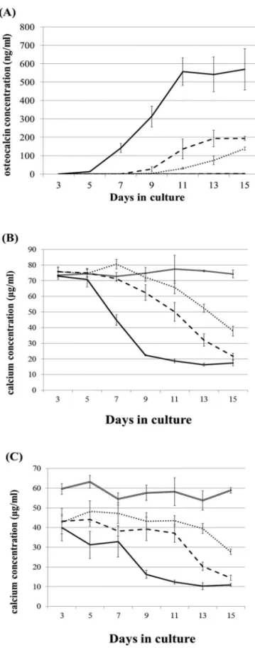
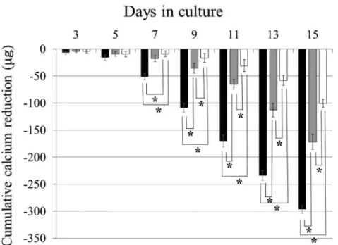
Calcium Concentration in Culture Medium as a Nondestructive and Rapid Marker of Osteogenesis
10
0
0
全文
(2)
(3)
(4)
(5)
(6)
(7)
(8)
(9)
(10)
図


関連したドキュメント
One reason for the existence of the current work is to produce a tool for resolving this conjecture (as Herglotz’ mean curvature variation formula can be used to give a simple proof
We show that a discrete fixed point theorem of Eilenberg is equivalent to the restriction of the contraction principle to the class of non-Archimedean bounded metric spaces.. We
Instead an elementary random occurrence will be denoted by the variable (though unpredictable) element x of the (now Cartesian) sample space, and a general random variable will
Two numerical examples are described to demonstrate the application of the variational finite element analysis to simulate the hydraulic heads and free surface in a porous medium..
Two numerical examples are described to demonstrate the application of the variational finite element analysis to simulate the hydraulic heads and free surface in a porous medium..
In this work we give definitions of the notions of superior limit and inferior limit of a real distribution of n variables at a point of its domain and study some properties of
This problem becomes more interesting in the case of a fractional differential equation where it closely resembles a boundary value problem, in the sense that the initial value
An explicit expression of the speed of the oil- water interface is given in a pseudo-2D case via the resolution of an auxiliary Riemann problem.. The explicit 2D solution is
