埼玉医科大学腎臓内科学教室 〔平成 16 年 11 月 23 日 受付〕 原 著
動脈硬化抑制に関わるAT
1受容体拮抗薬 とエストロゲンの作用機序:
酸化ストレスに対する相互作用を中心に
津田 昌宏
Mechanism of Inhibitory Action of AT1 Receptor Blocker and Estrogen on Atherosclerosis
Masahiro Tsuda (Department of Nephrology, Saitama Medical School, Moroyama, Iruma - gun, Saitama, 350 - 0495, Japan)
In the present study, we investigated a gender difference of atherosclerotic changes induced in apoE-deficient (ApoEKO)mice, focusing on oxidative stress, and the possible interaction between olmesartan, an AT1 (angiotensin type 1 receptor)blocker(ARB), and estrogen.After treatment with high cholesterol diet(HCD) for 6 weeks, apparent atherosclerotic lesion formation including lipid deposition and increase in superoxide production and p47phox expression were observed in ApoEKO mice. These changes were significantly greater in male than in female mice, although plasma cholesterol level was not different. Preceding ovariectomy enhanced atherosclerotic lesion and oxidative stress 6 weeks after HCD. The changes in ovariectomized mice were reversed by 17β-estradiol(80μg/ kg/day)replacement. On the other hand, olmesartan(3 mg/kg/day)inhibited both atherosclerosis and oxidative stress observed in ApoEKO mice treated with HCD. The inhibitory effect of olmesartan on atherosclerosis was significantly stronger in female than in male and ovariectomized ApoEKO mice. Neither the smaller dose of estrogen(20μg/kg/day)nor olmesartan(0.5 mg/kg/day)influenced atherosclerosis and oxidative stress. However, co - administration of olmesartan and estrogen at these smaller doses attenuated atherosclerosis as well as oxidative stress. We further investigated interaction between AT1 receptor stimulation and estrogen on NADPH oxidase activityusing cultured VSMC, which mainly was expressed AT1 receptor. The NADPH oxidase activity in cultured VSMC was increased by Angiotensin II.17β-estradiol attenuated NADPH oxidase activity induced by angiotensin II without affecting the expression of AT1 receptor. These results indicate that estrogen enhances the inhibitory effect of AT1 receptor blocker on atherosclerosis. Our results also suggest that estrogen and ARB act synergistically to inhibit oxidative stress, by attenuating NADPH oxidase activity.
Keywords: atherosclerosis, estrogen, AT1 receptor blocker, NADPH oxidase
J Saitama Med School 2005;32:1 - 11
(Received November 23, 2004) 緒 言 高脂血症,糖尿病および喫煙は心血管危険因子と してよく知られている1, 2).それらは血管壁での細胞 レドックス状態を変化させ内皮細胞機能不全や動脈 硬化を促進する.これら心血管危険因子による共通 の機序として初期に酸化ストレスが関与し,血管の 炎症を引き起こすと考えられている.活性酸素種の 過剰産生を含む酸化ストレスは,血管平滑筋細胞の 肥大や増殖,細胞間物質の遊走及び調節にかかわっ ており3 - 5),動脈硬化症,血管再狭窄,糖尿病や高血 圧に伴う血管障害を引き起こす原因の一つである6). NADPH oxidaseは,NADHあるいはNADPHを電子供 与体として用い,酸素に一電子還元する化学反応を触 媒することでsuperoxideを産生させる7).また,血管平 滑筋細胞において,NADPH oxidaseは細胞膜サブユ
ニットであるnox1, nox4, p22phoxと細胞質内サブユニッ
エストロゲンが心血管保護作用を有することを示して いる12 - 14).エストロゲン補充療法は更年期女性での心 血管疾患の発生を抑制し14),血中LDLコレステロール 値を低下させHDLコレステロール値を増加させる15). しかし,その作用機序については充分には明らかにさ れておらず,脂質代謝以外作用を有する可能性も考え られる. 本研究においてはこれまでの報告をもとに,動脈硬 化性病変形成におけるエストロゲンの役割について, 特にAT1受容体拮抗薬(ARB)との相互作用に注目しな がら,動脈硬化症のモデルであるapolipoprotein E遺伝 子欠損(ApoEKO)マウスを用いて酸化ストレスへの影 響を中心に検討すると共に,NADPH oxidase活性に対 するエストロゲンの作用機序と,AT1受容体刺激との 関連について,培養血管平滑筋細胞を用いて検討した. 方 法 動物及び薬物投与 本実験では動脈硬化症モデルである6週齢成熟 ApoEKOマウス(The Jackson Laboratory, Bar Harbor, ME)を用いた.全ての動物実験は埼玉医科大学動 物実験委員会の承諾を得,愛媛大学動物実験指針 に準拠して行った.動物は,6週齢から標準食(MF, Oriental Yeast Co. Ltd.)あるいは高コレステロール食 (1.25 % cholesterol, 10 % coconut oil in MF) で6週 間
飼育した.両側卵巣摘出は高コレステロール食開始 48時間前に施行した.17β-estradiol (Sigma Chemial Co., St. Louis, MO)(20 あるいは 80μg/kg/day)は propylene glycolで溶解し皮下投与した.ARBである Olmesartan (RNH-6270, 三 共 株 式 会 社 よ り 供 与 )は osmotic minipump (Alzet model 1002, DURECT Corporation, Cupertino, CA) を用いて 0.5あるいは 3.0 mg/kg/dayの 用 量 で 腹 腔 内 投 与 し た16).血 中 コレステロール値はcholesterol oxidase法(Cholesterol E-test, WAKO Chemical Industries, Ltd., Tokyo)を 用いて,組織 Ang II は Peninsula Laboratories Kit (Peninsula Laboratories, Inc) を用いてそれぞれ測定
した17).
動脈硬化性病変の測定
近 位 大 動 脈 の 凍 結 切 片 を 作 成 し,Elastica van
Gieson染色を用い,病変サイズは画像解析ソフト
Densitograph Imaging Software(ATTO Corporation, Tokyo, Japan)を用いて算出し,脂肪沈着は,oil red-O
染色後同じく画像解析ソフトを用いて算出した18 - 20).
Superoxide産生
Superoxide産生は,近位大動脈の新鮮凍結切片を作 成し,これをdihydroethidium (DHE; 10μmol/L) と共 に遮光した加湿チェンバー内で37℃,30分間インキュ ベートし,superoxideをethidiumの赤色螢光として検 出した21, 22).
NADPH oxidase 活性
大動脈サンプルは内皮を傷つけないように迅速に 摘 出 し, 氷 冷 し たTris-surcrose buffer(10 mM Tris pH 7.1, 340 mM surcrose, 1 mM EDTA, 1 mM PMSF, 10μg/mL aprotinin)500 mLでホモジナイズし,30分 間抽出した.さらにサンプルを遠心(15,000 g, 10 min, 4℃)し,上清(20μg protein) に78μM cytochrome C (Sigma -Aldrich, St. Louis, MO), 100μM NADPH (Sigma -Aldrich)を加え,1000 U/mL superoxide dismutase(SOD)(Sigma-Aldrich)存在下及び,非存 在下で37℃,60分間インキュベート後,吸光度を測定 した.NADPH oxidase 活性はSODの存在,非存在下
での吸光度より算出した23).
Real Time RT-PCR
大動脈サンプルよりRNAを抽出し,reverse transcription を行ったのち,定量的polymerase chain reactionをSYBR green I キット(MJ Research, Inc., Waltham, MA) を用い て行った.
PCRプライマーは,
p47phox : 5’ - GTCCCTGCATCCTATCTGGA - 3’(forward) 5’ - GGGACATCTCGTCCTCTTCA - 3’(reverse), AT1受容体: 5’-GTTCCTGCTCACGTGTCTCA-3’(forward) 5’ - CATCAGCCAGATGATGATGC - 3’(reverse), β-actin: 5’-CCTGTATGCCTCTGGTCGTA-3’(forward) 5’-CCATCTCCTGCTCGAAGTCT-3’(reverse)を用いた. 血管平滑筋細胞培養と受容体結合能測定 成熟Sprague-Dawleyラット大動脈血管平滑筋細胞 (VSMC)は以前の報告のように準備し培養を行った24-26). 今回の実験では 5 から 7 継代したsubconfluent VSMCを 用いた.AT1やAT2のreceptor binding はHayashidaらの 報告に従い測定した21).すなわち,1μmol/L valsartan あるいは 1μmol/L PD123319(Research Biochemical International)存在,非存在下で 125I -[Sar1, Ile8] Ang II (Du Pont NEN Research Product)と伴に37℃ ,1時
間培養した後で,結合した放射活性を測定した.AT1 receptor bindingは総カウント量とvalsartanと一緒に 培養したサンプルのカウント量間の差を用いて,AT2 receptor bindingは総カウント量からPD123319存在下 でのカウント量を差引きして算出した. 培養VSMCにおけるNADPH oxidase活性測定 VSMCは24時 間 無 血 清 で 培 養 し て 静 止 状 態 と した後,培養液とAng II (Sigma Chemical Co., St. Louis, MO) ,17β-estradiol (Sigma Chemical Co.)を添加 した.NADPH oxidase活性は 5μmol/L のlucigeninを 用いて測定した5).化学発光はluminometer (AB-2200, ATTO Corporation, Tokyo, Japan)で 5 分間測定した. 統計処理
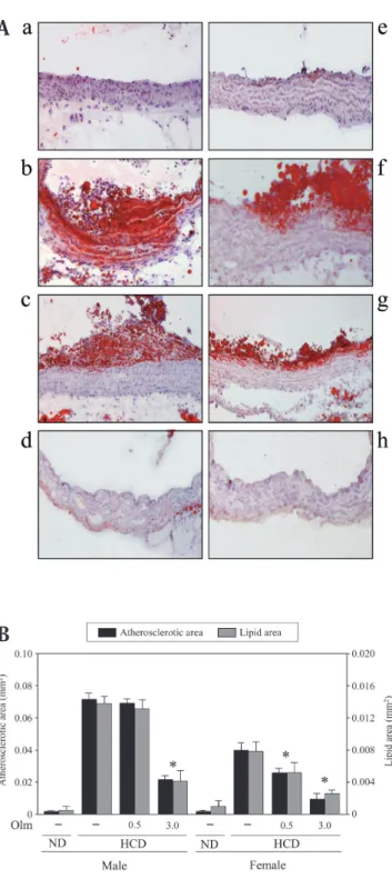
ある時は,グループ間の差を検出するためNewman-Keuls検定を施行した.危険率は5%未満を統計学的に 有意とみなした. 結 果 ApoEKOマウスにおける動脈硬化性病変の性差 高コレステロール食投与6 週間後で,対照群に比 べ著しい血中コレステロール値の上昇が認められた (雄性マウス 対照群: 46±3.6 mg/dL, 比較群: 1684± 96.0 mg/dL, 雌性マウス 対照群: 46.6±3.6 mg/dL, 比較 群: 1592±79.0 mg/dL).近位大動脈においては,高コ レステロール食投与6 週間で脂肪沈着を伴う動脈硬化 性病変形成が認められた.この動脈硬化性病変は雄性 マウスに比べ雌性マウスにおいて有意に減弱してい た(Fig. 1).さらに,雌性マウスでは卵巣摘出により 近位大動脈での脂肪沈着を伴う動脈硬化性病変形成は 対照群に比べ増加を認めた(Fig. 2).卵巣摘出による 変化は17β-estradiol投与により,用量依存性に抑制さ れた(Fig. 2).血中エストロゲン濃度は対照群で224.6 ±16.8 pg/mLであり,卵巣摘出により著明な血中エス トロゲン濃度の低下を認めた(卵巣摘出群: 54.6±2.6 pg/mL).この血中エストロゲン濃度は,雄性マウス (55.0±5.1 pg/mL)とほぼ同レベルまで低下していた. さらに,エストロゲン投与は,血中エストロゲン濃度 を85.5±2.8 pg/mL(20μg/kg/day投与),193.3±3.7 pg/mL(80μg/kg/day投 与 ) と 増 加 さ せ,80μg/kg/ day投与で正常雌性マウスとほぼ同レベルまでの回復 を認めた.血中コレステロール値は両側卵巣摘出とエ ストロゲン投与による影響は認めなかった(卵巣摘出 群: 1536±65 mg/dL, エストロゲン投与群: 1557±57 mg/dL).また,卵巣摘出及びエストロゲン投与は血 圧には影響を与えず(対照群: 118.6±6.1 mmHg, 卵巣 摘出群: 111.2±3.9 mmHg, エストロゲン投与群: 112.1 ±6.8 mmHg ),組織Ang IIについても,卵巣摘出とエ ストロゲン投与共に正常雌性マウスと同レベルであっ た(雌性マウス: 59±5.0 pg/mg protein, 卵巣摘出群: 57 ±7.0 pg/mg protein, エストロゲン投与群(80μg/kg/ day): 58±1.0 pg/mg protein). 動脈硬化に対する17β-estradiolとolmesartanの相 互作用 3 mg/kg/dayのolmesartan投与により,雌雄とも に明らかな脂肪沈着を伴う動脈硬化性病変形成の抑 制を認めた (Fig. 1).比較的低用量であるOlmesartan (0.5 mg/kg/day) の投与では雌性マウスにおいての み明らかな減少を認めたが,雄性マウス,卵巣摘出雌 性マウスではこれらの変化はほとんど認めなかった. (Fig. 1).さらに単独投与では脂肪沈着と動脈硬化性 病変に影響を与えない比較的低用量のolmesartan(0.5 mg/kg/day)とエストロゲン(20μg/kg/day)の併用 により有意な抑制を認めた (Fig. 2).なお今回用いた
Fig. 1. Sex difference in atherosclerotic area and effect of olmesartan in apolipoprotein - deficient (ApoEKO) mice. ApoEKO mice were treated with a high - cholesterol diet or standard normal diet for 6 weeks as described in “Materials and Methods”. Olmesartan was administered at a dose of 0.5 or 3 mg/kg/day for 2 weeks before sampling. Aortic samples were taken and cross sections were prepared as described in “Methods”. (A) Representative results of oil red - O staining of proximal aorta. Magnification x 200. (B) Morphometric analysis of atherosclerotic area and lipid area in cross sections of proximal aorta. Values are mean±SE of 7-8 experiments. *p<0.05 vs. HCD. ND: standard normal diet, HCD: high -cholesterol diet, Olm: olmesartan, (a∼d) male mice, (e∼h) female mice. a and e: ND, b and f: HCD, c and g: olmesartan (0.5 mg/kg/day), d and h: olmesartan (3.0 mg/kg/day).
A
量のolmesartanでは,血圧に影響を与えず (雄性マウ ス 対照群: 114.5±4.2 mmHg, olmesartan投与群: 114.6 ±1.6 mmHg, 雌 性 マ ウ ス 対 照 群: 118.6±6.1 mmHg, olmesartan投与群: 115.7±9.2 mmHg, 卵巣摘出群 併用 投与群: 117.1±6.3 mmHg),血中コレステロール値に も影響を与えなかった (olmesartan投与群: 1628±98 mg/dL, 併用群: 1654±68 mg/dL). 動脈硬化性病変に対するolmesartanとエストロゲン の相互作用効果 Fig. 3に示すように,superoxide産生は大動脈にお いて高コレステロール食投与で増加を示したが,この 増加は雄性マウスや卵巣摘出マウスに比べ,雌性マ ウスでは減弱をしていた.また,NADPH oxidase活性 も高コレステロール食投与で増加を認めたが,その 増加も雄性マウスや卵巣摘出マウスに比べ,雌性マ ウスでの抑制が認められた (Fig. 5).NADPH oxidase
のサブユニットであるp47phoxのmRNA発現について
も 検 討 し た と こ ろNADPH oxidaseと 同 様 の 結 果 が 認められた (Fig. 6A).卵巣摘出マウスにおいては, 80μg/kg/dayの17β-estradiol投与でsuperoxide産生,
NADPH oxidase活性とp47phox mRNAレベルの上昇が
有意に抑制された(Fig. 4, 5, 6A).一方,3 mg/kg/day のolmesartanはsuperoxide産生,NADPH oxidase活性
とp47phox mRNA発現を抑制したが,この抑制効果は雌
Fig. 2. Effect of estrogen and olmesartan on atherosclerotic area in ovariectomized ApoEKO mice. Female ApoEKO mice were ovariectomized and treated with HCD as described in “Methods”. 17β-estradiol was administered at a dose of 20 or 80μg/kg/day for 14 days before sampling. Olmesartan was administered at a dose of 0.5 or 3.0 mg/kg/day 14 days before sampling. Aortic samples were taken as described in Figure 1. (A) Representative results of oil red - O staining of proximal aorta. Magnification x 200. (B) Morphometric analysis of atherosclerotic area and lipid area in cross sections of proximal aorta. Values are mean±SE of 7 -8 experiments. †p<0.05 vs. HCD, **p<0.01 vs. HCD+OVX. OVX: ovariectomy, Olm: olmesartan, E: 17β-estradiol. a: control (OVX mice), b: olmesartan (0.5 mg/kg/day), c: olmesartan (3.0 mg/kg/day), d: 17β-estradiol (20μg/kg/day), e: 17β-estradiol (80μg/kg/day), f: olmesartan (0.5 mg/kg/ day) and 17β-estradiol (20μg/kg/day).
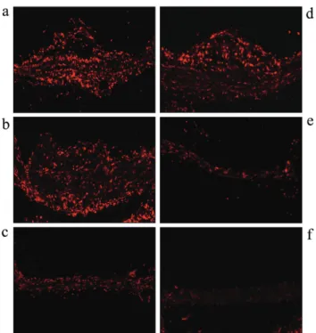
Fig. 3. Sex difference on superoxide production in ApoEKO mice. Freshly frozen sections of proximal aor ta were prepared and superoxide was detected with dihydroethidium as described in “Materials and Methods”. Olmesartan and 17 β-estradiol were administered as described in Figure 2. (a∼d) male mice, (e∼h) female mice. a and e: ND, b and f: HCD, c and g: olmesartan (0.5 mg/kg/day), d and h: olmesartan (3.0 mg/kg/day).
A
性マウスでより強く認められた (Fig. 3, 5, 6A).特に, 比較的低用量のolmesartan (0.5 mg/kg/day)は,雄性 マウスや卵巣摘出マウスにおけるsuperoxide産生,
NADPH oxidase活性,p47phox mRNA発現上昇に影響
を与えなかったが,雌性マウスにおいては,明らかに これらの変化を抑制した (Fig. 4, 5, 6A).比較的低用 量の17β-estradiol (20μg/kg/day)とolmesartan (0.5 mg/kg/day) は,単独では上記の変化に影響を及ぼさ なかったが,これら両者を併用すると,superoxide産
生,NADPH oxidase活性,p47phox mRNA発現が有意
に抑制された (Fig. 4, 5, 6A).近位大動脈におけるAT1 受容体 mRNA発現は高コレステロール食により増加 したが,その増加は雌性マウスに比べ,雄性マウス や卵巣摘出マウスでは大きかった.また,AT1受容体 mRNA発現は17β-estradiol (80μg/kg/day) では発現 が抑制されたが,olmesartanによる影響は認められな かった (Fig. 6B). 培養血管平滑筋細胞におけるNADPH oxidase活性に 対するエストロゲンの効果
Fig. 7に示すように,Ang IIはVSMCにおけるNADPH oxidase活性上昇を引き起こした (15分後にピーク). 17β-estradiol (10- 8 M)単独ではNADPH oxidaseに影
響を与えなかったが, Ang IIによるNADPH oxidase活
性上昇を抑制した.さらに17β-estradiolはAT1受容体 発現には影響せず,一方AT2 受容体はこのVSMCにお いては検知されなかった (データ不提示). 考 察 これまでの報告において,17β-estradiolはApoEKO マウスにおいてで抗動脈硬化作用をしめしている27). これは血中リポ蛋白に対する17β-estradiolの効果で 一部説明できる.本研究では,高コレステロール食 投与による脂肪沈着を伴った動脈硬化性病変形成が, 雄性マウスや卵巣摘出マウスに比して,雌性マウス において抑制され,卵巣摘出マウスにおける変化は 17β-estradiol投与により回復した (Fig. 2).今回用い た17β-estradiol投与量で,卵巣摘出により,血中エ ストロゲン濃度の著しい低下を認め (対照群: 224.6± 16.8 pg/mL, 卵巣摘出群: 54.6±2.6 pg/mL),エストロ ゲン投与 (80μg/kg/day) にて血中エストロゲン濃度 の回復を認めた.この血中エストロゲン濃度は正常雌 性ApoEKOマウスと同様であり,有効な量であると考 えられる28).また,本研究においては,脂肪沈着と動 脈硬化性病変の変化が平行しており,一般的にはこれ
Fig. 4. Effect of olmesartan and estrogen on superoxide production in ovariectomized ApoEKO mice. Superoxide was detected with dihydroethidium as described in “Materials and Methods”. Olmesartan and 17β-estradiol were administered as described in Figure 2. a: control (OVX mice), b: olmesartan (0.5 mg/kg/day), c: olmesar tan (3.0 mg/kg/day), d: 17β-estradiol (20μg/kg/day), e: 17β-estradiol (80μg/ kg/day), f: olmesartan (0.5 mg/kg/day) and 17β-estradiol (20μg/kg/day).
らが一致することはすくないが,12週齢という脂肪沈 着が主の時期の動脈硬化を検討しているためと考えら れる29).さらに,血中コレステロール値は雌雄差を認め ず (雄性マウス: 1684±96 mg/dL, 雌性マウス: 1592±79 mg/dL),エストロゲンやolmesartanによる影響は認め なかったので脂質代謝以外の作用が関与していると考 える (エストロゲン投与群: 1557±57 mg/dL, olmesartan 投 与 群 : 1628±98 mg/dL).Cathapermal ら30)は, 17β-estradiolがエストロゲン受容体を介してsuperoxide に刺激されたVSMC増殖を抑制すると報告しており, Strehlowら31)は17β-estradiolがVSMCに お い てAng II によるフリーラジカル産生増加を抑制すると報告し ている.本実験においては,superoxide産生,NADPH
oxidase活性とp47phox mRNA発現が雄性マウスに比べ雌
性マウスで有意に抑制されており,また雌性マウスに おいては,卵巣摘出による増強と17β-estradiol投与で 改善することを明らかにした (Fig. 3, 4, 5, 6A).
Superoxide産生とNADPH oxidaseの活性化は動脈硬
化性病変形成に重要な役割を担っており32 - 35),Ang II
はAT1受容体を介してNADPH oxidaseを活性化するこ
とで酸化ストレスの主要な調節を行っている10, 11).こ れに対して,エストロゲンはレニン−アンジオテンシン 系に拮抗し36),また肝細胞でのアンジオテンシノー ゲン合成はエストロゲンにより調節されると報告さ れている37).エストロゲンはレニンとアンジオテンシ ン変換酵素を低下させ,その結果Ang IIの産生を抑制 するといわれている38, 39).さらにエストロゲン投与に より大動脈組織あるいはVSMCでAT1受容体発現が抑 制されるという報告もある40, 41).我々はこれまでに,
17β-estradiolがAT1受容体由来のextracellular
signal-Fig. 7. Effect of estrogen on NADPH oxidase activity induced by Ang II in cultured VSMC. Subconfluent VSMC were incubated with Ang II (10- 7 M) and 17β-estradiol (10- 8 M) for 0, 5, 15, 30 minutes, and 1, 3, 6, 12, 24 hours. NADPH oxidase activity was measured by the chemiluminescence method using lucigenin as described in “Methods”. Values are expressed as mean±SE of 4 experiments. ○: Ang II (10- 7 M), △: 17β-estradiol (10- 8 M), ●: Ang II with 17β-estradiol. Fig. 6. Effect of olmesartan and estrogen on expression of
p47phox and AT1 receptor in ApoEKO mice. Aortic samples were taken from ApoEKO mice as described in Figure 1. Levels of p47phox mRNA and AT1 receptor mRNA were assayed by quantitative real time RT - PCR as described in “Methods”. (A) Effect of combination of olmesartan and estrogen on expression of p47phox mRNA in ApoEKO mice. (B) Effect of combination of olmesartan and estrogen on expression of AT1 receptor mRNA in ApoEKO mice. Values are mean±SE of 5 experiments. *p<0.05 vs. ND, †p<0.05 vs. HCD, **p<0.05 vs. HCD+OVX.
A
regulated kinase (ERK)とc-fos発現を抑制し,VSMC の増殖を阻害する事を報告した25).これらの結果から ARBが動脈硬化に対するエストロゲンの抑制効果を さらに増強する可能性が考えられる.本研究の結果で は,動脈硬化及び酸化ストレスに対するolmesartanの 抑制効果は雄性マウスや卵巣摘出マウスと比べ,雌 性マウスで強く認められ (Fig. 1, 2, 3, 4, 5, 6A),卵巣 摘出マウスにおいては,比較的低用量のolmesartanと 17β-estradiolの併用により脂肪沈着,動脈硬化性病 変,superoxide産 生,NADPH oxidase活 性 とp47phox mRNA 発現を抑制するのを認めた (Fig. 2, 4, 5及び 6A).Lavignerら42)は,VSMCからのsuperoxide産生
はNADPH oxidaseがp47phox成分を必要とすることを
報告しており,これらの結果は我々の結果とあわせる と,エストロゲンが少なくとも一部は酸化ストレスへ の抑制を介してARBの抗動脈硬化作用に貢献してい ること,AT1受容体とエストロゲンのクロストークが 血管障害の進展に対して重要な影響を与えることを示 している.また,活性酵素種の一つである一酸化窒素 (NO) は,血管壁において通常NO合成酵素(NOS) に より産生され,血管拡張や,血小板凝集抑制,平滑 筋増殖抑制,単球付着抑制などの作用を介し抗動脈 硬化作用を有すると考えられている.Oemarら43)は, 人の動脈硬化性病変において,NOの産生とNOSの発 現が低下しており,NO産生を刺激すると,動脈硬化 性病変の進行を抑制すると報告している44).従って, 本研究における動脈硬化モデルマウスの病変形成や, エストロゲンとARBによる抑制効果についても,NO 産生調節が関与する可能性も残っている.さらに, 最近 ARB である olmesar tan が endothelin を介した
superoxide産生を抑制することが報告されており53), olmesartanの動脈硬化抑制作用にはendothelinを介 するsuperoxide産生を抑制する経路が含まれる可能性 も考えられ,今後さらなる検討が必要と思われる. 本研究ではAT1受容体mRNA発現は,雌性マウス に比べ雄性マウスや卵巣摘出マウスで著しい増加を 認め,それらは17β-estradiol (80μg/kg/day)投与で 抑制を認めたが,olmesartan投与では変化がみられな かった(Fig. 6B).この結果はエストロゲンによるAT1 受容体mRNA発現の抑制作用がエストロゲンの抗動 脈硬化作用を増強する可能性を示している.しかし, 卵巣摘出群において,比較的低用量のエストロゲン とolmesartanの併用による明らかな動脈硬化性病変, superoxide産生とNADPH oxidase活性の抑制を認め たが(Fig. 2, 4及び5),このときAT1受容体発現には 変化が認められなかった(Fig. 6B).また,粥状動脈 硬化病変の炎症巣においては,アンギオテンシン変 換酵素(ACE) およびAngII, AT受容体の発現が認め
られており45),本研究では,卵巣摘出やエストロゲン による組織Ang IIへの影響を認めなかった.これらの 結果は,エストロゲンとAT1受容体拮抗薬 (ARB) 併用 による抗動脈硬化作用には,AT1受容体発現調節以外 の要因が働いていることを示している.Ang IIはAT1 受容体を介して,NADPH oxidase活性によるLDLコ レステロールの酸化46),macrophage chemoattractant protein I (MCP -1) やvascular cell adhesion molecule
(VCAM)の発現を増加させ47, 48),血管平滑筋細胞を増 殖し6)動脈硬化を促進させる. Xuら49)は,ARB投与によるAT 2受容体活性が心血管 保護作用に重要であると示しており,Wuら50)は,AT 2 受容体刺激でMCP-1の発現を低下させることを示し ている.一方,エストロゲンは,MCP-1を低下させ51),
extracellular signal-regulated kinase(ERK)活性を抑制
する52).以上より,ARBによるAT
2受容体刺激作用と
エストロゲンの刺激作用が相乗的に脂肪沈着と動脈硬 化性病変を抑制している可能性が考えられた. また,VSMCを用いた結果よりエストロゲンが直接 Ang IIによるNADPH oxidase活性を阻害する可能性も 考えられ,VSMCを用いた検討では,Fig. 7に示すよ うにAng IIはNADPH oxidase活性を時間依存性に増加 させ,エストロゲン単独では活性に変化を与えなかっ たが,Ang IIとエストロゲンの同時投与により5分 以内にAng IIによる活性上昇が抑制された.このとき
AT1受容体は変化しなかった.このことはVSMCにお
いて,エストロゲンがAng IIを介したNADPH oxidase 活性を直接抑制することを示しており,その作用は, 発現が比較的短時間で生じている.
さらに,細胞質サブユニットと細胞膜サブユニッ
トの複合体がNADPH oxodaseとして働き8),AT
1受容 体を介したAng IIの刺激により,細胞質サブユニッ トの細胞膜サブユニットへの移動と結合することが NADPH oxidase活 性 を引 き 起 こす こ と がい わ れ て いる54, 55).以上のことより,NADPH oxidaseの細胞質 サブユニットが細胞膜サブユニットへの移動をエスト ロゲンが比較的短時間に抑制するnon-genomic作用も もつ可能性が考えられる. 今回の研究より,動脈硬化性病変に対して,少なく とも一部は酸化ストレスを抑制することで,エストロ
ゲンがAT1受容体を介したAng II刺激を抑制すること,
動脈硬化モデルマウスにおいて,エストロゲンが動脈 硬化性病変及び酸化ストレスに対し抑制的に働くこ とを示唆する結果が得られた.この結果は,エストロ ゲン製剤が動脈硬化抑制作用を有する可能性を示して いるが,この点に関しても,今後臨床の場でさらに検 討を重ねることが重要であると考える.さらに,実験 的には,雄性動脈硬化マウスに17β-estradiol投与を 行う検討が有用であると思われるが,この点につい ては,過去にも検討されておらず,今後の研究の課題 であると考える. 結 論
1. apolipoprotein E遺伝子 欠 損 (ApoEKO) マウス の おける動脈硬化形成に対し,エストロゲン及び アンジオテンシンIIタイプI (AT1) 受容体拮抗薬, olmesartan, の作用について酸化ストレスとの関連 性から検討した. 2. ApoEKOマウスの卵巣摘出は動脈硬化及び酸化ス トレスを増強させ,エストロゲン(80μg/kg/day) 投与により改善を認めた. 3. olmesartan(3 mg/kg/day) 投与は酸化ストレスと 動脈硬化性病変を著明に抑制したが,その抑制作 用は雄性マウスや卵巣摘出マウスに比べ,雌性マ ウスでより強く認められた.一方それぞれ単独で は影響を与えない比較的低用量のolmesartan(0.5 mg/kg/day) あ る い は エ ス ト ロ ゲ ン(20μg/kg/ day) の併用投与では,動脈硬化性病変や酸化スト レスの減少を認めた. 4. 以上の結果からエストロゲンとARBが相乗的に酸 化ストレスの抑制と共に動脈硬化を改善する事が 明らかとなった. 5. さらに,AT1受容体刺激作用とエストロゲン作用と の関連性について明確にするため,アンジオテン シンII受容体サブタイプのうち,AT1受容体を主に 発現している培養血管平滑筋細胞を用い以下の実 験を行った. 6. 培養血管平滑筋細胞をアンジオテンシンIIにより 刺激するとNADPH oxidase活性の上昇が認めら れ,この上昇はエストロゲンを同時に投与するこ とにより減弱した.この際,エストロゲンによる AT1受容体発現への影響を認めなかった. 7. 以上の結果より,エストロゲンは動脈硬化性病変 に対し,AT1受容体拮抗薬の作用を増強させること が明らかとなった.培養血管平滑筋細胞を用いた 実験から,この作用は血圧や血中コレステロール の変化を介したものではなく,エストロゲンの直 接的な作用であると考えられた.これらの結果は ARBとエストロゲン補充の併用療法が,更年期に みられる心血管疾患に対する効果的な治療となる 可能性を示唆するものと思われた. 謝 辞 本 研 究 に あ た り, 御 指 導, 御 協 力 を 頂 き ま し た 愛 媛 大 学 医 学 部 医 化 学 心 血 管 生 物 学 分 野 堀内正嗣教授,岩井將助教授,埼玉医科大学腎臓内科 学教室 鈴木洋通教授および教室員各位に深く感謝 致します.なお本研究の一部は第27回日本高血圧学会 総会において発表した. 引用文献
1) Pyorala K, Laakso M, Uusitupa M. Diabetes and atherosclerosis: an epidemiologic view. Diabetes Metab Rev 1987;3:463 - 524.
2) Steenland K, Thun M, Lally C, Heath C Jr. Environ-mental tobacco smoke and coronary heart disease in the American Cancer Society CPS-II cohort. Circulation 1996;94:622 - 8.
3) Weber DS, Taniyama Y, Rocic P, Seshiah PN, Dechert MA, Gerthoffer WT, et al. Phosphoinositide-dependent kinase 1 and p21-activated protein kinase mediate reactive oxygen species-dependent regulation of platelet-derived growth factor-induced smooth muscle cell migration. Circ Res 2004;94: 1219 - 26.
4) Rajagopalan S, Meng XP, Ramasamy S, Harrison DG, Galis ZS. Reactive oxygen species produced by macrophage-derived foam cells regulate the activity of vascular matrix metalloproteinases in vitro. Implications for atherosclerotic plaque stability. J Clin Invest 1996;98:2572 - 9.
5) Zafari AM, Ushio-Fukai M, Akers M, Yin Q, Shah A, Harrison DG, et al. Role of NADH/NADPH oxidase-derived H2O2 in angiotensin II-induced vascular hypertrophy. Hypertension 1998;32:488 - 95.
6) Taniyama Y, Griendling KK. Reactive oxygen species in the vasculature: molecular and cellular mechanisms. Hypertension 2003;42:1075 - 81.
7) Griendling KK, Sorescu D, Ushio-Fukai M. NAD(P)H oxidase: role in cardiovascular biology and disease. Circ Res 2000;86:494 - 501.
8) G r i e n d l i n g K K , S o r e s c u D , L a s s e g u e B , Ushio-Fukai M. Modulation of protein kinase activity and gene expression by reactive oxygen species and their role in vascular physiology and pathophysiology. Arterioscler Thromb Vasc Biol 2000;20:2175 - 83.
9) Dzau VJ. Tissue angiotensin and pathobiology of vascular disease: a unifying hypothesis. Hyper-tension 2001;37:1047 - 52.
Alexander RW. Angiotensin II stimulates NADH and NADPH oxidase activity in cultured vascular smooth muscle cells. Circ Res 1994;74:1141 - 8.
11) Seshiah PN, Weber DS, Rocic P, Valppu L, Taniyama Y, Griendling KK. Angiotensin II stimulation of NAD(P)H oxidase activity: upstream mediators. Circ Res 2002;91:406 - 13.
12) Lerner DJ, Kannel WB. Patterns of coronary heart disease morbidity and mortality in the sexes: a 26 - year follow - up of the Framingham population. Am Heart J 1986;111:383 - 90.
13) Stampfer MJ, Colditz GA, Willett WC, Manson JE, Rosner B, Speizer FE, et al. Postmenopausal estrogen therapy and cardiovascular disease. Ten - year follow - up from the Nurses' Health Study. N Engl J Med 1991;325:756 - 62.
14) Rossouw JE. Hormones, genetic factors, and gender differences in cardiovascular disease. Cardiovasc Res 2002;53:550 - 7.
15) Walsh B, Schif f I, Rosner B, Greenberg L, Ravinkar V, Sacks FM. Effects of postmenopausal estrogen replacement on the concentrations and metabolism of plasma lipoprotein. N Engl J Med 1991;325:1196 - 204.
16) Liu HW, Iwai M, Takeda-Matsubara Y, Wu L, Li JM, Okumura M, et al. Effect of estrogen and AT1 receptor blocker on neointima formation. Hypertension 2002;40:451 - 7.
17) Leri A, Liu Y, Wang X, Kajstura J, Malhotra A, Meggs LG, et al. Overexpression of insulin-like growth factor-1 attenuates the myocyte renin-angiotensin system in transgenic mice. Circ Res 1999;84:752 - 62.
18) Paigen B, Morrow A, Holmes PA, Mitchell D, Williams RA. Quantitative assessment of athero-sclerotic lesions in mice. Atherosclerosis 1987;68: 231 - 40.
19) Zhang SH, Reddick RL, Burkey B, Maeda N. Diet-induced atherosclerosis in mice heterozygous and homozygous for apolipoprotein E gene disruption. J Clin Invest 1994;94:937 - 45.
20) Ozaki M, Kawashima S, Yamashita T, Hirase T, Namiki M, Inoue N, et al. Overexpression of endo-thelial nitric oxide synthase accelerates athero-sclerotic lesion formation in apoE-deficient mice. J Clin Invest 2002;110:331 - 40.
21) Szocs K, Lassegue B, Sorescu D, Hilenski LL, Valppu L, Couse TL, et al. Upregulation of nox - based NAD(P)H oxidases in restenosis after carotid injury. Arterioscler Thromb Vasc Biol 2002;
22:21 - 7.
22) Iwai M, Liu HW, Chen R, Ide A, Okamoto S, Hata R, et al. Possible inhibition of focal cerebral ischemia by angiotensin II type 2 receptor stimulation. Circulation. 2004, in press.
23) Kolbeck RC, She ZW, Callahan LA, Nosek TM. Increased superoxide production during fatigue in the perfused rat diaphragm. Am J Respir Crit Care Med 1997;156:140 - 5.
24) Hayashida W, Horiuchi M, Dzau VJ. Intracellular third loop domain of angiotensin II type - 2 receptor. Role in mediating signal transduction and cellular function. J Biol Chem 1996;271:21985 - 92.
25) Horiuchi M, Hayashida W, Akishita M, Tamura K, Daviet L, Lehtonen JY, et al. Stimulation of different subtypes of angiotensin II receptors, AT1 and AT2 receptors, regulates STAT activation by negative crosstalk. Circ Res 1999;84: 876 - 82.
26) Takeda - Matsubara Y, Nakagami H, Iwai M, Cui TX, Shiuchi T, Akishita M, et al. Estrogen activates phosphatases and antagonizes growth - promoting effects of angiotensin II. Hypertension 2002;39:41 - 5. 27) Bourassa PA, Milos PM, Gaynor BJ, Breslow JL,
Aiello RJ. Estrogen reduces atherosclerotic lesion development in apolipoprotein E - deficient mice. Proc Natl Acad Sci U S A 1996;93:10022 - 7.
28) Edwards MW, Bain SD, Bailey MC, Lantry MM, Howard GA. 17 beta estradiol stimulation of endosteal bone formation in the ovariectomized mouse: an animal model for the evaluation of bone - targeted estrogens. Bone 1992;13:29 - 34. 29) Jawien J, Nastalek P, Korbut R. Mouse models of
experimental atherosclerosis. J Physiol Pharmacol 2004;55:503 - 17.
30) Cathapermal S, Lavigne MC, Leong-Son M, Alibadi T, Ramwell PW. Stereoisomer - specific inhibition of superoxide anion - induced rat aortic smooth - muscle cell proliferation by 17beta - estradiol is estrogen receptor dependent. J Cardiovasc Pharmacol 1998;31:499 - 05.
31) Strehlow K, Rotter S, Wassmann S, Adam O, Grohe C, Laufs K, et al. Modulation of antioxidant enzyme expression and function by estrogen. Circ Res 2003;93:170 - 7.
33) Barry - Lane PA, Patterson C, van der Merwe M, Hu Z, Holland SM, Yeh ET, et al. p47phox is required for atherosclerotic lesion progression in ApoE( - / - ) mice. J Clin Invest 2001;108:1513 - 22.
34) Lassegue B, Sorescu D, Szocs K, Yin Q, Akers M, Zhang Y, et al. Novel gp91phox homologues in vascular smooth muscle cells: nox1 mediates angiotensin II - induced superoxide formation and redox - sensitive signaling pathways. Circ Res 2001;88:888 - 94.
35) Fukui T, Ishizaka N, Rajagopalan S, Laursen JB, Capers QIV, Taylor WR, et al. p22phox mRNA expression and NADPH oxidase activity are in-creased in aortas from hypertensive rats. Circ Res 1997;80:45 - 71.
36) Kuroski de Bold ML. Estrogen, natriuretic peptides and the renin - angiotensin system. Cardiovasc Res 1999;41:524 - 31.
37) Klett C, Ganten D, Hellmann W, Kaling M, Ryffel GU, Weimar-Ehl T, et al. Regulation of angio-tensinogen synthesis and secretion by steroid hormones. Endocrinology 1992;130:3660 - 8.
38) Schunkert H, Danser AH, Hense HW, Derkx FH, Kurzinger S, Reigger GA. Ef fects of estrogen replacement therapy on renin - angiotensin system in postmenopausal women. Circulation 1997;95:39 - 45. 39) Sanada M, Higashi Y, Nakagawa K, Sasaki S,
Kodama I, Sakashita T, et al. Estrogen replacement therapy in postmenopausal women augments reactive hyperemia in the forearm by reducing angio-tensin converting enzyme activity. Atherosclerosis 2001;158:391 - 7.
40) Nickenig G, Baumer AT, Grohe C, Kahler t S, Strehlow K, Rosenkranz S, et al. Estrogen modulates AT1 receptor gene expression in vitro and vivo. Circulation 1998;97:2197 - 201.
41) Nickenig G, Strehlow K, Wassmann S, Baumer AT, Albor y K, Sauer H, et al. Differential effects of estrogen and progesterone on AT(1) receptor gene expression in vascular smooth muscle cells. Circulation 2000;102:1828 - 33.
42) Lavigne MC, Malech HL, Holland SM, Leto TL. Genetic demonstration of p47phox - dependent superoxide anion production in murine vascular smooth muscle cells. Circulation 2001;104:79 - 84. 43) Oemar BS, Tschudi MR, Godoy N, Brovkovich V,
Malinski T, Luscher TF. Reduced endothelial nitric oxide synthase expression and production in human atherosclerosis. Circulation 1998;97:2494 - 8.
44) B o g e r R H , B o d e - B o g e r S M , B r a n d e s R P,
Phivthong-ngam L, Bohme M, Nafe R, et al. Dietar y L-arginine reduces the progression of atherosclerosis in cholesterol-fed rabbits: com-parison with lovastatin. Circulation 1997;96: 1282 - 90. 45) Diet F, Pratt RE, Berry GJ, Momose N, Gibbons GH,
Dzau VJ. Increased accumulation of tissue ACE in human atherosclerotic coronar y arter y disease. Circulation 1996;94:2756 - 67.
46) Keidar S, Kaplan M, Hof fman A, Aviram M. Angiotensin II stimulates macrophage - mediated oxidation of low density lipoproteins. Atherosclerosis 1995;115:201 - 15.
47) Chen XL, Tummala PE, Olbrych MT, Alexander RW, Medford RM. Angiotensin II induces monocyte chemoattractant protein - 1 gene expression in rat vascular smooth muscle cells. Circ Res 1998;83: 952 - 9.
48) Tummala PE, Chen XL, Sundell CL, Laursen JB, Hammes CP, Alexander RW, et al. Angiotensin II induces vascular cell adhesion molecule-1 expression in rat vasculature: A potential link between the renin - angiotensin system and atherosclerosis. Circulation 1999;100:1223 - 9.
49) Xu J, Carretero OA, Liu YH, Shesely EG, Yang F, Kapke A, et al. Role of AT2 receptors in the cardio- protective effect of AT1 antagonists in mice. Hyper-tension 2002;40:244 - 50.
50) Wu L, Iwai M, Li Z, Shiuchi T, Min LJ, Cui TX, et al. Regulation of inhibitory protein - kappaB and monocyte chemoattractant protein - 1 by angiotensin II type 2 receptor - activated Src homology protein tyrosine phosphatase - 1 in fetal vascular smooth muscle cells. Mol Endocrinol 2004;18:666 - 78.
51) J a n i s K , H o e l t k e J , N a z a r e t h M , F a n t i P, Poppenberg K, Aronica SM. Estrogen decreases expression of chemokine receptors, and suppresses chemokine bioactivity in murine monocytes. Am J Reprod Immunol 2004;51:22 - 31.
52) Hwang KC, Lee KH, Jang Y. Inhibition of MEK1,2/ERK mitogenic pathway by estrogen with antiproliferative properties in rat aortic smooth muscle cells. J Steroid Biochem Mol Biol 2002; 80:85 - 90.
53) Yao L, Kobori H, Rahman M, Seth DM, Shokoji T, Fan Y, et al. Olmesar tan improves endothelin-induced hypertension and oxidative stress in rats. Hypertens Res 2004;27:493 - 500.
oxidase in smooth muscle cells from human resist-ance arteries: regulation by angiotensin II. Circ Res 2002;90:1205 - 13.
55) Wassmann S, Laufs U, Muller K, Konkol C, Ahlbory K, Baumer AT, et al. Cellular antioxidant effects of atorvastatin in vitro and in vivo. Arterio-scler Thromb Vasc Biol 2002;22:300 - 5.
56) Kannel WB, Hjor tland MC, McNamara PM,
Gordon T. Menopause and risk of cardiovascular disease: the Framingham study. Ann Intern Med 1976;85:447 - 52.
57) Grady D, Applegate W, Bush T, Furberg C, Riggs B, Hulley SB. Heart and Estrogen/progestin Replacement Study (HERS): design, methods, and baseline characteristics. Control Clin Trials 1998;19: 314 - 35.