Acat med. Nagasaki. 13 : 183-186
Congenital Invagination of the Penis into the Urinary Bladder
Jack D. KIRSHBAUM, M.S., M.D.
Department of Pathology, Atomic Bomb Casualty Commission, Nagasaki, Japan
Received for publication, March 3, 1969
Although malformations in the new born are common, congenital anomalies of the penis are extremely rare and the case to be reported is unique in that the penis was located within the urinary bladder and associated with other malformations. A similar case has not been found in the literature, and such authorities as POTTER(1) and STOWERS(2) do not mention this condition in their text books. However, congenital abscence of the penis and the presence of a scrotum is recorded.(1)
EMBRYOLOGY
The earliest manifestation of the penis in the embryo appears as the genital tubercle which develops from the genital eminence. The genital eminence appears divided by a central groove and forms two genital folds. Lateral to these two folds are the genital swellings which later form the scrotal folds. The penis appears to develop more rapidly than the genital swellings, and the latter eventually are pouched out by the descent of the testicles. The finding of the penis within the urinary bladder probably is related to the failure of the urogenital sinus to develop normally in view of the other anomalies present.
HISTORY
The infant was full term and lived one and a half hours. The mother age 35 had three previous abortions and two normal children.
The labor was short and the delivery was spontaneous. There was no history of previous illness or intake of drugs. The infant weighed 2,910 grams and measured 49 cm. in length.
ESSENTIAL AUTOPSY FINDINGS
The body was that of a full term Caucasian male infant. The
*Senior Pathologist
, Department of Pathology, A.B.C. C.
scrotum appeared edematous and the penis was absent (See Figure 1).
The anus was absent and the remaining external examination was normal.
Internal Examination:
Heart: The right ventricle was hypertrophied and was thicker than the left. The foramen ovale was widely patent up to 6.0
mm. and the ductus arteriosus was widely patent. The
large vessels were in normal relationship to one another.
Lungs: Were firm and atelectatic.
Abdominal Cavity: The entire colon was markedly dilated up to 8.0 cm. in circumference and filled with dark meconium. The
rectum ended as a blind pouch, and communicated with the
posterior wall of the dilated urinary bladder. The mucosa
was stained by meconium. On the right lateral side of the
bladder there was a second opening 2.0 cm. in diameter
and communicated with the sigmoid colon.
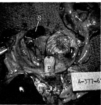
Penis: The penis protruded into the urinary bladder and measured 10.0mm. in length and 5.0mm. in diameter completely covered
by skin and had an external meatus which was stenosed
(See Figure 2). The testicles were situated in the scrotum.
Kidneys: The left kidney was one and a half times normal size and appeared normal on sectioning. The right kidney was absent.
Ureters: The left was twice the normal thickness and was dilated.
The right ureter was absent.
Prostate Gland: Was present, but poorly developed.
Microscopic examination of the penis revealed the normal structures of the penis and an abscence of the urethral canal. Only buds of ure- thral squamous epithelium were present, which extended the entire penis to the stenotic meatus. At the base of the penis the skin fused with the bladder mucosa and the normal squamous egitheliumn of the skin fused with transitional epithelium of the bladder.
Anatomical diagnosis was congenital invagination of the penis into the lumen of the urinary bladder, recto-vesical communication, imper- forated anus, atresia of the penile urethra, congenital abscence of the right kidney and ureter, hydroureter left side, and widely patent foramen ovale and ductus arteriosus with hypertrophy of the right ventricle.
DISCUSSION
Imperforated anus is frequently associated with other malformations of the genito-urinary tract as in the case presented. The terminal rectum opened into the urinary bladder as did the sigmoid colon. The above defects usually result from abnormal down growth of the urore- ctal septum.
Although this case presented numerous congenital malformations, the most unusual finding was the presence of the penis within the urinary bladder.
REFERENCES
1) POTTER Edith L.: p. 392, Pathology of the Fetus and the New Born, 2nd Edition. The Year Book Publishers, Chicago.
2) STOWERS, Daniel: p. 676, Pediatric Pathology, 2nd Edition. The Williams and Wilkins Co., Baltimore.
Fig. 1. Photograph shows abscence of penis and edematous scrotum.
Fig. 2. Photograph shows: P, invaginated penis into the urinary bladder; R, rectum; S, sigmoid
colon. , Note abscence of right kidney and
ui eter.