Acta Med. Nagasaki 36:206-212
Effects of Low Dose Radiation on Mammals 1
Yutaka OKUMURA, Mariko MINE
and Masao KISHIKAWA
Scientific Data Center for Atomic Bomb Disaster, Nagasaki University School of Medicine,
Sakamoto-machi, Nagasaki 852, Japan
Received for publication, June 15, 1991
SUMMARY : Radiation has been applied widely to clinics, researches and industries nowadays. Irradiation by atomic bomb produced many victims in Hiroshima and Nagasaki. Radiation effects on animals and human belings have been reported extensively, especially at a dose range of high amount of radiation. As radiation effects at low dose have not been well studied, it is believed that even a small amount of radiation produces hazardous effects. However, it might not be true. Beneficial effects of a low dose of radiation are summarized here.
INTRODUCTION
Radiation is a useful too in clinics, researches and industries. However, it is hazardous when its large amount is exposed to human beings.
It has been thought that even a small amount of radiation produces damages in proportion to its amount. However, we don't know how a low dose of radiaton affects human beings. The beneficial effects of low dose radiation on mam- mals are summarized here.
IMMUNE RESPONSE
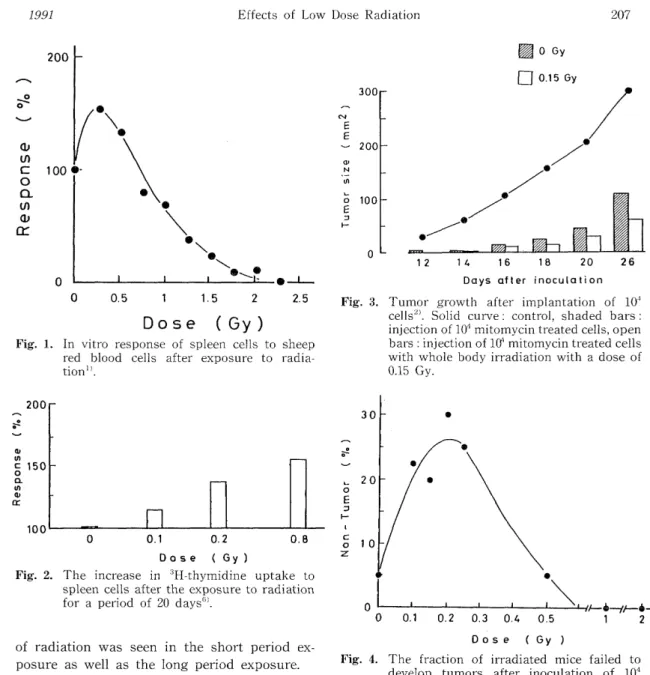
The immune suppressive effects of radia- tion have been known when a large amount of radiation is exposed. However, radiation acts as immune augmentation when a small dose of radiation is exposed" 3'. In vitro response of spleen cells to sheep red blood cells after ex- posure to radiation is shown in Fig. 1." The number of plaque-forming cells is indicated as
a percentage of the control against radiation dose. Low doses of radiation, 0.25 to 0.5 Gy, showed augmented response. Above 0.75 Gy, the response was reduced less than control.
This augmentation might be due to injury of highly radiosensitive suppressor T cells or their subpopulation. It can be said that the low dose of radiation results in an enhancement of the immune mechanism leading to increased antibody formation. When radiation exposure is performed with a low dose rate for a extend- ed period of time, it become clear whether cell delation and radiation damage are accumula- tive, or whether homeostatic adaptation to radiation is generated. Radiation exposure to mice at a low dose rate was extended for a period of 25 days''. Proliferative response of spleen cells from irradiated mice was analyz- ed by phytohemagglutinin stimulation. The in- crease in 3H-thymidine uptake is shown in Fig.
2. Radiation exposure with a dose to 0.8 Gy
This paper was presented at the Japan-China Symposium on the Effects of Radiation in Human Beings, held on June 20, 1990 at Beijing, China.
during a period of 20 days increased prolifera- tive response of spleen cells. Stimulative effect
Fig. 1. In vitro response of spleen cells to sheep red blood cells after exposure to radia-
tion".
Fig. 3. Tumor growth after implantation of 104 cells". Solid curve : control, shaded bars : injection of 104 mitomycin treated cells, open
bars : injection of 10' mitomycin treated cells
with whole body irradiation with a dose of
0.15 Gy.
Fig. 2. The increase in 3H-thymidine uptake to spleen cells after the exposure to radiation
for a period of 20 days".
of radiation was seen in the short period ex- posure as well as the long period exposure.
TUMOR GROWTH
Irradiation with a low dose augments the immune function of animals and affects sup- pression of the grpwth of transplanted tumors2'.
A dose of 0.15 Gy was exposed to whole body of mice and 104 mitomycin-treated tumor cells were injected subcutaneously into mice. After 21 days, mice received 104 viable tumor cells.
The tumor size after the inoculation of the viable tumor cells is shown in Fig. 3. A solid curve with closed circles is of control. Shaded bars are of mitomycin-treated cells. Open bars are
Fig. 4. The fraction of irradiated mice failed to develop tumors after inoculation of 104
tumor cells 3).
of mitomycin-treated cells with whole body irradiation. ' With 0.15 Gy. By the injection of mitomycin-treated cells, the tumor growth was inhibited, and the irradiation suppressed the tumor growth further. These effects might be caused by the immune augmentation.
The low dose irradiation suppressed the
transplantability of tumors'). Radiation was
exposed to mice just before the inoculation of
104 tumor cells. The exposure with low doses,
from 0.1 Gy to 0.3 Gy, resulted that 20 % to 30 %
Fig. 5. Tumor dose 50, TD50, of mice irradiated with a dose of 0.1 Gy"'
of mice failed to develop tumors as seen in Fig.
4. This low dose augmentation is less pro- nounced in animals that have undergone sple- nectomy. Very radiosensitive suppressor T cells might be implicated in this phenomenon.
The tumor transplantability was assayed by tumor dose 50 %, or TD50, which indicates the number of tumor cells forming a tumor in transplanted mice in 50 %1". The larger TD50 means the more difficulty of tumor formation.
Irradiation with a dose of 0.1 Gy increased TD5o value than nonirradiated control (Fig. 5). This means that a low dose suppressed the trans- plantability of tumor cells in mice. The result is associated with the suppression of the tumor transplantation which is shown in the above study.
It was examined whether the low doe affected radiosensitivity or tumor cells growing in mice"
104 tumor cells were transplanted into mice, and after 12 days of the transplantation, a dose of 0.1 Gy was exposed to the whole body of the tumor bearing mice. The radiosensitivity after 12 hours of irradiation is show in Fig. 6. Solid curve with solid circles in figure is of control, nonirradiation. The curve has a steep decreas- ing part of the surviving fraction at a low dose region, and a tail part at a high dose region.
The steep part corresponds to the surviving fraction of aerobic cells in tumors, and the tail part dose to that of hypoxic cells. A dotted curve with open circles is of tumor bearing mice irradiated with the dose of 0.1 Gy. The initial
Fig. 6. Radio sensitivity of tumor cells in mice after the whole body irradiation with a dose of
0.1 Gy'"
decreasing part was the same of the control.
However, the tailing part of the surviving fac- tion was lower than the control. This means irradiation changed immune function resulting
Fig. 7. The incorporation of 3H-thymidine into spleen cells isolated from tumor bearing mice by the stimulation by mitogens"'. Con-A:
concanavalin-A, PHA : phytohemagglutinin, IL-2: interleukin 2,
MLR : mixed lymphoid reaction
in as if the decrease in the fraction of hypoxic cells.
Irradiation with the dose of 0.1 Gy changed immune response assayed by the stimulation by mitogens"'. Fig. 7 shows the incorporation of 3H-thymidine into spleen cells isolated from tumor bearing mice in the stimulation by concanavalin-A (Con-A), phytohemagglutinin (PHA), interleukin 2 (IL-2) and mixed lymphoid reaction (MLR). The percentage of the incor- poration into spleen cells from nontumor bear- ing mice was indicated. Shaded bars are of tumor bearing mice without irradiation, and open bars are of tumor bearing mice for 0.1 Gy whole body irradiation. In any stimulation, irradiation with the dose 0.1 Gy augumented immune response.
Table 1. Mean Survival time of LAF, mic'
Sex Control Irradiated Difference
days days days
Male 684 ± 14 783 ± 14 99
Female 803 ± 16 820 ± 18 17
SURVIVAL TIME
Mice were irradiated with a low dose rate, 0.14 mGy per hour, every days for 8 hours a day until mice die'. And the mean survival time was examined (Table 1). For male mice, the mean survival time was 684 days for control and 783 days for irradiated mice. The mean survival time was increased by 99 days by irradiation.
However, for female mice, the mean survial time was 803 days for control and 820 days for irradiated mice, the difference was 17 days. This
difference was not significant statistically. Male mice would be affected by the environmental conditions such as irradiation, although female mice would not.
CHROMOSOME ABERRATION
Chromosome aberration of human lympho- cytes was produced by the exposure with a dose of 1.5 Gy. The exposure with a low dose of 0.01 Gy before 1.5 Gy irradiation decreased the chromosome aberration 12'. Fig. 8 shows the decrease of chromosome aberration with the interval between two doses, 0.01 Gy and 1.5 Gy.
This decrease in the chromosome aberration
might be adaptive response to irradiation. A
low dose might change the structure of chro-
mosome, and this change suppreassed the chro-
mosome aberration induced by successive ir-
radiation.
Fig. 8. The decrease of the chromosome aberration irradiation with a low dose"'. The time
interval between a dose of 0.01 Gy and a
test dose of 1.5 Gy is indicated on abscissa.
A-BOMB SURVIVORS
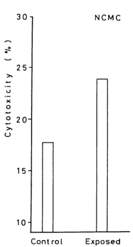
Cellular immune functions were analyzed among victims who survived the atomic bomb in Hiroshima and who lived in the United States of Americas' Radiation doses of the exposed group were from 0.01 Gy to 0.5 Gy. Lymphocytes were isolated from the peripheral blood of those individuals, and natural cell-mediated cyto- toxicity (NCMC) was assessed and the results are shown in Fig. 9. The difference of cy- totoxicity was significant statistiaclly. Other immune function was analysed by mitogen responsr to phytohemagglutinin and to allo- geneic lymphocytes and interferon production.
Although the same trend of the increase was observed, the differences were not significant statistically. The same study has been per- formed on the atomic bomb survivors living in Hiroshima". But the study did not confirm this difference. The different environmental con- ditions between the United States and Japan
Fig. 9. Natural cell-mediated cytotoxicity of A-bomb survivors".
might have affected the immune function.
The rate of death from leukemia in atomic bomb survivors in Nagasaki and Hiroshima is shown in Fig. 107'. A solid curve is for Hiroshima, and a dotted curve is for Nagasaki.
The death rate increases with dose for both cities, although the rate for Hiroshima was higher than that for Nagasaki. From this figure it is clear that there is a threshold dose before the rate increases with dose. Above the threshold dose, the increase in death rate is observed. Below the threahold the rate is nearly the same or smaller than control.
It is widely belived that cancer death is observed even at a low dose range in propor- tion to radiation dose. Howere, the threshold dose of cancer mortality analyzed for the atomic bomb surviors is observed as shown in Table 2. The threshold dose was observed in many cancer deaths. From the table, we can under- stand that thre is no increase in the inci- dence of cancer death by irradiation under the threshold dose in some cancer.
Table 2. Threshold of Cancer Mortality"
Cancer Nagasaki Hiroshima
Gy Gy
Leukemia 0.36 0.12
Lung 0.28 ND
Breast 0.08 ND
Colon 0.54 0.31
Stomach ND 0.12
NT)- Nnt detected
Fig. 10. The Rate of death of leukemia in A-bomb survivors in Nagasaki and Hiroshima".
The risk of death, or the ratio of death rate to that of control, for male survivors is shown in Fig. 1110). For total death, the risk was about unity for the dose range examined. The risk of cancer increased with dose. We confirmed that the irradiation causes cancer. However, the risk of noncancer disease decreased to 0.65 around 1 Gy of radiation dose, and this decrase was significant statistically. Radiation from atomic bombing decreased the mortality from noncancer disease at a low dose range. This beneficial effect of low dose radiation is called radiation hormesis.
In conclusion, it can be said that animal and human beings have systems to respond to the low dose radiation. One of main response is of immune system. And the stimulation by the low dose radiation resulted in the low martality
Fig. 11. The risk of death for male A-bomb survivors : total death, cancer and non-cancer diseases'°
from noncancer disease for male survivors. The low dose of radiation is not necessarily harmful, but rather beneficial under certain conditions.