1 Dokkyo Journal of Medical Sciences
42(1):1〜10,2015
Mechanism of Airway Remodeling Induced by Repeated Inhalation of Methacholine in a Mouse Model of Asthma
Ryosuke Souma M.D., Kumiya Sugiyama M.D., Naoto Fueki M.D., Kentaro Nakano M.D., Hideyuki Satoh M.D., Hironori Sagara M.D., Yasutsugu Fukushima M.D.
Department of Respiratory Medicine Dokkyo Medical University Koshigaya Hospital Conflict of interest:The authors have declared that they have no conflicts of interest.
SUMMARY
Background:Increased severity of asthma is contributed by airway tissue remodeling, which may be as- sociated with chronic allergic inflammation. A recent study revealed the potential capacity of repeated bron- choconstriction, e.g. induced by a muscarine agonist, methacholine(Mch)challenge, to involve in airway re- modeling, even though allergic inflammation is not implicated. We have evaluated the influence of repeated bronchoconstriction induced by Mch inhalation on airway remodeling in a murine model of asthma and have examined its machanisms.
Methods:Mice were immunized with ovalbumin(OVA), and consequently, challenged by either daily OVA inhalation(the OVA group;a model of asthma with allergic inflammation)or daily Mch inhalation(the Mch group;a model of asthma without allergic inflammation). Lung tissues were obtained and were evalu- ated histologically after 5, 10, and 15 consecutive inhalation challenges of both OVA and Mch.
Results:Eosinophilia in the airway observed only on the OVA group. Subepithelial collagen-band thick- ness increased also in the OVA group(p<0.01)after 15 challenges, but not in the Mch group. Significant increase in thickness of airway smooth muscle layer and the number of goblet cells were revealed in both the OVA and Mch group after 10(p<0.05 and p<0.01, p<0.01 and p<0.05, respectively, for the compari- son of the two challenge groups with the control group)and 15 challenges(p<0.05 and p<0.01, both p<
0.01, respectively, for the comparison with control), further, all these measurements were greater in the OVA group than in the Mch group after both 10 and 15 challenges(both p<0.05 and p<0.01, respective- ly). An increase in mast cell counts within the airway wall was shown in the OVA group after 10 challenges
(p<0.01 compared with control), not in the Mch group at all. Epithelia expression of transforming growth factor b (TGF-b)increased in both challenge groups after 15 challenges(both p<0.05 compared with control), and was higher than in Mch(p<0.05).
Conclusion:Repeated Mch inhalation may induce airway remodeling, while comparatively mild, poten- tially resulting in progressive severity of asthma. The results implicate that the potential risk associated with Mch challenge should be considered.
Key Words: airway remodeling, animal model, asthma, eosinophils, methacholine
Original
Received October 10, 2014;accepted October 31, 2014 Reprint requests to:Ryosuke Souma M.D.
Department of Respiratory Medicine Dokkyo Medical University Koshigaya Hospital 2-1- 50 Minami-Koshigaya, Koshigaya, Saitama 343-8555, Japan
Ryosuke Souma
INTRODUCTION
Asthma is a chronic inflammatory disease of the air- ways, characterized by airway hyperresponsiveness
(AHR)and reversible airflow limitation caused by bronchoconstriction and is associated with proliferative structural changes of the airway walls, referred to as airway remodeling1〜4). Whereas, these structural changes contribute to the AHR and the limitation of reversible airflow obstruction, and consequently lead to an increase in severity of the disease in asthmatic pa- tients. Airway remodeling represents structural chang- es of the airways, such as subepithelial fibrosis, hyper- trophy and hyperplasia of airway smooth muscle mass, and goblet cell proliferation, and has been considered a consequence of eosinophilic inflammation, i.e. allergic inflammation, in the airways, induced by inhaled-aller- gen challenge in atopic asthma5).
Recently a study has demonstrated that repeated in- halation of methacholine(Mch), a muscarine agonist, which causes bronchoconstriction without eosinophilic inflammation, possibly induce the similar structural changes of the airways6). To develop our understand- ing of the pathophysiology of the airway remodeling in asthma should be most effective measures for investi-
gating potential targets for therapeutic intervention.
Accordingly, we examined the influence of repeated inhalation of Mch on pathology and pathogenesis of the airway disease by means of employing a mouse model of asthma. The results have clarified whether repeated Mch inhalation, namely recurrent bronchoconstriction without non-allergic inflammation, can lead the similar structural modification of the airways, i.e. airway re- modeling, in asthma, and therefore discussed the po- tential mechanisms of forming that airway remodeling.
MATERIAL and METHODS
Animals and Study designMouse models of asthma were established from OVA-sensitized mice according to methods reported previously with some modifications7〜10). Male BALB/c mice(Clea, Japan)of 8 weeks of age were immunized by intraperitoneal(IP)injected with 8 mg ovalbumin
( OVA;Sigma-Aldrich, St. Louis, MO)in 4 mg Al
(OH)3 gel(alum;Sigma-Aldrich)on days 0 and 7
(Figure 1), and was consecutively challenged by 20- min daily inhalation of 1 mg/ml OVA for 5, 10, or 15 consecutive days(OVA group). To induce bronchocon- striction, as mechanical stress alone, without allergic inflammation, mice were challenged by 15-min daily
2 DJMS
Figure 1 Protocols for sensitization and challenge
Closed arrows indicate inhalation(IH), gray arrows indicate intraperitoneal administration, and open arrows indicate sacrifice. N=21 for each group and n=7 for each measurement.
Airway remodeling by methacholine inhalation
inhalation of acetyl-b-methylcholine chloride, metha- choline(Sigma-Aldrich)(Mch group)to establish an inflammation-independent mechanical stress asthma model. The control group received intraperitoneal sa- line injections instead of OVA-alum on days 0 and 7, and inhaled saline daily for 20 min for 5, 10, or 15 con- secutive days. For inhalation, mice were placed in sealed chambers and drugs were aerosolized by an ul- trasonic nebulizer(Omron, Kyoto, Japan). On days 19, 24, and 29(after 5, 10, and 15 inhalation treatments, respectively), lung tissues were collected for histologi- cal analyses. The above protocol was approved by the Committee of Laboratory Animal Research Center at Dokkyo Medical University(#02-261).
Histological preparation
Lungs were removed, fixed in 4% paraformaldehyde, embedded in paraffin, and sliced into 3-mm thick sec-
tions. Sections were stained histochemically with Han- sel(Eosinostain, Torii Pharmaceutical, Tokyo, Japan)
Elastica-Masson, and alcian-blue and periodicacid- Schiff(PAS)for the detection of eosinophils, the colla- gen band, and goblet cells, respectively, and were stained immunohistochemically with the use of anti- mouse mast cell tryptase mAb(Abcam, Cambridge, UK)and with the use of anti-mouse actin and a- smooth muscle mAb(Sigma-Aldrich), each of which combined with N-Histofine MOUSESTAIN KIT®
(Nichirei, Tokyo, Japan)for the detection of mast cells and smooth muscle mass, respectively. To evaluate the expression of TGF-b, tissues also were stained with anti-mouse TGF-b mAb(Abcam), combined with the VECTASTAIN Elite ABC Kit®(Vector Laboratories, CA), and N-Histofine Simple Stain DAB solution®
(Nichirei)
42(1) (2015) 3
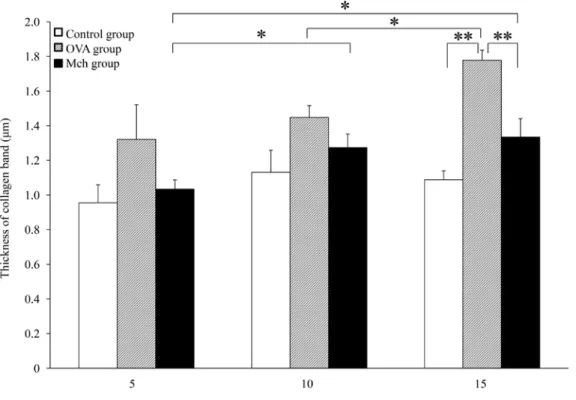
Figure 2 Expansion of the airway collagen band in the OVA group but not in the Mch group Change in collagen band thickness in OVA-injected/OVA-boosted(OVA group)mice, OVA- injected/methacholine(Mch)-challenged mice(Mch group), and saline-injected control group mice after 5, 10, or 15 challenges. The allergic inflammation model(OVA group)showed greater collagen band thickness after 15 challenges than either the Mch or control groups. **p<0.01 and
*p<0.05. Significant differences by number of challenges were observed between 10 and 15 challenges in the OVA group(p<0.05), between 5 and 10 challenges in the Mch group(p<
0.05), and between 5 and 15 challenges in the Mch group(p<0.05). N=21 for each group, and n
=7 for each measurement. Data expressed as mean ± SEM.
Ryosuke Souma
RESULTS
Airway remodeling by methacholineCollagen band thickness, which is a marker of in- flammatory fibrosis, increased progressively as a fre- quency of OVA challenge( Fig. 2). Collagen band thickness increased significantly in the OVA group be- tween 10 and 15 challenges(1.45±0.07 mm vs. 1.78±
0.06 mm, p<0.05), while collagen band thickness in Mch group increased significantly between the five and ten challenge(1.03±0.05 mm vs. 1.27±0.08 mm, p
<0.05)and between the five and fifteen(p<0.05), but not between the ten and fifteen(1.27±0.08 mm vs.
1.33±0.11 mm). The collagen band was significantly thicker in the OVA group after 15 daily OVA chal- lenges than in either the Mch group after the same number of challenges(p<0.01)or in the control group
(p<0.01). In contrast, the collagen band thickness was no greater in the Mch group compared to the control group(p>0.05). Thus, Mch-induced remodeling was Measurement of airway thickness, collagen band thick-
ness, infiltrating cell numbers, and TGF-b expression Airway and collagen band thicknesses were estimat- ed along the main bronchus. The number of goblet cells in the main bronchus was also counted during es- timation of airway and collagen band thicknesses. The number of infiltrating eosinophils in the main bronchus was estimated by counting Hansel-stained cells and expressed per one millimeter square11). Similarly, the number of infiltrating mast cells was estimated by counting the number of tryptase-positive cells. The expression of TGF-b in trachea was estimated by inte- gration of the immunostaining signal.
Statistical Analysis
Statistical analyses were performed using Microsoft Excel® or JMP®(for the Wilcoxon signed rank test). A P value<0.05 was considered significant. Results are expressed as mean±standard error of the mean
(SEM).
4 DJMS
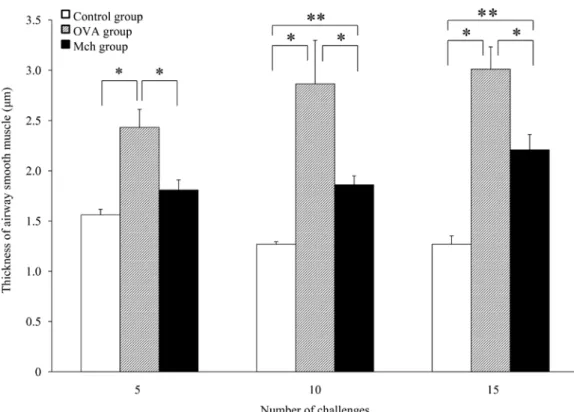
Figure 3 Expansion of the airway smooth muscle layer in both the OVA and Mch group
**p<0.01 and *p<0.05. Both the OVA and Mch groups exhibited smooth muscle layer expansion compared to the control group. Expansion was greater in the OVA mice, indicating more substantial remodeling. No significant differences by number of challenges were observed in any of the groups. N=21 for each group and n=7 for each measurement. Data expressed as mean ± SEM.
Airway remodeling by methacholine inhalation
in the OVA group than the Mch and control groups
(both p<0.01)and greater in the Mch group com- pared to the control group(p<0.01 or p<0.05). Thus, Mch-induced remodeling includes mild goblet cell hy- perplasia.
Immune cell infiltration
The numbers of eosinophils infiltrated into the air- way was significantly higher in the OVA group at all measurements(Fig. 5;p<0.01 vs. Mch and control groups), but there was no effect of challenge number.
In contrast, no significant eosinophil infiltration was ob- served in the Mch group as cell numbers were no higher than in the control group. The numbers of mast cells infiltrated into the airway(Fig. 6)was also sig- nificantly higher in the OVA group compared to the Mch group after 5 and 10 challenges(5 challenges:4.0
±1.0 cells vs. 1.3±0.4 cells, p<0.05;10 challenges:8.7
±0.7 cells vs. 4.5±1.0 cells, p<0.05)and compared to the control group after both 5 and 10 challenges(p<
not associated with fibrosis.
Airway smooth muscle thickness increased progres- sively with the number of challenges in both the OVA and Mch groups( Fig. 3), although the differences among the three measurements were not significant.
At all measurements, however, airway smooth muscle thickness was greater in the OVA group compared to both the Mch group(p<0.05)and control group(p<
0.05). After 10 and 15 challenges, smooth muscle thick- ness was also greater in the Mch group compared to the control group(p<0.01). Thus, Mch-induced re- modeling appears to include smooth muscle hyperpla- sia and/or hypertrophy.
The OVA group exhibited a significantly higher number of goblet cells(Fig. 4), although these num- bers did not increase significantly with the number of challenges. In contrast, the number of goblet cells did increase progressively with the number of challenges in the Mch group(five vs. ten p<0.01). At all mea- surements, goblet cell number was significantly greater
42(1) (2015) 5
Figure 4 Goblet cell hyperplasia in both the OVA and Mch group
The OVA group showed substantial goblet cell hyperplasia. The Mch group showed more modest goblet cells hyperplasia. **p<0.01 and *p<0.05. No significant differences by number of challenges were observed in the OVA group, but there were significant differences between 5 and 10 challenges(p<0.01)and between 5 and 15 challenges(p<0.01)in the Mch group. N=
21 for each group and n=7 for each measurement. Data expressed as mean±SEM.
Ryosuke Souma
6 DJMS
Figure 5 Eosinophilia in the OVA but not Mch group
The OVA group showed substantial eosinophil infiltration into the airway, while the Mch group did not. **p
<0.01. No significant differences by number of challenges were observed in any of the three groups. N=21 for each group and n=7 for each measurement. Data expressed as mean±SEM.
Figure 6 Mast cell infiltration
The numbers of mast cells increased in the airway of the OVA group compared to the Mch and control groups after 5 and 10 challenges. **p<0.01 and *p<0.05. No significant differences by number of challenges were observed in the OVA group. However, significant differences were observed in the Mch group(5 vs. 10 challenges;p<0.05, 5 vs. 15 challenges;p<0.01)and in the control group(5 vs. 10 challenges;p<0.05, 5 vs.
15 challenges;p<0.01). N=21 for each group and n=7 for each measurement. Data expressed as mean ± SEM.
Airway remodeling by methacholine inhalation
0.01), but not after 15 challenges(7.7±2.1 cells vs. 5.6
±1.1 cells vs. 3.3±0.5). Expression of TGF-b(Fig. 7)
was significantly higher in the OVA group compared to the control group after 10 challenges(22.7±10.4 vs.
3.7±1.1, p<0.01)and compared to both the Mch group and control groups after 15 challenges(56.4±
17.7 vs. 7.1±0.4 vs. 4.3±0.9;p<0.05 vs. both). TGF- b expression was significantly higher in the Mch group compared to the control group after 15 challenges(p<
0.05). Unlike OVA-associated remodeling, Mch-in- duced remodeling does not include eosinophilia, but does include mild mast cell infiltration and TGF-b ex- pression.
DISCUSSION
Both the subepithelial collagen band and airway smooth muscle layer were expanded significantly by OVA immunization and subsequent booster sensitiza-
tion, verifying successful induction of airway remodel- ing. However, frequent inhalation of the non-inflamma- tory agent MC also induced airway remodeling, albeit more restricted than that induced by OVA. These find- ings support results from patients with asthma and suggest that overuse of Mch may exacerbate asthma severity6).
Airway remodeling induced by Mch may result from two pathogenic pathways, bronchoconstriction-depen- dent stress and Mch-induced inflammation. Airway smooth muscles may become hypertrophied by fre- quent Mch-induced bronchoconstriction even if it were without airway inflammation(stress-induced hypertro- phy). For example, arterial smooth muscles were hy- pertrophied and synthesis of matrix components stimu- lated by mechanical pressure12,13). Actually, the effects of frequent mechanical stress alone have not been con- sidered in airway, because it is difficult to separate
42(1) (2015) 7
Figure 7 Substantial overexpression of TGF-b by the OVA group and more modest overexpression by Mch group
The OVA group expressed substantially higher levels of TGF-b than the control group after 10 and 15 challenges and higher levels than the Mch group after 15 challenges. The Mch group expressed higher levels of TGF-b than the control group after 15 challenges. **p<0.01 and *p<0.05.
Significant differences by number of challenges were observed between 5 and 10 challenges(p<
0.05)and between 5 and 15 challenges(p<0.05)in the OVA group and between 5 and 15 challenges(p<0.01)in the Mch group. N=21 for each group and n=7 for each measurement. Data expressed as mean ± SEM.
Ryosuke Souma
these effects from those of airway inflammation2〜5). In the present study, airway smooth muscles treated with Mch were significantly hypertrophied. In contrast to the OVA group, however, collagen band thickness was not significantly expanded by Mch, indicating that air- way inflammation induces more uniform remodeling than frequent mechanical bronchoconstriction. None- theless, frequent mechanical bronchoconstriction can induce uneven airway remodeling that may impede respiration.
These results have two major implications for the diagnosis and treatment of asthma. First, examination of airway hyperresponsiveness using Mch or acetyl- choline should be limited to use. Namely the airway hyperresponsiveness examination should be performed only for the initial diagnosis of asthma. Second, bron- chodilators like LABA may contribute to inhibit re- modeling. The US-FDA recommendation against regu- lar use except by patients not successfully treated by ICS is based on the view that inhibition of airway in- flammation is the most important treatment goal for asthma medication, and we agre14). Nonetheless, regu- lar use of LABA may lead to prevention airway re- modeling caused by frequent bronchoconstriction.
The other possible Mch-induced remodeling path- way is mimic allergic inflammation. In one study, Mch increased Ki67-positive cell population in both the epi- thelium and the submucosa of asthma patients6). The authors suggested that Mch could damage the airway epithelium and that upregulation of the proliferation regulator, Ki67 is a reparative response. If Mch does cause mimic allergic inflammation, it likely occurs with- out eosinophil infiltration as neither the aforementioned clinical study6)nor our study found a significant in- crease in tissue eosinophil number. In contrast, we did not evaluate the number of Ki67-positive cells. We cannot eliminate the possibility of Mch-induced cellular damage leading inflammation as no study has yet in- vestigated Mch toxicity on isolated airway epithelial cells. It has also been reported that chronic intermit- tent mechanical stress from bronchoconstriction can increase mucin protein expression and goblet cell pro- liferation1 5), consistent with our observation of in- creased numbers of alcian-blue PAS-positive cells. The other possibility is that Mch or mechanical stress on the airway may induce mimic allergic inflammation
though a pathway not involving activation of eosino- phils. In such a case, mast cells could be the principal mediator cell, because Mch enhanced mast cells infil- tration and infiltration increased with the number of challenges16). Mast cells express receptors for IL-3, IL- 5, and granulocyte-macrophage-colony stimulating fac- tor(GM-CSF), and they regulate allergic airway in- flammation together with T-cells17). Also, mast cells activate fibroblasts to produce fibrogenic cytokines such as TGF-b18), thereby contributing to pulmonary fibrosis. Epithelial TGF-b was increased by Mch in both a previous study on asthma patients6)and in our study, and again expression increased with Mch chal- lenge number. Therefore, it appears possible that Mch caused mimic allergic inflammation without eosinophil- ia. However, Mch caused no significant expansion of the collagen band. Further studies are required to dis- tinguish Mch-induced remodeling via inflammation from remodeling via mechanical stress.
Eosinophilic inflammation induced by OVA had the strongest effect on airway remodeling, but frequent in- halation of Mch also induced several remodeling re- sponses without airway eosinophilia. Eosinophilia is im- portant in the pathogenesis of asthma, but may not be essential key factor because mepolizumab, a humanized monoclonal antibody against IL-5 that inhibits airway eosinophilia, did not improve airway hyperresponsive- ness19). Subsequent studies reported conflicting results on the efficacy of mepolizumab for asthma treat- ment20〜25). A more recent study reported that anti- IL-5 receptor a mAb, not anti-IL-5 mAb, inhibited air- way hyperresponsiveness in a mouse asthma model10). Airway smooth muscles express IL-5 receptor-a, and IL- 5 contributes to airway remodeling2 6). Thus, IL-5 may be an important signaling factor for airway remodeling, although the expression of IL-5 was not measured in our study.
In summary, frequent inhalation of Mch may be a risk factor for airway remodeling. Airway remodeling induced by Mch may arise from frequent mechanical bronchoconstriction or mast cell-dependent, eosino- phil-independent mimic allergic inflammation. We rec- ommend that frequent inhalation of Mch should be limited to use. This test should be performed for initial asthma diagnosis but not for regular treatment moni- toring.
8 DJMS
Airway remodeling by methacholine inhalation
Acknowledgment We thank Ms. Kyoko Inui, Ms.
Emi Matsuda, and Ms. Risako Seki for technical assis- tance.
REFERENCES
1) Bousquet J, Chanez P, Lacoste JY, et al:Eosinophilic inflammation in asthma. N Engl J Med 323:1033- 1039, 1990.
2) Bousquet J, Jeffery PK, Busse WW, et al:Asthma.
From bronchoconstriction to airways inflammation and remodeling. Am J Respir Crit Care Med 161:
1720-1745, 2000.
3) Fredberg JJ:Airway smooth muscle in asthma:flirt- ing with disaster. Eur Respir J 12:1252-1256, 1998.
4) Mauad T, Bel EH, Sterk PJ:Asthma therapy and air- way remodeling. J Allergy Clin Immunol 120:997- 1009, 2007.
5) Kumar RK:Understanding airway wall remodeling in asthma:a basis for improvements in therapy?
Pharmacol Ther 91:93-104, 2001.
6) Grainge CL, Lau LC, Ward JA, et al:Effect of bron- choconstriction on airway remodeling in asthma. N Engl J Med 364:2006-2015, 2011.
7) Nakajima H, Iwamoto I, Tomoe S, et al:CD4+ T- lymphocytes and interleukin-5 mediate antigen-in- duced eosinophil infiltration into the mouse trachea.
Am Rev Respir Dis 146:374-377, 1992.
8) Tanaka H, Masuda T, Tokuoka S, et al:The effect of allergen-induced airway inflammation on airway re- modeling in a murine model of allergic asthma. In- flamm Res 50:616-624, 2001.
9) Cheng G, Arima M, Honda K, et al:Anti-interleu- kin-9 antibody treatment inhibits airway inflamma- tion and hyperreactivity in mouse asthma model. Am J Respir Crit Care Med 166:409-416, 2002.
10) Obara K, Sugiyama K, Hirata H, et al:Monoclonal antibody against IL-5 receptor alpha, but not IL-5, inhibits airway hyperresponsiveness associated with airway remodeling. Int Med J 20:579-583, 2013.
11) Kikkawa Y, Sugiyama K, Obara K, et al:Interferon- alpha inhibits airway eosinophilia and hyperrespon- siveness in an animal asthma model. Asia Pac Allergy 2:256-263, 2012.
12) Dobrin PB:Mechanical factors associated with the development of intimal and medial thickening in vein
grafts subjected to arterial pressure. A model of ar- teries exposed to hypertension. Hypertension 26:38- 43, 1995.
13) Leung DY, Glagov S, Mathews MB:Cyclic stretching stimulates synthesis of matrix components by arterial smooth muscle cells in vitro. Science 191:475-477, 1976.
14) FDA Drug Safety Communication:New safety re- quirements for long-acting inhaled asthma medica- tions called Long-Acting Beta-Agonists(LABAs).
U.S. Food and Drug Administration. Feb 18, 2010.
15) Park JA, Tschumperlin DJ:Chronic intermittent me- chanical stress increases MUC5AC protein expres- sion. Am J Respir Cell Mol Biol 41:459-466, 2009.
16) Mark C. Siracusa, Brian S. Kim, Jonathan M. Spergel, et al:Basophils and allergic inflammation. J Allergy Clin Immunol 132:789-801, 2013
17) Dahl C, Hoffmann HJ, Saito H, et al:Human mast cells express receptors for IL-3, IL-5 and GM-CSF;
a partial map of receptors on human mast cells cul- tured in vitro. Allergy 59:1087–1096, 2004.
18) Veerappan A, O’Connor NJ, Brazin J, et al:Mast Cells:A pivotal role in pulmonary fibrosis. DNA and Cell Biol 32:206-218, 2013.
19) Leckie MJ, ten Brinke A, Khan J, et al:Effects of an interleukin-5 blocking monoclonal antibody on eosino- phils, airway hyper-responsiveness, and the late asth- matic response. Lancet 356:2144-2148, 2000.
20) O’Byrne PM, Inman MD, Parameswaran K:The tri- als and tribulations of IL-5, eosinophils, and allergic asthma. J Allergy Clin Immunol 108:503-508, 2001.
21) van Rensen EL, Stirling RG, Scheerens J, et al:Evi- dence for systemic rather than pulmonary effects of interleukin-5 administration in asthma. Thorax 56:
935-940, 2001.
22) Flood-Page PT, Menzies-Gow AN, Kay AB, et al:
Eosinophil’s role remains uncertain as anti-interleu- kin-5 only partially depletes numbers in asthmatic airway. Am J Respir Crit Care Med 167:199-204, 2003.
23) Menzies-Gow A, Flood-Page P, Sehmi R, et al:Anti- IL-5(mepolizumab)therapy induces bone marrow eosinophil maturational arrest and decreases eosino- phil progenitors in the bronchial mucosa of atopic asthmatics. J Allergy Clin Immunol 111:714-719, 2003.
42(1) (2015) 9
Ryosuke Souma
24) Haldar P, Brightling CE, Hargadon B, et al:Mepoli- zumab and exacerbations of refractory eosinophilic asthma. N Engl J Med 360:973-984, 2009.
25) Nair P, Pizzichini MM, Kjarsgaard M, et al:Mepoli- zumab for prednisone-dependent asthma with spu-
tum eosinophilia. N Engl J Med 360:985-993, 2009.
26) Rizzo CA, Yang R, Greenfeder S, et al:The IL-5 re- ceptor on human bronchus selectively primes for hy- perresponsiveness. J Allergy Clin Immunol 109:404- 409, 2002.
10 DJMS