Nickel (Ni) is contained in various alloys, which are widely used in accessories and biomaterials, and causes inflammation and allergy. However, little is known about the regulation of Ni release from alloys in vivo. In this study, we established a novel in vivo model in which the release of Ni from metal wire could be analyzed quantitatively. A Ni wire (99.9 %; ø0.8 mm×5 mm) was implanted subcutaneously in the dorsum of mice. Ni ions in the skin tissue (diameter : 14 mm) on the wire were extracted and quantified by the fluorometry using Newport Green. The content of Ni ion in the tissue was significantly increased from 8 h. When lipopolysaccharide (LPS, 1 µg) was injected into the same site immediately after the implantation of Ni wire, the content of Ni ions in the tissue was significantly increased. These results suggest that the release of Ni from metal wires in vivo was enhanced by LPS-induced inflammation. The findings were confirmed by in vitro model using a macrophage-like cell line RAW264. RAW 264 cells (1 ×10⁵ cells/mL, 0.2 mL) were seeded on a Ni plate (5 mm square) and incubated for 24 h in the medium containing LPS (0.1, 0.3 and 1.0 µg/mL). LPS enhanced the release of Ni by RAW 264 cells in a concentration-dependent manner. Interestingly, the enhancement was observed only when the cells were attached with Ni plate. The inhibitor of lysosome chloroquine (10 µM) and the V-ATPase inhibitor bafilomycin A1 (1 nM) but not the Na+/H+ exchanger (NHE) inhibitor amiloride inhibited partially the elution of Ni by unstimulated RAW264 cells. In contrast, chloroquine, bafilomycin A1 and amiloride potently inhibited that induced by the LPS-stimulated RAW264 cells. These results suggested that LPS-stimulated RAW264 cells caused the release of Ni ions via the release of lysosome and via the activation of V-ATPase and NHE. In conclusion, we established the novel models for the release of Ni from metals in vivo and in vitro and demonstrated that the activation of inflammatory cells apparently enhanced the release of Ni.
Elution of Ni ions from materials and enhancement by inflammation
Noriyasu Hirasawa
Laboratory of Pharmacotherapy of Life- style Related Diseases Graduate School of Pharmaceutical Sciences, Tohoku University
1.緒 言
肌に直接接する装飾品に含まれるニッケルは、汗で溶出 されやすくアレルギーや炎症を誘発しやすい金属である。
ひとたび金属アレルギーを発症すると、ニッケル以外の金 属にも過敏になることが知られており、種々の装飾品によ り発赤、かゆみなどが生じて、装飾品を身につけることが 制限される。また金属イオンにより強い接触皮膚炎を起こ し、色素沈着を誘発する場合もあり、コスメトロジーの観 点からも大きな問題となっている。ニッケルが金属アレル ギーを最も引き起こしやすい金属であることは周知の事実 であり、そのアレルギー誘発機構については多くの報告が なされてきた1, 2)。従来、ニッケルは組織蛋白に結合しハ プテンとなると考えられてきたが、最近ニッケルは直接リ ンパ球上のMHC 複合体に結合して、細胞を活性化するこ とが明らかにされ3)、組織蛋白との結合による抗原の形成 は必ずしも必要ではないことも示唆された。一方、金属ア レルギーの初期段階はニッケルを含む装飾品や金属医用材 料からのニッケルイオンの溶出である。しかしこのニッケ
ルが溶出される分子機構や炎症が生じて金属周囲組織の環 境が変化した場合のニッケル溶出の変化の定量的な解析は これまでなされていない。その最も大きな理由は優れた実 験モデルがなかった点にある。
申請者はニッケル線をマウス背部皮下に埋稙させる新規 モデルを開発し4)、ニッケルによる炎症反応の誘発とニッ ケルの溶出を定量的に解析することを可能にした。本研究 は、本モデルを用いて、ニッケル線からのニッケル溶出が、
その周囲で感染を模倣した炎症を誘発したときに増大する ことを示した。また、炎症細胞を用いた細胞培養系でのニ ッケルイオン溶出評価系も確立し、ニッケル溶出の分子機 序について解析した。
2.実験方法
2 - 1.ニッケル線誘発炎症モデルの作製4)
Diethyl ether麻酔下、マウス(22−25g)の背部皮下に 移植針(13G)を用いて、UV照射滅菌処理したニッケル線
(99.98% ニッケル, ニラコ)(φ0.8 mm× 5mm)を埋稙 した。その直後に、lipopolysaccharide(LPS, 5µg/mL)
0.2mLをニッケル線近傍に皮下投与した。対照群として salineを0.2 ml皮下投与した。
2 - 2.組織中のニッケル量の測定
一定時間後に、エーテル麻酔下マウスを脱血死させ、ニ ッケル線を中心に、皮打ち抜き用パンチ(内径1.4 cm)で 皮膚(表皮、真皮及び皮下組織)を打ち抜き、ニッケル 東北大学大学院薬学研究科生活習慣病治療薬学分野
平 澤 典 保
線を除いた後、皮膚をEppendorf tube(1.5mL)に入れ た。その後、Milli Q 水500µL加えて、ミンスし、4℃
で24時間ニッケルイオンを抽出した。この上清を希釈し、
Newport Green DCF dipotassium saltを最終濃度 1µM となるように加え、分光蛍光光度計F-2000(日立ハイテク)
を用いてEx 505nm/Em535nmの蛍光を測定した。
2 - 3.マウスマクロファージ様細胞株 RAW-264 細胞に よるニッケル溶出の測定
ニッケル板(99.98% , 厚さ0.05 mm, ニラコ;5×5 mm)
を96 well plateの1wellにつき1枚ずつ設置し、RAW264 細 胞(1.0−5.0×10⁵cells/ml 10 %(v/v)FBS-EMEM)を 200µlずつ播種し、5% CO2存在下37℃で一定時間培養 した。ニッケル板非接着群は96-well plateにあらかじめ RAW264細胞を播種し、2時間後にニッケル板を細胞と 接触しないように斜めに設置した。
2 - 4.薬物処理
細 胞 を 播 種 し た 2 時 間 後、chloroquine5)( 最 終 濃 度 10µM)、amiloride6)( 最 終 濃 度 100µM)、 あ る い は bafilomycin A17)
(最終濃度 1nM)を加え1時間前処理し た後、LPS(最終濃度1µg/mL)で一定時間刺激した。
2 - 5.統計処理
棄却検定はTompson’s F-testにより行った。有意差検 定はStudent-Newman-Keuls testにより行った。
3. 結 果 3 - 1.ニッケル線誘発炎症
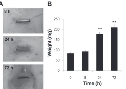
マウス背部皮下にニッケル線を埋稙し、ニッケル線によ り生じる炎症を観察した。8時間後ではニッケル線周囲の 組織において、弱いながらも発赤が認められ、24時間以 後においては出血を伴う強い炎症が認められた(Fig. 1A)。
また、浮腫のパラメータとして皮膚重量を測定した結果、
8時間後の皮膚重量は100mg前後であり有意な増加は認 められなかったが、24時間後においては170−180mg、72 時間後においては200−220mgとなり、有意な増加が認め られた(Fig. 1B)。
3 - 2.ニッケル線表面の観察
埋稙して72時間後のニッケル線の表面を走査型電子顕 微鏡を用いて観察した。未処置のニッケル線の表面と比較 して埋稙後のニッケル線の表面は腐食が確認され、ニッケ ルが溶出していることが示唆された(Fig. 2)。
Figure 1. Ni wire-induced inflammation
(A) A Ni wire was implanted subcutaneously in the dorsum of each mouse. The mice were sacrificed 8, 24 and 72 h after the implantation, and the skin around the wire was photographed.
(B) The skin tissue (diameter: 14 mm) on the wire was excised and the weight of tissue was measured. The values are the means of four mice with S.E.M. shown by vertical bars. Statistical significance;
**P<0.01 vs. the 0 h group.
Figure 2. Erosion of the surface of Ni wire
The surfaces of untreated and implanted Ni wire (72 h) were analyzed by scanning electron microscope.
3 - 3.Lipopolysaccharide によるニッケル線周囲組織 へのニッケル溶出量の増大
ニッケル線からのニッケル溶出が周囲の炎症反応によ り影響を受けるか明らかにするため、ニッケル線の埋稙 と 同 時 に 起 炎 剤lipopolysaccharide(LPS)を 用 い て 炎 症 反応を誘発した場合の、ニッケル溶出量の変化を解析し た。埋稙24時間後のニッケル線周囲組織の切片を作製 後、Hematoxylin-Eosin染色し組織形態を観察した。ニッ ケル線の埋稙によりニッケル線周囲組織の皮膚の肥厚及び 好中球やマクロファージなどの炎症細胞の浸潤が認めら れ、LPSを投与した場合はさらに炎症細胞の浸潤が増大し た(Fig. 3A)。このような条件下でニッケル溶出について 比較したところ、埋稙8時間後及び24時間後においてLPS を注射した群(LPS群)では、vehicle(Saline)を注射した 場合(Saline群)に比べてニッケル溶出が有意に増加した
(Fig. 3B)。従ってLPSで炎症反応を誘発すると、ニッケ ル線からのニッケル溶出が促進することが明らかになった。
3 - 4.LPS 刺激マウスマクロファージ様細胞株 RAW264 によるニッケル板からのニッケル溶出の促進
LPSによる炎症反応によりニッケル溶出が促進する現象 がin vitroにおいても認められるか明らかにするため、マ
ウス腹腔由来マクロファージ細胞株であるRAW264細胞 を用い、培養細胞によるニッケル溶出を検討した。96- well plateの各wellに5mm角のニッケル板を置き、10%
FBSを含むEMEM培地を加えて24時間incubateした場合 には顕著なニッケル溶出は見られなかった。しかし、ニッ ケ ル 板 上 にRAW264細 胞(1.0及 び3.0×10⁵ cells/mL)を 播種した場合には細胞数依存的にニッケル溶出が促進した。
さらにRAW264細胞をLPSで刺激したところ、ニッケル の溶出量が有意に増加した(Fig. 4A)。
ま た 細 胞 播 種 数 を1.0×10⁵cells/mLと し、RAW264細 胞を各濃度のLPSで刺激したところ、LPS濃度依存的に ニッケルの溶出量が増加した(Fig. 4B)。以上の結果から、
RAW264細胞によりニッケル板からのニッケル溶出は促 進され、この溶出作用はLPS刺激により細胞を活性化す ることにより著しく促進されることが明らかになった。
3 - 5.LPS 刺激 RAW264 細胞の培養上清の pH 解析 LPS刺激後のRAW264細胞の培養上清中のpHを解析し た。その結果、培養上清中のpHは細胞数依存的に低下す る傾向を示したものの、pH値7.2−7.5程度とその変動は ごくわずかであった(Fig. 5A)。また、LPS刺激による培 養上清のpH値の変化は認められず(Fig. 5B)、培養上清
Figure 3. Increase in release of Ni by lipopolysaccharide- induced inflammation
(A) Ni wire was implanted subcutaneously in the dorsum of each mouse. Lipopolysaccharide (LPS) was injected into the same site immediately after the implantation. Twenty- four hours after the implantation, the mice were sacrificed and the skin on the wire was dissected. Paraffin section of the tissue was prepared and stained with hematoxylin- eosin.
(B) The skin tissue (diameter: 14 mm) on the wire was excised 8, 24 and 72 h after the implantation of Ni wires and Ni in the tissue was quantified by the fluorometry. The values are the means of five mice with S.E.M. shown by vertical bars. Statistical significance; *P<0.05, **P<0.01 vs. the Saline group at the corresponding point.
Figure 4. Enhancement of elution of Ni ions from Ni plate by lipopolysaccharide-stimulated RAW 264 cells
(A) RAW 264 cells (1 and 3×10⁵ cells/mL, 0.2 mL) seeded on a Ni plate (5 mm square) and incubated for 24 h in the presence (closed columns) or absence (open columns) of LPS (1 µg/mL). The values are the means of five samples with S.E.M shown by vertical bars. Statistical significance; *P<0.05 vs. the corresponding unstimulated group.
(B) RAW 264 cells (1 ×10⁵ cells/mL, 0.2 mL) seeded on a Ni plate (5 mm square) and incubated for 24 h in c o n t a i n i n g L P S ( 0 . 1 , 0 . 3 a n d 1 . 0 µ g / m L ) . T h e concentration of Ni in the supernatant was determined after the 24 h incubation. The values are the means of five samples with S.E.M. shown by vertical bars. Statistical significance; *P<0.05 vs. the unstimulated group.
中のpHはLPS刺激の有無に関わらず、一定に保たれてい ることが明らかになった。従って、LPSによるニッケル溶 出の増大は、培養上清のpHが低下したためではないこと が示唆された。
3 - 6.LPS 刺激 RAW264 細胞によるニッケル溶出促 進の経時変化
RAW264細胞をニッケル板上に播種し、LPSで刺激した 時のニッケル溶出の経時変化を解析した。Mediumのみで incubateした場合、及びニッケル板上でRAW264細胞を 培養した場合では、培養上清中のニッケル濃度は、24時 間経過後もわずかに増加した程度であった。しかし、LPS でRAW264細胞を刺激した場合には培養液のニッケル濃 度は播種4時間後から増加し始め、8時間以後ではLPS で刺激していない群と比較して有意に増加した(Fig. 6)。
3 - 7.RAW264 細胞によるニッケル溶出促進における ニッケル板と細胞間の接着
RAW264細胞をLPSで刺激すると、ニッケル溶出が促 進するメカニズムとして細胞とニッケル板の接着部位の微 小環境において酸性化が生じニッケルが溶出する可能性が 考えられる。この点を明らかにするために、RAW264細
胞をニッケル板上に播種しニッケル板と細胞を接着させ た場合、あるいはニッケル板と細胞を接着させずに培養し た場合のニッケル板からのニッケル溶出を比較した。そ の結果、LPSで刺激していない条件下ではRAW264細胞 とニッケル板との接着及び非接着に関わらず、RAW264 細胞のニッケル溶出量に差は認められなかった。しかし、
RAW264細胞をニッケル板上に播種しニッケル板に接着 させた場合では、RAW264細胞をLPSで刺激すると、ニ ッケル溶出が有意に促進したのに対し(P < 0.01)、ニッケ ル板がRAW264細胞と接着しない条件下においては、LPS で刺激を加えてもニッケル溶出の促進は認められなかった
(Fig. 7)。従ってRAW細胞におけるLPS刺激によるニッ ケル溶出促進作用の発現には、ニッケル板と細胞との接着 が必要であることが示唆された。
3 - 8.リソソーム阻害薬及びプロトン輸送阻害薬の RAW264 細胞によるニッケル溶出抑制作用
RAW264細胞によるニッケル溶出効果のメカニズム を明らかにするため、LPS非存在下リソソーム阻害薬 chloroquine(10µM)、Na+/H+ exchanger(NHE) 阻 害 薬 amiloride(100 µM)、vacuolar-type(H+)-ATPase
(v-ATPase)阻 害 薬bafilomycin A1(1nM)の 各 薬 物
Figure 5. pH in the culture medium of lipopolysaccharide-stimulated RAW 264 cells
(A) RAW 264 cells (1 and 3×10⁵ cells/mL, 0.2 mL) seeded on a Ni plate (5 mm square) and incubated for 24 h in the presence (closed columns) or absence (opened columns) of LPS (1 µg/mL).
The pH of the culture medium was determined with pH meter. The values are the means of five samples with S.E.M shown by vertical bars.
(B) RAW 264 cells (1×10⁵ cells/mL, 0.2 mL) seeded on a Ni plate (5mm square) and incubated for 24 h in containing LPS (0.1, 0.3 and 1.0 µg/mL). The pH in the culture medium was determined with pH meter. The values are the means of five samples with S.E.M. shown by vertical bars.
F i g u r e 6 . R e q u i r e m e n t o f c e l l a t t a c h m e n t Enhancement of elution of Ni ions from Ni plate by lipopolysaccharide-stimulated RAW 264 cells RAW 264 cells (1×10⁵ cells/mL, 0.2 mL) seeded
on a Ni plate (5 mm square) and incubated for 4, 8, and 24 h in the presence or absence of LPS (1.0 µg/mL). The concentration of Ni in the culture medium was determined. The values are the means of five samples with S.E.M. shown by vertical bars.
Statistical significance; *P<0.05, **P<0.01 vs. the corresponding unstimulated group.
を添加し、これらの薬物の影響を解析した。その結果、
chloroquine添加により非刺激RAW264細胞による24時間 培養でのニッケル溶出は部分的に抑制され、bafilomycin A1も有意差はなかったもののニッケル溶出を抑制する傾 向が認められた。一方、amilorideにはニッケル溶出抑制 効果が認められなかった(Fig. 8)。
Figure 9. Inhibition of elution of Ni ions from Ni plate by lipopolysaccharide-stimulated RAW 264 cells by chloroquine, amiloride and bafilomycin A1
RAW 264 cells(1×10⁵cells/mL, 0.2 mL) were seeded on a Ni plate(5 mm square)and incubated for 1 h in the medium in the presence of chloroquine(CQ ; 10 µM),
amiloride(Ami ; 100 µM)and bafilomycin A1 (Baf ; 1 nM). Then the cells were stimulated with LPS(1.0 µg/mL)
for 8 h. The concentration of Ni in the supernatant was then determined. The values are the means of five samples with S.E.M. shown by vertical bars. Statistical significance;
**P<0.01 vs. unstimulated RAW 264 cells, ##P<0.01 vs.
LPS control.
また、RAW264細胞をLPSで刺激した場合の8時間で のニッケル溶出促進効果に対する効果を同様に解析した。
その結果、chloroquine、amiloride及びbafilomycin A1は いずれもLPSによるNi溶出促進を有意に抑制した(Fig.
9)。
Figure 7. Enhancement of elution of Ni ions from Ni plate by lipopolysaccharide-stimulated RAW 264 cells on Ni plate
(A) Experimental scheme of culture condition of RAW 264 cells.
(B) RAW 264 cells (1×10⁵cells/mL, 0.2 mL) seeded on (attached condition) or under a Ni plate (non- attached condition) were incubated for 24 h in presence or absence of LPS (1.0 µg/mL). The concentration of Ni in the culture medium was determined. The values are the means of five samples with S.E.M. shown by vertical bars. Statistical significance; **P<0.01 vs. the corresponding LPS (-) group.
Figure 8. Effects of drugs on elution of Ni ions from Ni plate by unstimulated RAW 264 cells
RAW 264 cells(1×10 ⁵ cells/mL, 0.2 mL)were seeded on a Ni plate(5 mm square)and incubated for 24 h in the medium containing chloroquine(CQ; 10 µM),
amiloride(Ami ; 100 µM)and bafilomycin A1(Baf ; 1 nM). The concentration of Ni in the supernatant was then determined. The values are the means of five samples with S.E.M. shown by vertical bars. Statistical significance;
**P<0.01 vs. medium alone(None)group, #P<0.05 vs.
unstimulated RAW 264 cells group.
4. 考 察
種々の装飾品に対する金属アレルギーは皮膚の健康を考 える上で,極めて大きな問題である。金属アレルギー誘発 の初期段階として当然ながら金属イオンの溶出が必須であ る。したがって生体内での金属からの金属イオンの溶出を 評価し、その溶出に伴う生体応答を明らかにすることは極 めて重要である。しかしこれまで,生体内において金属溶 出を詳細に解析できる実験系はなかった。そこで私たちは、
ニッケル線をマウス背部皮下に埋稙する炎症モデルを確立 し、ニッケルの溶出を定量的に解析できるモデルを構築し た。
ニッケル線をマウスの背部皮下に埋稙し、LPSにより炎 症反応を誘発するモデルは、金属製のバイオデバイスを外 科的に体内に埋稙した際に感染が生じた状態を模倣してい る。このような条件下で、金属からのニッケル溶出が増大 することが明らかになったことは、医療材用の安全性評価 の上で極めて重要なことである。
炎症細胞の活性化によりニッケル溶出が促進される現象 はin vitroにおいても認められた。すなわち、マウスマク ロファージ様細胞株RAW264細胞がニッケルを溶出する 作用をもつこと、さらに同細胞が起炎物質LPSにより活 性化されると、ニッケル溶出効果が増大することが明らか になった。ニッケルに限らず、金属が腐食しイオンを溶出 する原因として最も一般的なものが金属表面での酸性化で ある。本研究ではLPSでニッケル溶出が促進した場合で も培養液中のpHは低下しなかったことと、ニッケル板上 に播種したRAW264細胞をLPSで刺激するとニッケル溶 出が促進したのに対し、ニッケル板と接着しない条件下に おいては、LPSで刺激を加えてもニッケル溶出の促進は認 められなかったことから、RAW264細胞におけるLPS刺 激によるニッケル溶出の促進作用はニッケル板と細胞との 接着面で生じ、この局所環境においてLPS刺激により酸 性化が生じていることが示唆された。
マクロファージは体内に侵入した異物を除去する機構を 備えており、貪食した分解すべき異物を含むファゴソーム の形成と、細胞由来の様々なタンパク分解酵素を含むリソ ソームを融合しファゴリソソームを形成して消化分解する。
リソソーム内のタンパク分解酵素は至適pHが酸性である ため、リソソーム膜にあるV-ATPaseにより細胞質内プロ トンを積極的に取り込んでいる。Chloroquineは弱塩基性 の化合物で細胞内に取り込まれリソソーム内に取り込まれ 中和する作用がある。無刺激のマクロファージによるニッ ケルの溶出はリソソームを中和するchloroquine及びリソ ソームを酸性化を抑制するV-ATPase阻害薬bafilomycin A1により抑制されたことから、未活性状態のマクロファ ージによるニッケル溶出はリソソームの放出が関与して
いると考えられる。一方、マクロファージがLPS刺激に より活性化すると、細胞膜にあるV-ATPaseやNa+−H+ Exchanger(NHE)などのイオン輸送系が活性化すること が明らかになっている8−9)。Amiloride は細胞膜上に発現 しているNHEの阻害薬であり、bafilomycin A1はリソソ ーム膜上のV-ATPaseと同様に細胞膜上に発現している V-ATPaseも阻害する。今回の実験ではLPSにより活性化 されたマクロファージのNi溶出促進効果をbafilomycin A1
及びamilorideが強く抑制した。以上のことから、マクロ ファージは金属表面ではこれを異物と認識してプロトンを 含むリソソームのエキソサイトーシスにより、またLPSで 活性化するとリソソームの放出に加え細胞膜のV-ATPase やNHEが活性化され、細胞外へのH+放出をさらに増大さ せて著しくニッケル溶出が促進されると考えられる。
5.総 括
本研究において、生体内でのニッケル溶出は炎症細胞の 活性化により促進されることが明らかになった。そのため 装飾品や医用金属材料の安全性評価には従来の試験管内で の耐汗性試験、耐唾液性試験では不十分である可能性が示 唆された。特にLPS刺激したマクロファージ様細胞株を 用いたニッケル溶出系は、金属性装飾品等の安全性評価に 有用であると考えられる。また、コスメトロジーの観点か らは、皮膚のバリア機能をはじめ、健康な皮膚を維持する ことが金属アレルギーの低減につながるものと考えられる。
謝 辞
本研究を遂行するにあたり,ご支援を頂きました財団法 人コスメトロジー研究振興財団に深く感謝いたします。ま た、本研究にご協力くださいました東北大学大学院工学研 究科大津浩教授ならびに成島尚之教授に深く御礼いたしま す。
(引用文献)
1) Basketter, D.A., Briatico-Vangosa, G., Kaestner, W., Lally, C., Bontinck, W.J. (1993) Nickel, cobalt and chromium in consumer products: a role in allergic contact dermatitis? Contact Dermatitis, 28:15-25.
2) Sato, N., Kinbara, M., Kuroishi, T., Kimura, K., Iwakura, Y., Ohtsu, H., Sugawara, S., Endo, Y. (2007) Lipopolysaccharide promotes and augments metal allergies in mice,dependent on innate immunity and histidine decarboxylase. Clin. Exp. Allergy, 37: 743-751.
3) Thierse, H.J., Moulon, C., Allespach, Y., Zimmermann, B., Doetze, A., Kuppig, S., Wild, D., Herberg, F., Weltzien, H.U. (2004) Metal-protein complex-mediated transport and delivery of Ni2+ to TCR/MHC contact sites in nickel-
specific human T cell activation. J. Immunol. 172 : 1926- 1934.
4) Hirasawa, N., Goi, Y., Tanaka, R., Ishihara, K., Ohtsu, H., Ohuchi, K. (2010) Involvement of prostaglandins and histamine in Nickel wire-induced acute inflammation in mice. J. Biomedical Mtaerials Res. Part A, in press.
5) Schneider, P., Korolenko, T.A., Busch, U. (1997) A review of drug-induced lysosomal disorders of the liver in man and laboratory animals. Micros. Res. Tech., 36 : 253- 75.
6) Kleyman, T.R., Cragoe, E.J. Jr. (1988) Amiloride and its analogs as tools in the study of ion transport. J. Membr.
Biol., 105 : 1–21.
7) Bowman, E.J., Siebers, A. and Altendorf, K. (1988)
Bafilomycins: a class of inhibitors of membrane ATPases from microorganisms, animal cells, and plant cells. Proc.
Natl. Acad. Sci. USA 85 : 7972–7976.
8) Brisseau, G.F., Grinstein, S., Hackam, D.J., Nordström, T., Manolson, M.F., Khine, A.A., Rotstein, O.D. (1996) Interleukin-1 increases vacuolar-type H+-ATPase activity in murine peritoneal macrophages. J. Biol. Chem. 271:
2005-2011.
9) Vairo, G., Royston, A.K., Hamilton, J.A. (1992) Biochemical events accompanying macrophage activation and the inhibition of colony-stimulating factor-1-induced macrophage proliferation by tumor necrosis factor-a, interferon-g, and lipopolysaccharide. J. Cell. Physiol., 151:630–641.