T itle
A ssociation between atrial fibrillation, atrial enlargement, and
left ventricular geometric remodeling
A uthor(s )
S eko, Y uta; K ato, T akao; Haruna, T etsuya; Izumi, T oshiaki;
Miyamoto, S hoichi; Nakane, E isaku; Inoko, Moriaki
C itation
S cientific R eports (2018), 8
Is s ue D ate
2018-04-23
UR L
http://hdl.handle.net/2433/230864
R ig ht
©
T e A uthor(s) 2018.; T his article is licensed under a C reative
C ommons A ttribution 4.0 International L icense, which permits
use, sharing, adaptation, distribution and reproduction in any
medium or format, as long as you give appropriate credit to the
original author(s) and the source, provide a link to the C reative
C ommons license, and indicate if changes were made. T e
images or other third party material in this article are included
in the article’
s C reative C ommons license, unless indicated
otherwise in a credit line to the material. If material is not
included in the article’
s C reative C ommons license and your
intended use is not permitted by statutory regulation or exceeds
the permitted use, you will need to obtain permission directly
from the copyright holder. T o view a copy of this license, visit
http://creativecommons.org/licenses/by/4.0
T ype
J ournal A rticle
T extvers ion
publisher
Association between atrial
ibrillation, atrial enlargement,
and left ventricular geometric
remodeling
Yuta Seko
, Takao
Kato
, Tetsuya
Haruna
, Toshiaki Izumi , Shoichi Miyamo
to
,
Eisaku Nakane & Moriaki Inoko
This study investigated the relationship between atrial ibrillation (AF) and left ventricular (LV) geometric patterns in a hospital-based population in Japan. We retrospectively analyzed patients who had undergone simultaneous scheduled transthoracic echocardiography (TTE) and
electrocardiography during . A total of patients who had indings of previous myocardial
infarctions (n = ) and without the data on body surface area (n = ) were excluded from the study.
We calculated the LV mass index (LVMI) and relative wall (RWT) and categorized patients into four
groups as follows: normal geometry (n = ); concentric remodeling (normal LVMI and high RWT,
n = ); concentric hypertrophy (high LVMI and high RWT, n = ); and eccentric remodeling (high
LVMI and normal RWT, n = ). The mean left atrial volume indices (LAVI) were . , . , . , and
. mm/m in patients with normal geometry, concentric remodeling, concentric hypertrophy, and eccentric hypertrophy, respectively. The mean LV ejection fractions (LVEF) were . , . , . , and
. %, respectively, whereas the prevalence of AF was . %, . %, . %, and . % in patients with normal geometry, concentric remodeling, concentric hypertrophy, and eccentric hypertrophy, respectively. In conclusion, the prevalence of AF was increasing according to LV geometric remodeling patterns in association with LA size and LVEF.
Let ventricular (LV) hypertrophy (LVH) may be considered a compensatory efect since increasing LV wall thick-ness reduces LV wall stress1. However, as LVH progresses, it is associated with considerable cardiovascular (CV)
morbidity and mortality2,3. Recent studies have focused on the prognostic impact of more subtle LV
geomet-ric abnormalities. Patterns of LVH and geometgeomet-ric remodeling have previously been investigated extensively in patients with hypertension and valvular heart diseases4,5. Concentric LVH has a high mortality risk with
pre-served ejection fraction (EF)6,7 or a high mortality risk in patients without regression of abnormal LV geometry8;
other studies have reported that relative wall thickness has less impact on prognosis in patients with coronary heart disease3.
LVH and LV dilatation cause an in increase in end-diastolic pressure, followed by enlargement of the let atrium9. Atrial ibrillation (AF) is more prevalent among older people with pronounced morbidity and
mortal-ity10–13. AF can cause heart failure, afect quality of life, lengthen the hospitalization period, and increase
mortal-ity14. Let atrial (LA) remodeling, LA enlargement, and LV remodeling are related to AF development15. However,
the association between LV geometric patterns, LA enlargement, and AF remains unknown. herefore, in this study, we investigated the association between LV geometric patterns, LA enlargement, and AF.
Methods
Study population.
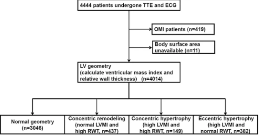
We retrospectively analyzed 4444 patients who had undergone simultaneous scheduled transthoracic echocardiography (TTE) and electrocardiography (ECG) at the Cardiovascular Center of Kitano Hospital during 2013. A lowchart of the study population is shown in Fig. 1. A total of 430 patients who had indings of previous myocardial infarctions (n = 419) and without the data on body surface area (n = 11) wereCardiovascular Center, The Tazuke Kofukai Medical Research Institute, Kitano Hospital, Osaka, Japan. Department of Cardiovascular Medicine, Kyoto University Graduate School of Medicine, Kyoto, Japan. Correspondence and requests for materials should be addressed to T.K. (email: tkato7 @kuhp.kyoto-u.ac.jp)
Received: 9 November 2017 Accepted: 11 April 2018 Published: xx xx xxxx
www.nature.com/scientificreports/
excluded from the study, because the old myocardial infarction afected their wall thickness and other data from the TTE. On the basis of the data from the TTE and ECG examinations in addition to the catheter database, we identiied patients who had a previous myocardial infarction. he ECG and TTE were ordered by each physician.
he research protocol was approved by the institutional review board of Kitano Hospital (approval no.: P16-02-005). Informed consent was not obtained from each patient since this was a retrospective study. he study pro-tocol conforms to the ethical guidelines of the 1975 Declaration of Helsinki as relected in a priori approval by the institution’s human research committee. Patients’ records and information were anonymized and de-identiied before analysis.
Data collection.
From the TTE database, we extracted data for patients’ wall thickness, LV diastolic diameter (LVDd), E/e’, LA diameter (LAD), LA volume index (LAVI), and LV ejection fraction (LVEF). We also extracted the body surface area from the TTE report. From the ECG database, we extracted cardiac rhythm and recorded it as it was documented. herefore, we could not determine whether the AF was paroxysmal or persistent.he LV mass was calculated using the formula recommended by the American Society of Echocardiography (ASE), and it was indexed to the body surface area as follows: LV mass = 0.8 × 1.04 [(LVDd + LVPWTd + IVST d)3− (LVDd)3] + 0.6, where LVDd was the LV diastolic diameter, IVSTd was the diastolic interventricular septal
wall thickness, and LVPWTd was the diastolic LV posterior wall thickness. In line with the ASE recommenda-tions, a high LV mass index (LVMI) was deined as >115 g/m2 for male patients and >95 g/m2 for female patients.
Relative wall thickness (RWT) was calculated using the following formula: (2 × LVPWTd)/(LVDd), which per-mits physicians to categorize an increase in the LV mass as either concentric (RWT >0.42) or eccentric (RWT
≤0.42) hypertrophy and identify concentric remodeling (a normal LV mass with an increased RWT)16,17. he
LAVI was calculated using the biplane area-length method and body surface area and deined high as a value
>42 mL/m214,15. Data from two-dimensional TTE were analyzed at baseline. he LVEF was measured using the
Teichholz method or the modiied Simpson rule method and deined low as LVEF <50%.
We then categorized 4014 patients into four groups as follows (Fig. 1): normal geometry (n = 3046), concen-tric remodeling (normal LVMI and high RWT, n = 437), concentric hypertrophy (high LVMI and high RWT, n = 149), and eccentric hypertrophy (high LVMI and normal RWT, n = 382).
We extracted patients’ data from the electronic medical data at our institution, including age, sex, and type of disease, i.e., hypertension (International Statistical Classiication of Diseases and Related Health Problems, Tenth Edition [ICD-10] codes I10, I11, I12, I13, I14, and I15), hyperlipidemia (ICD-10 code E78), diabetes mellitus (ICD-10 codes E10, E11, E12, E13, and E14), and chronic kidney disease (CKD) (ICD-10 code N18), from elec-tronic medical data at our institution.
Data availability.
he datasets generated during and/or analyzed during the current study are available from the corresponding author on reasonable request.Statistical analysis.
Categorical variables are presented as numbers and percentages, and were compared using the chi-square test or Fisher exact test. Continuous variables are expressed as a mean ± standard deviation or median (interquartile range). Based on their distributions, continuous variables were compared using the Student t-test or Wilcoxon rank-sum test. To analyze factors associated with AF, we used a multivariable logistic regression model (model 1) involving the following potential independent, clinically relevant variables: age >70 years; sex; LV geometric remodelings that was deined as ordered variables (normal geometry, concentric remod-eling, concentric hypertrophy, and eccentric hypertrophy); and the presence of comorbidities such as ishemic heart disease, hypertension, diabetes mellitus, hyperlipidemia, CKD and overweight (body mass index >25 kg/ m2). he adjusted odds ratios and 95% conidence intervals were calculated. To show the association of twovaria-bles (LAD and LVMI, LAD and RWT, LVEF and LVMI, and LVEF and RWT), we presented scatter plots, lines of best it, and correlation coeicients. To visualize the efect size of each variable, the LogWorth (−log10(p-value))
scales were presented18. In LogWorth sclaes, a highly signiicant p-value had a large LogWorth value and a
non-signiicant p-value had a low LogWorth value18. We generated the second multivariable regression model (model
2) including the LA size and LVEF to visualize the efect size of the LA size and LVEF as the LogWorth scales. In addition, we generated a multivariable logistic regression model using the same variables in AF for adjustment to analyze factors associated with a high LAVI and a low LVEF. Finally, from a logistic regression model with nomi-nal responses (AF or not) using LVEF and LAVI, we generated a formula for linear combinations of the response levels (x = a + b x LVEF + c x LAVI) and prediction formulas for the response levels predicting the presence of AF (1/ (1 + Exp(x)) and the absence of AF (1/(1 + EXP(-x)), then we compared the two levels and predicted the presence or absence of AF according to the larger response level. We showed a receiver-operating curve with an area under the curve and provided the sensitivity and 1-speciicity of this prediction formulas.
All statistical analyses were performed using JMP, version 13.2 (SAS Institute Inc., Chicago, IL, USA).
Results
Patients’ characteristics according to the LV geometric patterns.
Baseline characteristics of patients are provided in Table 1. Among patients’ characteristics, patients with a normal geometry were signif-icantly younger than who had abnormal geometry. he ratio of men were 52.4%, 58.3%, 48.3%, and 45.0% in patients with a normal geometry, concentric remodeling, concentric hypertrophy, and eccentric hypertrophy, respectively. Patients with concentric remodeling, concentric hypertrophy, and eccentric hypertrophy had high rates of hypertension, hyperlipidemia, diabetes mellitus, aortic valve stenosis, and CKD (Table 1).Baseline characteristics of echocardiography.
Baseline characteristics of echocardiographic indings are provided in Table 2. he mean LVEFs were 62.7, 62.6, 60.8, and 53.8% in patients with a normal geometry, concentric remodeling, concentric hypertrophy, and eccentric hypertrophy, respectively. he mean LAVIs were 22.5, 23.8, 33.3, and 37.0 mm/m2 in patients with a normal geometry, concentric remodeling, concentrichyper-trophy, and eccentric hyperhyper-trophy, respectively.
Prevalence of AF.
Overall, 3460 patients had a normal sinus rhythm, 455 had AF, 95 had a pacemaker rhythm, and 15 had other rhythms. he prevalence rates of AF were 10.4, 10.5, 14.8, and 16.8% in patients with a normal geometry, concentric remodeling, concentric hypertrophy, and eccentric hypertrophy, respectively (p < 0.0001, Fig. 2).Association among LV geometry, LA size, and LVEF.
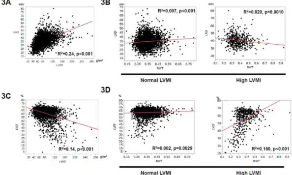
In order to investigate the underlying link between AF and LV geometry, we evaluated the relationship between LVMI, RWT, LVD, and LVEF. By using scatter plots, we visualized the association (Fig. 3). LAD was signiicantly related to the high LVMI (Fig. 3A). In contrast, LAD was small but positively related to high RWT when LVMI was normal (Fig. 3B), and negatively related to high RWT when LVMI was high (Fig. 3B). LVEF was signiicantly negatively correlated with LVMIAortic regurgitation, % 3.1 2.0 1.1 9.4 11.5 <0.0001 Mitral stenosis, % 0.2 0.2 0.2 0.7 0.5 0.340 Mitral regurgitation, % 3.6 2.5 1.8 4.7 13.0 <0.0001 Chronic kidney disease, % 14 10.5 20.1 38.9 25.9 <0.0001 Overweight, % 27.7 26.9 30.1 33.6 29.6 0.1419
Table 1. Baseline characteristics of the study subjects. Abbreviation: SD = standard deviation.
Total (n = 4,014)
Normal geometry (n = 3,046)
Concentric remodeling (n = 437)
Concentric hypertrophy (n = 149)
Eccentric hypertrophy (n = 382) p
LVDd, mm 46.7, 6.1 46.4, 4.8 40.7, 4.1 46.2, 4.9 55.6, 7.6 <0.0001 E/e’ 11.6, 4.8 10.8, 4.1 13.3, 5.4 17.3, 7.0 14.3, 5.7 <0.0001 LAD, mm 35.6, 7.0 34.7, 6.5 35.4, 6.8 40.0, 7.3 41.1, 7.7 <0.0001 LAVI, ml/m2 24.4, 14.8 22.5, 12.9 23.8, 12.5 33.3, 17.2 37.0, 21.6 <0.0001 LVEF, % 61.7, 7.6 62.7, 6.0 62.6, 4.9 60.8, 7.5 53.8, 14.5 <0.0001
www.nature.com/scientificreports/
(Fig. 3C). With regards to RWT, LVEF was only slightly correlated with RWT when LVMI was normal (Fig. 3D), but positively correlated with RWT when LVMI was high (Fig. 3D), showing that the relationship between LVEF and RWT was diferent for normal and high LVMI.
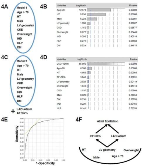
Factors associated with AF and underlying LA enlargement and a low LVEF.
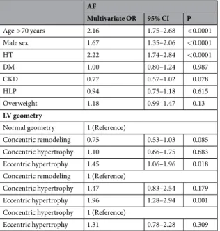
According to the results of the multivariate logistic regression analysis (model 1, Fig. 4A), the following variables were signiicantly associated with AF: age >70 years, male sex, hypertension, and LV geometric patterns (Table 3 and Fig. 4B). When we included two additional echocardiographic parameters, LA enlargement (LAD >40 mm) and a low LVEF (LVEF <50%), into a multivariate model for the presence of AF (model 2, Fig. 4C), LA enlargement was the echocardiographic parameter most associated with AF, and a low LVEF was the second most associated parame-ter (Fig. 4D), superior to the LV geometry. Finally, we sought the associated factors for LA enlargement and low LVEF. Table 4 shows the signiicant association with LV geometry and a high LAVI or LVEF. Finally, the presence of AF was well predicted by the values of LAVI and LVEF by receiver operating curve analysis (area under the curve 0.87; Fig. 4F). hese indicated that the LV morphology was linked to the LV function and LA size (sche-matic in Fig. 4F).Figure 2. Prevalence of atrial ibrillation (AF) with each geometry patients.
Figure 3. Association with LVMI, RWT, LA diameter, and LVEF. (A) Association between LVMI and LA diameter (LAD). Scatter plots show the positive correlation between LVMI and LAD. R2= 0.24, p < 0.001.
R = correlation coeicient. (B) Association between RWT and LAD. In patients with normal LVMI (let panel), scattered plots showed small correlation between RWT and LA diameter. R2= 0.007, p < 0.001. In high LVMI
(right panel), scatter plots showed small but, negative correlation between RWT and LA diameter. R2= 0.020,
p = 0.0010. (C) Association between LVMI and LVEF. Scatter plots showed the negative correlation between LVMI and LVEF. R2= 0.14, p < 0.001. (D) Association between RWT and LVEF. In normal LVMI (let panel),
scatter plots showed small correlation between RWT and LVEF. R2= 0.002, p = 0.0029. In high LVMI (right
Discussion
he main inding of the present study was that the prevalence of AF increased per the geometric remodeling pat-terns. he mechanism for why the prevalence of AF increased is unclear; however, cardiac remodeling manifests as changes in the cardiac size, shape, and function in response to aging, cardiac impairment, or an increased load9.
Cardiac remodeling causes an increase in end-diastolic pressure and expansion of end-diastolic volume with dias-tolic and sysdias-tolic dysfunction9. In the present study, patients with concentric remodeling, concentric hypertrophy,
and eccentric hypertrophy were older than those with a normal geometry. In addition, patients with concentric remodeling, concentric hypertrophy, and eccentric hypertrophy had a higher E/e’. E/e’ is highest in patients with concentric hypertrophy17.
Concentric remodeling and hypertrophy were oten caused by pressure overload that increased the RWT. his was an adaptation to normalize the systolic wall stress. However, LV was shown to be a substrate for LV diastolic dysfunction in concentric remodeling and an incremental risk for diastolic heart failure in concentric
Figure 4. (A) Model 1 in Table 3 includes factors included age >70 years; sex; LV geometric remodelings that were deined as ordered variables (normal geometry, concentric remodeling, concentric hypertrophy, and eccentric hypertrophy); and the presence of comorbidities such as ischemic heart disease (IHD), hypertension (HT), diabetes mellitus (DM), hyperlipidemia (HLP), CKD and overweight (body mass index
>25 kg/m2). (B) LogWorth scales of multivariate model 1 in Table 3. he p-values were transformed to the
www.nature.com/scientificreports/
hypertrophy. Direct underlying triggers of AF was mainly due to atrial remodeling. Atrial remodeling is caused by multiple factors19. he let atrium is a thin-walled structure that connects the pulmonary veins and LV in diastole
and pumps the LV (atrial kick) in end-diastole during sinus rhythm15. Previous work has highlighted the complex
mechanisms contributing to AF progression19–23, as the morphological, electrical, or neurohormonal remodeling
along with the ventricular responses were related. Diastolic impairment and an increasing diastolic illing pres-sure are related to the LA size. With an increase in LA illing prespres-sures, the atrial wall stretches and enlarges. In this study, overweight was not signiicantly associated with AF, which was inconsistent with the previous stud-ies24. One of the reasons may be the diferences in baseline characteristics in Japanese population and in Western
population. Body weights in Japan are much lower than those in the US and Europe. Other possible reason may be due to small numbers studied in our study, considering that a marginally signiicance existed.
AF
Multivariate OR 95% CI P Age >70 years 2.16 1.75–2.68 <0.0001 Male sex 1.67 1.35–2.06 <0.0001 HT 2.22 1.74–2.84 <0.0001
DM 1.00 0.80–1.24 0.987
CKD 0.77 0.57–1.02 0.078
HLP 0.94 0.75–1.18 0.615
Overweight 1.18 0.99–1.47 0.13 LV geometry
Normal geometry 1 (Reference)
Concentric remodeling 0.75 0.53–1.03 0.085 Concentric hypertrophy 1.10 0.66–1.75 0.683 Eccentric hypertrophy 1.45 1.06–1.96 0.018 Concentric remodeling 1 (Reference)
Concentric hypertrophy 1.47 0.83–2.54 0.179 Eccentric hypertrophy 1.96 1.28–2.94 0.001 Concentric hypertrophy 1 (Reference)
Eccentric hypertrophy 1.31 0.78–2.28 0.309
Table 3. Factors associated with AF according to multivariate logistic regression analysis. Abbreviations: OR = odds ratio; CI = conidence interval; RWT = relative wall thickness; LVMI = let ventricular mass index; HT = hypertension; DM = diabetes mellitus; CKD = chronic kidney disease, HLP = Hyperlipidemia.
A high LAVI A low LVEF
Multivariate OR 95% CI P Multivariate OR 95% CI p Age >70 years 3.29 2.43–4.47 <0.0001 0.85 0.63–1.14 0.288 Male sex 0.95 0.73–1.25 0.751 2.14 1.55–2.94 <0.0001 HT 2.14 1.53–2.99 <0.0001 4.60 2.93–7.23 <0.0001 DM 1.19 0.89–1.60 0.242 1.08 0.78–1.49 0.623 CKD 0.95 0.67–1.35 0.789 1.07 0.74–1.53 0.705 HLP 0.658 0.47–0.89 0.007 1.24 0.91–1.70 0.166 Overweight 0.91 0.68–1.24 0.574 0.72 0.51–0.99 0.045 LV geometry
Normal geometry 1 (Reference) 1 (Reference)
Concentric remodeling 1.03 0.66–1.61 0.872 0.41 0.19–0.86 0.018 Concentric hypertrophy 3.36 2.05–5.51 <0.0001 1.48 0.74–2.96 0.259 Eccentric hypertrophy 5.77 4.20–7.98 <0.0001 11.3 8.18–15.5 <0.0001 Concentric remodeling 1 (Reference) 1 (Reference)
Concentric hypertrophy 3.24 1.77–5.94 0.0001 3.59 1.37–9.37 0.008 Eccentric hypertrophy 5.56 3.44–8.99 <0.0001 27.5 13.1–57.9 <0.001 Concentric hypertrophy 1 (Reference) 1 (Reference)
Eccentric hypertrophy 1.71 1.02–2.87 0.040 7.66 3.83–15.3 <0.0001
Table 4. Factors associated with a high LAVI and a low LVEF according to multivariate logistic regression analysis. Abbreviations: OR = odds ratio; CI = conidence interval; RWT = relative wall thickness; LVMI = let ventricular mass index; HT = hypertension; DM = diabetes mellitus; CKD = chronic kidney disease,
non-valvular AF . Patel et al. reported that LAVI was associated only with LVMI, not RWT . his is consistent with our present study which showed that the relationship between LA size and RWT was diferent for normal and high LVMI.
In addition, LAVI and LVEF accurately predicted the presence of AF in our study; therefore, we performed multivariate analyses excluding these co-linear factors. A linear association with LVMI and LA enlargement was observed; however, the efect of RWT on a low LVEF was diferent between patients with a normal LVMI and those with a high LVMI. Because LV geometric remodeling was clearly associated with LA enlargement and a low LVEF, a schematic in which that LV morphology was linked to the LV function and LA size is provided (Fig. 4F). Furthermore, the LV morphology was linked to the prognosis7,26. hromboembolic events may be one of the
mechanisms for the higher mortality in LVH27. Although there are reportedly many factors related to AF and LA
enlargement, such as age, the presence of hypertension, and obesity24, LV geometric remodeling has an impact
on the presence of AF through the association with LA enlargement and a low LVEF. However, atrial ibrillation can develop without let atrial enlargement28. Atrial size increases with time in patients with atrial ibrillation,
without any structural or functional abnormality of LV and the valve28. Since our study is cross-sectional, we
could not identify the cause-efect relationship in each patient. Longitudinal follow-up could address this issue in each patient.
In clinical practice, LVH is a risk factor in hypertensive patients29 and severe AS patients30. In our study, we
provided the link between LV geometric patterns and AF prevalence through the contribution of atrial enlarge-ment and decreased LVEF. AF and LA enlargeenlarge-ment is a risk factor for ischemic stroke and may contribute to dou-bling all-cause31 and CV deaths14, and, in a speciic condition, non-CV deaths32. However, anticoagulants were
sub-optimally prescribed in these patients. Attention should be paid to paroxysmal AF in the LVH population to improve our daily practice.
here are several limitations to the present study. First, the subjects are heterogeneous because of the TTE and ECG indings. he ordering criteria for ECG and TTE were not set. Second, patients’ data were extracted from electronic medical data. A lack of information regarding exercise was an important limitation24. hird, we did
not consider the efect of valvular diseases such as mitral valve regurgitation. Fourth, patients with short-lasting paroxysmal AF were excluded, because we included patients with AF when they had an AF rhythm according to an ECG examination. Lastly, the data presented provide only hypothesis-generating associations between LV geometry and AF. Since this study had a cross-sectional design, a cause-efect relationship could not be assessed. Further prospective studies are needed to determine the association of LA remodeling, LV geometric changes, LV function, mortality, and the time-course for the development of AF.
Conclusions
he prevalence of AF was increasing according to LV geometric remodeling patterns in association with LA size and LVEF.
References
1. Zabalgoitia, M. et al. Impact of coronary artery disease on let ventricular systolic function and geometry in hypertensive patients with let ventricular hypertrophy (the LIFE study). Am. J. Cardiol.88, 646–650 (2001).
2. Casale, P. N. et al. Value of echocardiographic measurement of let ventricular mass in predicting cardiovascular morbid events in hypertensive men. Ann. Intern. Med.105, 173–178 (1986).
3. Ghali, J. K., Liao, Y. & Cooper, R. S. Inluence of let ventricular geometric patterns on prognosis in patients with or without coronary artery disease. J. Am. Coll. Cardiol.31, 1635–1640 (1998).
4. Koren, M. J., Devereux, R. B., Casale, P. N., Savage, D. D. & Laragh, J. H. Relation of let ventricular mass and geometry, to morbidity and mortality in uncomplicated essential hypertension. Ann. Intern. Med.114, 345–352 (1991).
5. Ganau, A. et al. Patterns of let ventricular hypertrophy and geometric remodeling in essential hypertension. J. Am. Coll. Cardiol.19, 1550–1558 (1992).
6. Patel, D. A., Lavie, C. J., Milani, R. V. & Ventura, H. O. Let atrial volume index predictive of mortality independent of let ventricular geometry in a large clinical cohort with preserved ejection fraction. Mayo Clin. Proc.86, 730–737 (2011).
7. Oktay, A. A. et al. Current Perspectives on Let Ventricular Geometry in Systemic Hypertension. Prog Cardiovasc Dis.59, 235–246 (2016).
8. Lavie, C. J. et al. Impact of echocardiographic let ventricular geometry on clinical prognosis. Prog Cardiovasc Dis.57, 3–9 (2014). 9. Cohn, J. N., Ferrari, R. & Sharpe, N. Cardiac remodeling–concepts and clinical implications: a consensus paper from an international
forum on cardiac remodeling. Behalf of an International Forum on Cardiac Remodeling. J. Am. Coll. Cardiol.35, 569–582 (2000). 10. Wolf, P. A., Abbott, R. D. & Kannel, W. B. Atrial ibrillation as an independent risk factor for stroke: the Framingham Study. Stroke.
22, 983–988 (1991).
11. Lin, H. J. et al. Stroke severity in atrial ibrillation: the Framingham Study. Stroke.27, 1760–1764 (1996).
12. Whisnant, J., P. et al. Comparison of population-based models of risk factors for TIA and ischemic stroke. Neurology.53, 532–536 (1999).
13. Benjamin, E. J. et al. Impact of atrial ibrillation on the risk of death: the Framingham Heart Study. Circulation.98, 946–952 (1998). 14. Wolf, P. A., Mitchell, J. B., Baker, C. S., Kannel, W. B. & D’Agostino, R. B. Impact of atrial ibrillation on mortality, stroke, and medical
costs. Arch. Intern. Med.158, 229–234 (1998).
www.nature.com/scientificreports/
16. Lang, R. M. et al. Recommendations for cardiac chamber quantification by echocardiography in adults: an update from the
American Society of Echocardiography and the European Association of Cardiovascular Imaging. J. Am. Soc. Echocardiogr.28, 1–39. e14 (2015).
17. Nagueh, S. F. et al. Recommendations for the evaluation of let ventricular diastolic function by echocardiography: an update from the American Society of Echocardiography and the European Association of Cardiovascular Imaging. J. Am. Soc. Echocardiogr.29, 277–314 (2016).
18. JMP 13.2 Online Documentation. https://www.jmp.com/support/help/13-2/Efect_Summary.shtml.
19. Kirchhof, P. et al. 2016 ESC guidelines for the management of atrial ibrillation developed in collaboration with EACTS. Europace. 18, 1609–1678 (2016).
20. El Aouar, L. M. et al. Relationship between let atrial volume and diastolic dysfunction in 500 Brazilian patients. Arq. Bras. Cardiol. 101, 52–58 (2013).
21. Patel, D. A. et al. Association of let ventricular geometry with let atrial enlargement in patients with preserved ejection fraction.
Congest. Heart Fail.8, 4–8 (2012).
22. Cioi, G., Mureddu, G. F., Stefenelli, C. & de Simone, G. Relationship between let ventricular geometry and let atrial size and function in patients with systemic hypertension. J. Hypertens.22, 1589–1596 (2004).
23. Melenovsky, V. et al. Let atrial remodeling and function in advanced heart failure with preserved or reduced ejection fraction. Circ. Heart Fail.8, 295–303 (2015).
24. Lavie, C. J. et al. Obesity and Atrial Fibrillation Prevalence, Pathogenesis, and Prognosis: Efects of Weight Loss and Exercise. J Am Coll Cardiol.70, 2022–2035 (2015).
25. Proietti, M., Raparelli, V., Basili, S., Olshansky, B. & Lip, G. Y. Relation of female sex to let atrial diameter and cardiovascular death in atrial ibrillation: he AFFIRM Trial. Int J Cardiol.207, 258–263 (2016).
26. Gerdts, E. et al. Relation of Let Ventricular Mass to Prognosis in Initially Asymptomatic Mild to Moderate Aortic Valve Stenosis.
Circ Cardiovasc Imaging.8, e003644 (2015).
27. Proietti, M. et al. Frequency of Let Ventricular Hypertrophy in Non-Valvular Atrial Fibrillation. Am J Cardiol.116, 877–882 (2015). 28. Sanilippo, A. J. Atrial Enlargement as a Consequence of Atrial Fibrillation A Prospective Echocardiographic Study. Circulation.82,
792–797 (1990).
29. Verdecchia, P. et al. Left ventricular hypertrophy as an independent predictor of acute cerebrovascular events in essential hypertension. Circulation.109, 2039–2044 (2001).
30. Minamino-Muta, E. et al. Impact of the let ventricular mass index on the outcomes of severe aortic stenosis. Heart.103, 1992–1999 (2017).
31. Patel, D. A. et al. Prediction of All-Cause Mortality by the Let Atrial Volume Index in Patients With Normal Let Ventricular Filling Pressure and Preserved Ejection Fraction. Mayo Clin Proc.90, 1499–505 (2015).
32. Minamino-Muta, E. et al. Causes of Death in Patients with Severe Aortic Stenosis: An Observational study. Sci Rep.7, 14723 (2017).
Author Contributions
Y.S. and T.K.: conceived the design, performed statistical analysis, and wrote manuscript. T.H., T.I., S.M., E.N., and M.I.: collected the data and made critical revision.
Additional Information
Competing Interests: he authors declare no competing interests.
Publisher's note: Springer Nature remains neutral with regard to jurisdictional claims in published maps and institutional ailiations.
Open Access This article is licensed under a Creative Commons Attribution 4.0 International License, which permits use, sharing, adaptation, distribution and reproduction in any medium or format, as long as you give appropriate credit to the original author(s) and the source, provide a link to the Cre-ative Commons license, and indicate if changes were made. he images or other third party material in this article are included in the article’s Creative Commons license, unless indicated otherwise in a credit line to the material. If material is not included in the article’s Creative Commons license and your intended use is not per-mitted by statutory regulation or exceeds the perper-mitted use, you will need to obtain permission directly from the copyright holder. To view a copy of this license, visit http://creativecommons.org/licenses/by/4.0/.