早稲田大学審査学位論文 博士(スポーツ科学)
Prevention of Anterior Cruciate Ligament Injury in Female Athletes during Maturation
成長期女子選手における 膝前十字靭帯損傷予防法の検討
2015 年 1 月
早稲田大学大学院 スポーツ科学研究科
大槻 玲子 OTSUKI, Reiko
研究指導教員:福林 徹 教授
ACKNOWLEDGEMENTS
I would like to thank many people who contributed in some way to this thesis.
First of all, I would like to express my deep and sincere gratitude to my supervisor, Dr. Toru Fukubayashi, for his knowledge and enduring support throughout this time.
He has been supportive and has given me freedom to pursue various projects without objection. He has always provided insightful discussions about the research.
I am also grateful to Dr. Norikazu Hirose whose support and guidance made my thesis work possible. He has provided me valuable guidance and scholarly inputs especially during the last year of my study. I would also gratefully acknowledge to my committee, Dr. Koji Kaneoka and Dr. Toshimasa Yanai for their helpful comments, orientation and valuable feedback.
My heartfelt thanks to my lab seniors and lab mates for all their personal and professional help that they have extended to me throughout. I could not have asked for more than what I got from them.
Finally, I would like to express my deep gratitude to my mother and father for their unconditional and continuous support and encouragement. I could not reach this far without their support.
TABLE OF CONTENTS
CHAPTER ONE : INTRODUCTION………. 1
1.1 Introduction and problem statement ………...…… 1
1.2 Objectives ………..………. 4
1.3 Hypotheses …….……….………...…...………. 5
1.4 Limitations …….……….………...…...………... 5
1.5 Delimitations ………..………..……….. 6
1.6 Ethical considerations ……….………..………. 6
1.7 Definition of terms ………..……... 7
CHAPTER TWO: LITERATURE REVIEW ………....……….…………... 9
2.1 Epidemiology ……....………... 9
2.1.1 Sex ……….. 9
2.1.2 Age ………. 10
2.1.3 Sports ……….. 10
2.2 Mechanisms of ACL injury ……….……...….………... 11
2.2.1 Playing situation ………….………..……….. 11
2.2.2 Video analysis ……….……….……….. 11
2.2.3 Cadaveric study ……….………..……….….……. 12
2.2.4 Bone bruises …….………..……….….…….….… 13
2.3 Risk factors of ACL injury ………….….….….….….….….….….….….….… 13
2.3.1 Anatomical risk factors ……….….….….….….….….….….…… 13
2.3.2 Hormonal risk factors ………….….….….….….….….….….….….…. 16
2.3.3 Neuromuscular risk factors ……….….….….….….….….…… 17
2.3.4 Biomechanical risk factors ……….….….….….….….….….….... 19
2.3.5 Developmental risk factors ……….….….….….….….….…… 21
2.4 Prevention of ACL injury ………….….….….….….….….….….….….….….. 23 2.4.1 Effects on biomechanical and neuromuscular functions ……… 23 2.4.2 Effects on ACL injury rate ……….….….….….….….….….…… 24 CHAPTER THREE: STUDY 1
ACL injury risk in female athletes during maturation ……….. 27 3.1 Introduction ………...……….. 27 3.2 Methods …….….….….….….….….….….….….….….….….….….….….….. 27 3.3 Results ……...……….….….….….….….….…… 32 3.4 Discussion ……….….….…... 35 3.5 Conclusion ……….….….….….….….….….….….….….….….….….. 38 CHAPTER FOUR: STUDY 2
Factors affecting knee abduction angle during a drop vertical jump ………….. 39 4.1 Introduction ………...……….. 39 4.2 Methods …….….….….….….….….….….….….….….….….….….….….….. 40 4.3 Results ……...……….….….….….….….….…… 48 4.4 Discussion ……….….….…... 53 4.5 Conclusion ……….….….….….….….….….….….….….….….….….. 56 CHAPTER FIVE: STUDY 3
Effect of skill training on lower-extremity kinematics and muscle function
in female pubertal athletes ……… 57 5.1 Introduction ………...……….. 57 5.2 Methods …….….….….….….….….….….….….….….….….….….….….….. 57 5.3 Results ……...……….….….….….….….….…… 61 5.4 Discussion ……….….….…... 68 5.5 Conclusion ……….….….….….….….….….….….….….….….….…. 70 CHAPTER SIX: STUDY 4
Effect of skill training on ACL injury risk in female pubertal athletes ………… 71
6.1 Introduction ………...……….. 71
6.2 Methods …….….….….….….….….….….….….….….….….….….….….….. 72
6.3 Results ……...……….….….….….….….….…… 74
6.4 Discussion ……….….….…... 75
6.5 Conclusion ……….….….….….….….….….….….….….….….….…. 78
CHAPTER SEVEN: GENERAL DISCUSSION AND CONCLUSIONS ……... 79
7.1 General discussion ……….……… 79
7.2 Relevance to the sports field ………..…… 83
7.3 Suggestions for future investigation ……….. 84
7.4 Conclusion ………. 85
REFERENCES ………...…...…..…………... 87
APPENDIX A Self-Administrated Rating Scale for Pubertal Development …….. 109
LIST OF TABLES
Table 2-1 Summary of risk factors of ACL injury 22
Table 2-2 Summary of ACL injury prevention intervention studies 26
Table 3-1 Subject demographics 33
Table 4-1 Means and standard deviations of isokinetic strength, EMG, and kinematics
50 Table 4-2 Regression model for isokinetic strength 51 Table 4-3 Regression model for preparatory muscle activity 51 Table 4-4 Regression model for knee abduction angle at initial contact 52 Table 4-5 Regression model for knee abduction angle 100ms before initial
contact
52
Table 5-1 Skill training program 60
Table 5-2 Subject demographics 62
Table 5-3 Comparison of kinematics between pre-test and post-test 1 63 Table 5-4 Comparison of preparatory muscle activity between pre-test and
post-test 1
63 Table 5-5 Comparison of kinematics between pre-test and post-test 2 64 Table 5-6 Comparison of preparatory muscle activity between pre-test and
post-test 2 64
Table 5-7 Comparison of strength between pre-test and post-test 2 65
Table 6-1 Subject demographics 74
Table 6-2 Changes in the knee valgus motion, knee flexion range of motion, and pKAM
75
LIST OF FIGURES
Figure 3-1 Experiment setup 29
Figure 3-2 Calculations of knee valgus motion and knee flexion range of motion
31 Figure 3-3 Example of use of the ACL injury prediction algorithm 31 Figure 3-4 Comparison of knee valgus motion (a), knee flexion range of
motion (b), and pKAM (c) among maturational stages
34 Figure 4-1 Knee extension/flexion testing position 43
Figure 4-2 Hip extension/flexion testing position 43
Figure 4-3 Hip abduction/adduction testing position 43 Figure 4-4 Experiment setup and drop vertical jump task 45
Figure 4-5 Marker placement 45
Figure 5-1 Flowchart of the study protocol 58
Figure 5-2 Kinematics in three testing sessions 66
Figure 5-3 Preparatory muscle activities in three testing sessions 67
Figure 5-4 Strength in pre-test and post-test 2 67
1
CHAPTER 1: INTRODUCTION
1.1 INTRODUCTION AND PROBLEM STATEMENT
Anterior cruciate ligament (ACL) injuries are frequently seen in the athletic population, especially among female athletes (Louw, Manilall, & Grimmer, 2008;
Renstrom et al., 2008; Shea, Grimm, Ewing, & Aoki, 2011). Although great progress in surgery and rehabilitation has made possible a faster and safer return to sport, ACL injuries are still recognized as one of the most devastating sport-related injuries. ACL injuries usually require surgery and extensive rehabilitation for at least 6 months. In addition, an ACL injury can triple the risk of osteoarthritis development regardless of the management (Caine & Golightly, 2011; Lohmander, Englund, Dahl, & Roos, 2007; Myklebust, Holm, Mæhlum, Engebretsen, & Bahr, 2003). Therefore, the risk management of ACL injuries in female athletes is critical.
During the last several decades, many researchers have made efforts to understand ACL injury. The research area covered the epidemiology, etiology, risk factor, treatment, and prevention of ACL injury. Especially in the last decade, the trend of research has shifted toward the prevention of this devastating injury.
Recently, several intervention studies indicated that training programs for ACL injury prevention might be effective in reducing the number of these injuries (Heidt Jr, Sweeterman, Carlonas, Traub, & Tekulve, 2000; Hewett, Lindenfeld, Riccobene, &
Noyes, 1999; Kiani et al., 2010; LaBella et al., 2011; Mandelbaum et al., 2005;
Myklebust, Engebretsen, et al., 2003; Olsen, Myklebust, Engebretsen, Holme, &
Bahr, 2005; Steffen, Myklebust, Olsen, Holme, & Bahr, 2008; Waldén, Atroshi, Magnusson, Wagner, & Hägglund, 2012). In contrast, Agel, Arendt, and Bershadsky (2005) reported that the rate of ACL injury had not been declining in high-risk sports,
2
such as basketball and soccer. Also, others reported that low participant compliance to injury prevention programs resulted in the low effectiveness of ACL injury prevention (Emery, Rose, McAllister, & Meeuwisse, 2007; Soligard et al., 2009; Steffen et al., 2008). Therefore, at this point, it is necessary to investigate how to effectively implement an ACL injury prevention program.
In this dissertation, it was first investigated when an injury prevention program should be initiated. The occurrence of ACL injuries significantly increases after the onset of growth spurt in female athletes (Granan, Bahr, Steindal, Furnes, &
Engebretsen, 2008). Around the same time, female athletes start to demonstrate high- risk movement patterns (Ford, Shapiro, Myer, Van Den Bogert, & Hewett, 2010;
Hewett, Myer, & Ford, 2004; Schmitz, Shultz, & Nguyen, 2009), probably owing to their rapid physical development. It is reasonable to modify the risk factor during or prior to the period when the number of ACL injury actually increases. Furthermore, learning proper movement skills might be easier for young athletes than for adults because pre-adolescents undergo significant developmental changes in the neural system (Malina, Bouchard, & Bar-Or, 2004). It is ideal for young athletes to learn proper movement skills as they learn other sport-specific skills. To date, no consensus has been reached on when to initiate an ACL injury prevention program for young female athletes. Therefore, the first study evaluated the ACL injury risk across the different maturational stages. Evaluation by maturational stage rather than by chronological age was performed because the development of structure and motor function depends more on physiological age (Malina et al., 2004). These
developmental changes in structure and motor function might increase the risk of ACL injury (Ford, Shapiro, et al., 2010; Hewett et al., 2004). ACL injury risk was investigated by using an ACL injury prediction algorithm that was developed to
3
estimate the probability of demonstrating high-risk movement patterns (Myer et al., 2010).
Second, in this dissertation, it was investigated what the focus of training should be to effectively reduce the risk of ACL injury. ACL injury prevention programs are multi-component programs that typically incorporate strength,
plyometric, balance, skill, and flexibility training (Heidt Jr et al., 2000; Hewett et al., 1999; Kiani et al., 2010; LaBella et al., 2011; Mandelbaum et al., 2005; Myklebust, Engebretsen, et al., 2003; Olsen et al., 2005; Petersen et al., 2005; Steffen et al., 2008;
Waldén et al., 2012). Although those programs seem effective, the important elements in an ACL injury prevention program are unknown. Furthermore, the volume of multi-component training might hinder the participants’ adherence to the program. To improve the efficacy of an ACL injury prevention program, a more targeted strategy is needed. The program should be refined to include elements that modify the injury risk. Recent studies have identified that ACL injuries are likely to occur with
excessive knee valgus motion within 40 ms after ground contact (Koga et al., 2010).
Thus, the prevention training program should be aimed at reducing this high-risk movement during this critical period. In the second study, it is investigated what factors influence the knee valgus motion at 40 ms after ground contact. The muscle strength, preparatory muscle activities, and landing skills were specifically examined, as these were considered the most influential factors on the knee valgus motion at 40 ms after ground contact. Preparatory muscle activity was examined rather than reflexive muscle activity. It was shown that reflexive muscle activity occurs at approximately 95 ms after a stimuli to the ACL, with an electro-mechanical delay about 40 ms (Dyhre-Poulsen & Krogsgaard, 2000). Therefore, the reflex response would not be fast enough to provide necessary constraint to stabilize the joint at 40 ms
4
after ground contact. Rather than the reflexive muscle activity, the preparatory muscle activity might be responsible for the control of the knee joint during dynamic activity.
Previous studies have demonstrated that the tensile stiffness of muscle was linearly proportional to the active tension developed by the muscle before perturbation (Dhaher, Tsoumanis, Houle, & Rymer, 2005; Zhang & Rymer, 1997). Also, it was shown that an increase in pre-activation level provides additional excitatory input to the motor pool and causes more motor neurons to fire (Fuglevand, Winter, & Patla, 1993). Therefore, the preparatory muscle activity might help reduce the strain on the ACL. The second study investigated how much of the knee valgus motion at 40 ms after ground contact could be explained by muscle strength, preparatory muscle activities, and landing skills.
Finally, on the basis of the findings of the first and second parts of the
dissertation, a training program that focused on improving landing mechanics during the critical time period was developed and implemented in pubertal female athletes.
The third study evaluated, in a laboratory setting, whether the training program would improve landing mechanics and what changes in strength, preparatory muscle activity and landing skill would be observed. In the last study, this training program was implemented for 6 months in pubertal female athletes to determine whether the training program could limit the movement changes associated with pubertal growth in young female athletes.
1.2 OBJECTIVES
The objectives of this study are as follows:
1. To evaluate when ACL injury risk increases in pubertal female athletes
5
2. To identify the factors (strength, muscle activity, skill) that affect biomechanical ACL injury risk
3. To evaluate whether these factors are modifiable through a training program 4. To evaluate whether the training program could limit the changes in
movement patterns associated with pubertal growth and reduce the risk of developing ACL injury
1.3 HYPOTHESES
The following hypotheses were investigated in the study:
1. ACL injury risk, measured with an ACL injury risk prediction algorithm, would increase after the onset of puberty in female athletes
2. The knee abduction angles at 40 ms after ground contact during a drop vertical jump would be affected by preparatory muscle activity and lower-extremity motion
3. Lower-extremity motion and preparatory muscle activity could be changed with a training program
4. The training program would be effective in limiting the movement changes associated with pubertal growth in female athletes and reducing the risk of ACL injury
1.4 LIMITATIONS
1. The results of this study are limited to female junior high school basketball players.
6
2. Pubertal stages were estimated by using a pubertal development scale. Other evaluations (e.g., skeletal age or time from peak height velocity) were not performed to determine pubertal stages.
3. Movement tasks were limited to the drop vertical jump. Single-leg landing or cutting tasks were not evaluated.
4. Other risk factors, such as morphological or hormonal factors, were not evaluated in this study.
1.5 DELIMITATIONS
1. The participants consisted of female junior basketball players between the ages of 10 and 14 years.
2. All participants were members of school basketball teams and had similar performance level.
3. ACL injury risk was evaluated by using an ACL injury prediction algorithm, which was shown to be valid and reliable.
1.6 ETHICAL CONSIDERATIONS
This project was approved by the Academic Research Ethical Review
Committee of Waseda University (application no.: 2012-242, 2012-068(1)). Informed consent was obtained from the participants and their guardians before enrollment in the study. Complete privacy and confidentiality was ensured throughout the study.
The names or other identifying information were removed from all test data. All data were kept in a safe area. The names of the subjects will never be used in any
presentation or publication related to the study results. All procedures were non-
7
invasive, and there was minimal risk of injury or harm with the measurements done according to a standardized protocol.
1.7 DEFINITIONS OF TERMS Contact injury:
A contact injury refers to an injury resulting from physical contact with other athletes, a moving object (e.g., ball), or a static object (e.g., goalpost).
Non-contact injury:
A non-contact injury refers to an injury sustained without contact with another athlete or object.
Athlete-exposure (AE):
The injury rate was reported as one athlete participating in one practice or competition.
Knee valgus:
The frontal plane angular deviation of the tibia away from the midline of the body relative to the femur (same as knee abduction).
Knee abduction:
The frontal plane angular deviation of the tibia away from the midline of the body relative to the femur (same as knee valgus).
8 Maturation stages:
Maturation stages were determined by using the self-administered rating scale for pubertal development (Carskadon & Acebo, 1993) in this study (Appendix).
Probability of high knee abduction moment (pKAM):
pKAM is the probability that a subject demonstrates a high knee abduction moment (> 21.74 Nm of knee abduction) during a drop vertical jump, which was shown to be a risk of ACL injury. The pKAM was obtained by using an ACL injury prediction algorithm (Myer et al., 2010). pKAM was used to assess the risk of ACL injury.
Initial contact:
The point at which the subject’s foot comes into contact with the ground and vertical ground reaction force exceed 10 N during the first landing of the drop vertical jump.
Preparatory muscle activity:
EMG activity for a period of a 200ms prior to initial contact during the drop vertical jump (Carcia & Martin, 2007; Zazulak et al., 2005).
Dominant leg:
The leg used to kick a ball for maximal distance.
9
CHAPTER 2: LITERATURE REVIEW
2.1 EPIDEMIOLOGY
Several national registries have been established to collect information on ACL surgeries. In Norway, the annual population incidence of ACL reconstruction
surgeries was 34 per 100,000 citizens (Granan et al., 2008). The incidence in the age group of 16-39 years was 85 per 100,000 citizens (Granan et al., 2008). In Germany, the incidence of ACL injuries in the active population was 70 per 100,000 citizens (Renstrom et al., 2008). In Sweden, the incidence in the age group of 10-64 years was 81 per 100,000 citizens. However, in Japan, there has been no nation-wide registry for monitoring the incidence of ACL injuries.
2.1.1 Sex
The rate of ACL injuries differs between the sexes. In a study including all age groups, the incidence of ACL injuries was found to be higher in males than in females (Granan et al., 2008). However, in the young population, females had a higher rate of ACL injuries than males (Granan et al., 2008). On the basis of the data from the National Collegiate Athletic Association (NCAA) Injury Surveillance System in the United States, the rate of ACL injuries were 2.8-3.4 times higher in female than in male soccer and basketball players (0.27 vs. 0.08/1000 AE in basketball; 0.31 vs.
0.11/1000 AE in soccer) (Agel et al., 2005). Similarly, the data from an American high school injury surveillance system demonstrated that the rate of ACL injuries was 3.4 times higher in girls than in boys in sex-comparable sports (0.089 and 0.026/1000 AE, respectively) (Joseph et al., 2013). In Japanese junior and senior high schools, the rate of ACL injuries was 2.7 times higher in girls than in boys (Okuwaki, 2012).
10
Although female athletes seem to have a higher rate of ACL injuries, when looking at the overall injury rate of ACL injuries, male athletes in certain sports such as football are at a higher risk of developing ACL injuries (Agel et al., 2005; Joseph et al., 2013).
2.1.2 Age
ACL injuries are frequently seen in young athletes. In female athletes, the rate of ACL injury seems to increase after puberty. In Norway, the highest number of ACL injuries is seen between the age of 15 and 19 years in females (Granan et al., 2008). In Japan, the number of ACL injuries gradually increases around the age of 14 years and peaks at the age of 17 years (Okuwaki, 2012).
2.1.3 Sports
The NCAA injury surveillance system reported the rate of ACL injuries in 15 sports. Women’s gymnastics (0.33/1000AE), women’s soccer (0.28/1000AE), women’s basketball (0.23/1000AE), and men’s spring football (0.33/1000AE) produced many ACL injuries (Hootman, Dick, & Agel, 2007). In high school, women’s soccer (0.117/1000AE), women’s gymnastics (0.114/1000AE), women’s basketball (0.107/1000AE), and men’s football (0.117/1000AE) demonstrated a high rate of ACL injuries. Japanese data also showed that women’s basketball, women’s soccer, and women’s gymnastics pose a high risk of ACL injuries among junior and senior high school sports (Okuwaki, 2012).
11 2.2 MECHANISMS OF ACL INJURY 2.2.1 Playing situations
Many of the ACL injuries are non-contact injuries. Boden, Dean, Feagin Jr, and Garrett Jr (2000) reported that 72% of all ACL injuries were non-contact. Arendt and Dick (1995) showed that non-contact ACL injuries accounted for 73% in women’s basketball, 55% in women’s soccer, 61% in men’s basketball, and 40% in men’s soccer. In soccer, 63% of the injured players were changing direction, while 25%
were landing at the time of injury (Arendt & Dick, 1995). In basketball, 59% of ACL injuries occurred during landing and 10% were performing cutting movements at the time of injury (Arendt & Dick, 1995).
2.2.2 Video analysis
A systematic video analysis of ACL injury situations was performed in team handball (Olsen, 2004). The common mechanisms of non-contact ACL injuries involved a forceful valgus collapse with the knee close to full extension combined with some tibial rotation (Olsen, 2004). Similarly, a video analysis of ACL injury in basketball showed that valgus collapse of the knee was observed in many cases (Krosshaug et al., 2007). Koga et al. (2010) performed a more detailed analysis of injury situations by using a model-based image-matching technique. They found that the main mechanisms of ACL injury involved increased knee valgus motion
combined with internal tibial rotation (Koga et al., 2010). A reduced knee flexion was also commonly observed (Koga et al., 2010). The authors considered that ACL injuries most likely occurred approximately 40 ms after the initial ground contact (Koga et al., 2010). In addition to the lower-extremity motion, Hewett, Torg, and Boden (2009) reported that lateral trunk motion toward the injured side was
12
commonly observed at the time of ACL injury. The lateral motion of the trunk shifts the ground reaction force vector laterally; as a result, it might increase the knee abduction moment (Hewett et al., 2009). Sheehan, Sipprell, and Boden (2012) also reported that ACL injuries occurred with the center of mas far posterior to the base of support.
2.2.3 Cadaveric study
Several researchers have investigated the mechanisms of ACL injury by using human cadavers. The hypothesized mechanisms of injury in these studies were anterior tibial translation with excessive quadriceps contraction, knee valgus, tibial internal rotation, combination of knee valgus and tibial rotation, and impingement under the femoral intercondylar notch. Recently, ACL strain was evaluated in physiologically relevant multi-planar loading conditions during high-risk activities (Levine et al., 2013). ACL failure was generated with a combination of anterior tibial shear force, and knee abduction and internal tibial rotation moments under axial impact. Furthermore, Kiapour et al. (2014) investigated the timing sequence of multi- planar knee kinematics and ACL strain during a simulated landing task. They reported that anterior tibial translation and ACL strain increased with quadriceps contraction before initial contact (Kiapour et al., 2014). Knee flexion, anterior tibial translation, knee abduction, and ACL strain were initiated and reached their maximum almost simultaneously (Kiapour et al., 2014). Approximately 40 ms later, internal tibial rotation was initiated and peaked (Kiapour et al., 2014). These findings suggested that the primary mechanism of ACL injury involved the anterior tibial translation and knee abduction moment. Internal tibial rotation might be a secondary mechanism of ACL injury.
13 2.2.4 Bone bruises
Bone bruises on the femoral condyle or tibia are commonly observed in ACL injuries. Large impacts between the femoral and tibial articular cartilage during ACL injuries result in bone bruises. As the distribution of the bone bruises provides critical information on how injury occurred, several studies have investigated the location of bone bruises. A systematic review reported that the most common locations were the lateral femoral condyle and the posterior lateral tibial plateau (Patel, Hageman, Quatman, Wordeman, & Hewett, 2014). Quatman et al. (2011) investigated the distributions of articular cartilage pressure similar to the bone bruise patterns in ACL injury by using a finite element knee model. Knee abduction combined with anterior tibial translation or internal or external tibial rotation resulted in increased articular cartilage pressure on the lateral femoral condyle and posterior lateral tibial plateau (Quatman et al., 2011). Therefore, bone bruises on the lateral femur and tibia might be the result of lateral joint compression induced by knee abduction motion, whereas the bone bruises on the posterior tibial plateau might occur as a result of anterior tibial translation or internal tibial rotation.
2.3 RISK FACTORS OF ACL INJURY 2.3.1 Anatomical risk factors
Body mass
Increased body mass index (BMI) might be a risk factor of ACL injury in females. A prospective study by Uhorchak et al. (2003) reported that female army recruits with a BMI >1 standard deviation above the mean had 3.5 times greater risk of ACL injury than did those with a lower BMI.
14 ACL structure
According to Chaudhari, Zelman, Flanigan, Kaeding, and Nagaraja (2009), ACL-injured subjects had smaller ACL volume than the non-injured controls in both men and women. When comparing by sex, the ACL in women was smaller in length, cross-sectional area, volume, and mass (Chandrashekar, Slauterbeck, & Hashemi, 2005). In addition, the ACL in women had lower mechanical properties, such as strain and stress at failure and modulus of elasticity, when compared with the ACL in men (Chandrashekar, Mansouri, Slauterbeck, & Hashemi, 2006). Furthermore, the ACL in women had lower fibril concentration and lower percentage area occupied by collagen fibrils (Hashemi, Chandrashekar, Mansouri, Slauterbeck, & Hardy, 2008). These differences in geometry, mechanical properties, and ultrastructure might contribute to the sex difference in ACL injury.
Intercondylar notch
Smaller intercondylar notch width or index seems to be a risk factor of ACL injury. Large prospective studies have generally reported that subjects with a smaller intercondylar notch were more likely to sustain an ACL injury (LaPrade, Burnett Ii, &
Daniel, 1994; Shelbourne, Davis, & Klootwyk, 1998; Souryal, Freeman, & Daniel, 1993; Uhorchak et al., 2003). Some researchers speculated that persons with narrower intercondylar notches had smaller, and thus weaker, ACLs (Dienst et al., 2007). In contrast, others reported that the size of the ACL did not vary in proportion to the size of the intercondylar notch (Anderson, Dome, Gautam, Awh, & Rennirt, 2001;
Muneta, Takakuda, & Yamamoto, 1997). They suggested that normal-sized ACLs were impinged in stenotic notches with rotational or translational movements.
15 Tibial slope
Tibial slope, the posterior inclination of the tibial plateau, contributes to sagittal plane laxity. An increased tibial slope produces anterior shear force during a
quadriceps contraction. Biomechanical studies indicated that anterior tibial translation and ACL loading increased as the tibial slope increases during standing, walking and squatting (Shelburne, Kim, Sterett, & Pandy, 2011). Furthermore, posterior tibial slope was correlated with both peak anterior tibial acceleration and peak anteromedial bundle strain during simulated landing (McLean et al., 2011). Several retrospective studies reported that a steeper tibial slope was observed in subjects with ACL injury compared with the control group (Hohmann, Bryant, Reaburn, & Tetsworth, 2011;
Todd, Lalliss, Garcia, DeBerardino, & Cameron, 2010). In addition, some studies with magnetic resonance imaging evaluated the geometry of both the lateral and medial tibial plateau. The lateral tibial slope was associated with ACL injury, whereas the medial tibial slope showed no difference between subjects with and those without ACL injury (Hashemi et al., 2010; Simon, Everhart, Nagaraja, & Chaudhari, 2010;
Stijak, Herzog, & Schai, 2008). These findings suggested that a steeper lateral tibial slope might cause the lateral femoral condyle to slide posteriorly over the lateral tibial plateau during joint loading. As a result, the femur externally rotates relative to the tibia, which increases the strain on the ACL.
Joint laxity and static alignment
Increased general joint laxity (Ramesh, Von Arx, Azzopardi, & Schranz, 2005), anterior knee laxity (Uhorchak et al., 2003; Woodford-Rogers, Cyphert L Fau - Denegar, & Denegar, 1994), genu recurvatum (Loudon, Jenkins, & Loudon, 1996;
Myer, Ford, Paterno, Nick, & Hewett, 2008; Ramesh et al., 2005), and tibial internal
16
rotation laxity (Branch et al., 2010) were reported to be risk factors of ACL injuries.
Increased quadriceps angle (Q-angle) may place increased dynamic knee valgus stress. A study has shown that female basketball players with knee injuries had increased Q-angle when compared with non-injured players (Shambaugh, Klein, &
Herbert, 1991). However, others did not find the Q-angle to be a predictor of ACL injuries (Söderman, Alfredson, Pietilä, & Werner, 2001). There is not enough evidence to support the hypothesis that the Q-angle is a risk factor of ACL injuries.
Foot pronation or an increased navicular drop might be associated with ACL injuries.
Several retrospective studies have found that subjects with an ACL injury had an increased subtalar pronation or navicular drop (Allen & Glasoe, 2000; Loudon et al., 1996; Woodford-Rogers et al., 1994). However, other studies did not find increased subtalar pronation in ACL-injured subjects (Jenkins, Killian, Williams Iii, Loudon, &
Raedeke, 2007; Smith, Szczerba, Arnold, Martin, & Perrin, 1997). To date, no consensus has been reached on whether foot pronation is a risk factor of ACL injury.
2.3.2 Hormonal risk factors
Sex differences in ACL injury might be partly explained by sex hormones.
However, the link between ACL injury and fluctuations of sex hormones during the menstrual cycle remains controversial. It seems that more studies have indicated that the preovulatory phase of the menstrual cycle poses an increased risk of ACL injury (Hewett, Zazulak, & Myer, 2007). A prospective study by Dragoo et al. (2011) found that female athletes with higher serum relaxin level were more likely to tear their ACL. These studies have directly associated the level of sex hormones with ACL injury.
17
Several studies have investigated the effects of sex hormones on ACL injury risks. Decreased collagen formation was observed with increased concentrations of estradiol, thereby reducing the mechanical properties of the ligament (Liu, Al-Shaikh, Panossian, Finerman, & Lane, 1997; Yu, Liu, Hatch, Panossian, & Finerman, 1999).
Changes in sex hormones influence the knee joint laxity across the menstrual cycle (Eiling, Bryant, Petersen, Murphy, & Hohmann, 2007; Heitz, Eisenman, Beck, &
Walker, 1999; Shultz, Kirk, Johnson, Sander, & Perrin, 2004); however, a large variability might exist among individuals. Furthermore, changes in joint laxity were reported to be associated with changes in knee kinematics during the menstrual cycle (Park, Stefanyshyn, Ramage, Hart, & Ronsky, 2009; Shultz et al., 2012).
2.3.3 Neuromuscular risk factors Strength
A prospective study by Myer et al. (2009) showed that female soccer and basketball players who sustained ACL injuries had increased relative quadriceps strength and decreased relative hamstring strength. Wild, Steele, and Munro (2013) reported that female adolescents with reduced hamstrings strength had significantly increased knee abduction alignment and decreased hip abduction moments. Weak hamstrings might be a risk factor of ACL injury.
Muscle activity
Many researches have investigated how muscle activation patterns around the knee joint are associated with ACL injury. Quadriceps contraction produces anterior translation of the tibia, and places strain on the ACL when knee flexion is < 30°
(Renstrom, Arms, Stanwyck, Johnson, & Pope, 1986; Withrow, Huston, Wojtys, &
18
Ashton-Miller, 2008). Therefore, co-contraction of the quadriceps and hamstrings is important in maintaining dynamic stability. Hamstrings recruitment limits the anterior drawer of the tibia and reduces the load on the ACL (Fleming et al., 2003; Li et al., 1999; More et al., 1993). During cutting or landing, high quadriceps activation occurs just before the foot strike and peaks in mid-eccentric motion (Colby et al., 2000). On the other hand, hamstrings activation was submaximal at and after the initial contact (Colby et al., 2000). Padua, Garcia, Arnold, and Granata (2005) found that female athletes demonstrated increased quadriceps and soleus activation and decreased hamstrings-to-quadriceps ratio. Moreover, several studies have investigated
preparatory muscle activation, as reflective or voluntary muscle activation is too slow for ACL injuries that occur approximately 40 ms after ground contact (Chappell, Creighton, Giuliani, Yu, & Garrett, 2007; Hanson, Padua, Blackburn, Prentice, &
Hirth, 2008; Palmieri-Smith, Wojtys, & Ashton-Miller, 2008). Female athletes typically prepare landing with increased quadriceps activation and decreased hamstrings activation when compared with male athletes (Chappell et al., 2007).
Similarly, female athletes demonstrated greater quadriceps to hamstrings co-
activation ratio than male athletes during the preparatory and loading phases of a side- step cutting maneuver (Hanson et al., 2008). In addition to the amount of muscle activation, a slower response of the hamstrings to the anterior tibial translation load was observed in female athletes (Huston & Wojtys, 1996).
Decreased joint compression on the medial side might contribute to an increase in dynamic knee valgus. Rozzi, Lephart, Gear, and Fu (1999) have shown that female athletes demonstrated increased lateral hamstrings activation during landing. Myer, Ford, and Hewett (2005) found that female athletes had a decreased ratio of medial- to-lateral quadriceps activation during high-risk maneuvers. Similarly, Palmieri-Smith
19
et al. (2008) reported that higher knee valgus angles in females were associated with increased preparatory muscle activity in the lateral hamstrings and vastus lateralis, whereas lower knee valgus angles were associated with increased preparatory activity of the vastus medialis muscle. Furthermore, Palmieri-Smith, McLean, Ashton-Miller, and Wojtys (2009) calculated the medial-to-lateral quadriceps-to-hamstrings co- contraction ratio. Female athletes had decreased medial-to-lateral quadriceps-to- hamstrings activation and increased external knee abduction moment (Palmieri-Smith et al., 2009).
The gluteus medius and maximus play an important role in controlling the hip kinematics. Insufficiency of these muscles might increase hip adduction and internal rotation, thereby increasing the strain on the ACL. Several studies have investigated sex differences in gluteus medius and maximus activities (Carcia & Martin, 2007;
Hanson et al., 2008; Zazulak et al., 2005; Zeller, McCrory, Kibler, & Uhl, 2003).
Surprisingly, only one study found that females demonstrated decreased activity of the gluteus maximus (Zazulak et al., 2005). Other studies did not find sex differences in gluteus medius or maximus activities (Carcia & Martin, 2007; Zeller et al., 2003).
Hanson et al. (2008) showed that female athletes demonstrated increased preparatory gluteus medius activity during side step cutting in contrast to their hypothesis.
All these studies on muscle activity were sex comparisons and were not tested on subjects with actual ACL injury. Sufficient activation of the hamstrings during landing might be important in ACL injury.
2.3.4 Biomechanical risk factors
A prospective study by Hewett et al. (2005) reported that ACL-injured athletes had increased knee abduction angle, knee abduction moment, and ground reaction
20
force during a drop vertical jump when compared with uninjured subjects. In sex comparison studies, female athletes demonstrated increased knee abduction angle during bilateral landing (Chappell, Yu, Kirkendall, & Garrett, 2002; Ford, Myer, &
Hewett, 2003; Hewett et al., 2005; Kernozek, Torry, Van Hoof, Cowley, & Tanner, 2005), unilateral landing (Pappas, Hagins, Sheikhzadeh, Nordin, & Rose, 2007) and cutting (Ford, Myer, Toms, & Hewett, 2005; McLean, Huang, & Van Den Bogert, 2005; McLean, Lipfert, & van den Bogert, 2004; Sigward, Pollard, Havens, &
Powers, 2012) compared with male athletes. The knee abduction moment was also increased in female athletes (Kernozek et al., 2005; Landry, McKean, Hubley-Kozey, Stanish, & Deluzio, 2007; McLean et al., 2005; Sigward et al., 2012).
Concerning the sagittal plane, no consensus has been reached. Some reported that a smaller knee flexion was observed in females during landing (Lephart, Ferris, Riemann, Myers, & Fu, 2002; Schmitz, Kulas, Perrin, Riemann, & Shultz, 2007; Yu et al., 2005). Female athletes also showed decreased knee flexion during the
preparation of the landing compared with male athletes (Chappell et al., 2007). A decreased knee flexion angle increases the anterior tibial shear force generated by the quadriceps, thereby straining the ACL. In addition, female athletes with decreased knee flexion angle during landing demonstrated an increased knee abduction angle and knee abduction moment, which increases the risk of ACL injury (Pollard, Sigward, & Powers, 2010). On the other hand, during cutting maneuvers, no sex difference in knee flexion angle was observed (Ford et al., 2005; Landry et al., 2007;
Pollard, Davis, & Hamill, 2004; Sigward et al., 2012)
Proximally, a prospective study identified significantly increased hip flexion and adduction moment in ACL injured subjects, but they were not significant
predictors of ACL injury (Hewett et al., 2005). In sex comparisons, no consensus has
21
been made in hip kinematics and kinetics. A prospective study by Zazulak, Hewett, Reeves, Goldberg, and Cholewicki (2007) reported that lateral trunk displacement after perturbation was greater in ACL-injured athletes than in uninjured athletes. Also, Dempsey et al. (2007) found that lateral trunk lean or rotation over the stance limb might increase the knee abduction load. Thus, the stability of the hip and trunk have a great effect on knee kinematics.
2.3.5 Developmental risk factors
Female adolescents develop increased knee valgus motion after the onset of the pubertal growth spurt. Hewett et al. (2004) investigated, in a cross-sectional study, whether musculoskeletal changes that accompany maturation were associated with reduced control of the knee joint. In the prepubertal and early pubertal stages, females and males were shown to have similar landing mechanics; however, in the late and post pubertal stages, females displayed more knee valgus motion than males (Hewett et al., 2004). This study also found that males, but not females, had increased leg strength after the growth spurt (Hewett et al., 2004). Therefore, biomechanical changes associated with maturation might be due to the changes in muscular
development. Ford, Shapiro, et al. (2010) conducted a longitudinal study investigating the changes in biomechanical risk factors during maturation. They found that pubertal females had increased knee abduction angle from the first year to the second year, whereas pubertal males did not show a similar change (Ford, Shapiro, et al., 2010).
The knee flexion angle also was reported to change during maturation. Yu et al.
(2005) investigated the age and sex effects on the lower-extremity kinematics of young soccer players in a stop-jump task. They found that female soccer players had reduced knee flexion angles at the initial foot contact and during the landing of the
22
stop-jump task compared with their male counterparts (Yu et al., 2005). This sex difference were observed after the age of 12 years, and increased until before age 16 years (Yu et al., 2005). The appearance of these high-risk movement patterns might influence the risk of ACL injury.
Summary
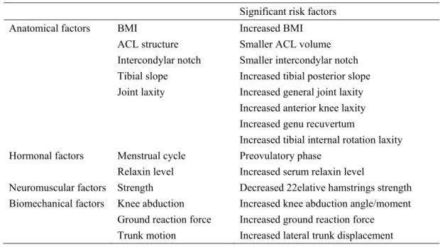
Although many studies have investigated the risk factors of ACL injuries, some were limited by their small sample sizes or retrospective design. Also, some factors were evaluated only with sex comparisons and were not directly tested on ACL- injured subjects. Table 2-1 summarizes the significant risk factors to date on the basis of prospective studies.
Significant risk factors
Anatomical factors BMI Increased BMI
ACL structure Smaller ACL volume Intercondylar notch Smaller intercondylar notch Tibial slope Increased tibial posterior slope Joint laxity Increased general joint laxity
Increased anterior knee laxity Increased genu recuvertum
Increased tibial internal rotation laxity Hormonal factors Menstrual cycle Preovulatory phase
Relaxin level Increased serum relaxin level
Neuromuscular factors Strength Decreased 22elative hamstrings strength Biomechanical factors Knee abduction Increased knee abduction angle/moment
Ground reaction force Increased ground reaction force Trunk motion Increased lateral trunk displacement
Table 2-1 Summary of risk factors of ACL injury
23 2.4 PREVENTION OF ACL INJURY
2.4.1 Effects on biomechanical and neuromuscular functions
Several studies have evaluated the effect of injury prevention training on lower extremity mechanics. Hewett et al. (1999) investigated the effect of a jump-training program on the landing mechanics in female adolescents. Female athletes had significantly decreased peak landing force and knee abduction and adduction moments after the training period (Hewett et al., 1999). Lephart et al. (2005) evaluated the effect of an 8-week plyometric and resistance training and found that increased hip and knee flexion angles and decreased hip and knee flexion moments were observed in both training groups. Myer, Ford, McLean, and Hewett (2006) investigated the effect of a 6-week neuromuscular training on the lower-extremity biomechanics in female adolescents. Their study showed that the neuromuscular training decreased the knee valgus and varus torques compared with the control group (Myer et al., 2006). Pollard, Sigward, Ota, Langford, and Powers (2006) investigated the influence of in-season injury prevention training, by using the Prevent Injury and Enhance Performance (PEP) program, on hip and knee kinematics during a landing task. After the season, the subjects demonstrated decreased hip internal rotation and increased hip abduction; however, there were no differences in knee valgus or knee flexion angles (Pollard et al., 2006). Lim et al. (2009) evaluated the effects of an 8- week injury prevention training in female high school basketball players. The training group showed increased knee flexion angle and inter-knee distances, and decreased maximum knee extension torques during the rebound-jump task after the training period (Lim et al., 2009).
The effects of injury prevention training on neuromuscular functions were also reported. Hewett et al. (1999) found that a jump-training program increased the
24
hamstrings-to-quadriceps muscle peak torque ratio on both dominant and non- dominant sides. Lim et al. (2009) also reported that an injury prevention program improved the strength of hip abductors, hip extensors, and knee flexors. Wojtys, Huston, Taylor, and Bastian (1996) investigated the effects of isokinetic, isotonic, and agility training on the muscle reaction time against the tibial anterior translation stimulus. Agility training improved the spinal reflex times of the medial and lateral quadriceps (Wojtys et al., 1996). Also, the cortical response time in the agility training group was improved in the gastrocnemius, medial hamstrings, and lateral quadriceps (Wojtys et al., 1996). Chimera, Swanik, Swanik, and Straub (2004) investigated the effects of plyometric training on muscle activation strategies in the lower-extremity. Plyometric training increased the preparatory hip adductor activity and adductor-to-abductor co-activation during landing (Chimera et al., 2004). Lephart et al. (2005) also evaluated the effect of plyometric or resistance training on muscle activation. Their results showed that both types of training improved the preparatory muscle activity of the gluteus medius during landing (Lephart et al., 2005).
As the methods of injury prevention training are different among studies, the results were not consistent. However, most of the studies have demonstrated that an injury prevention program was effective in improving the biomechanical and neuromuscular functions associated with ACL injury.
2.4.2 Effects on ACL injury rates
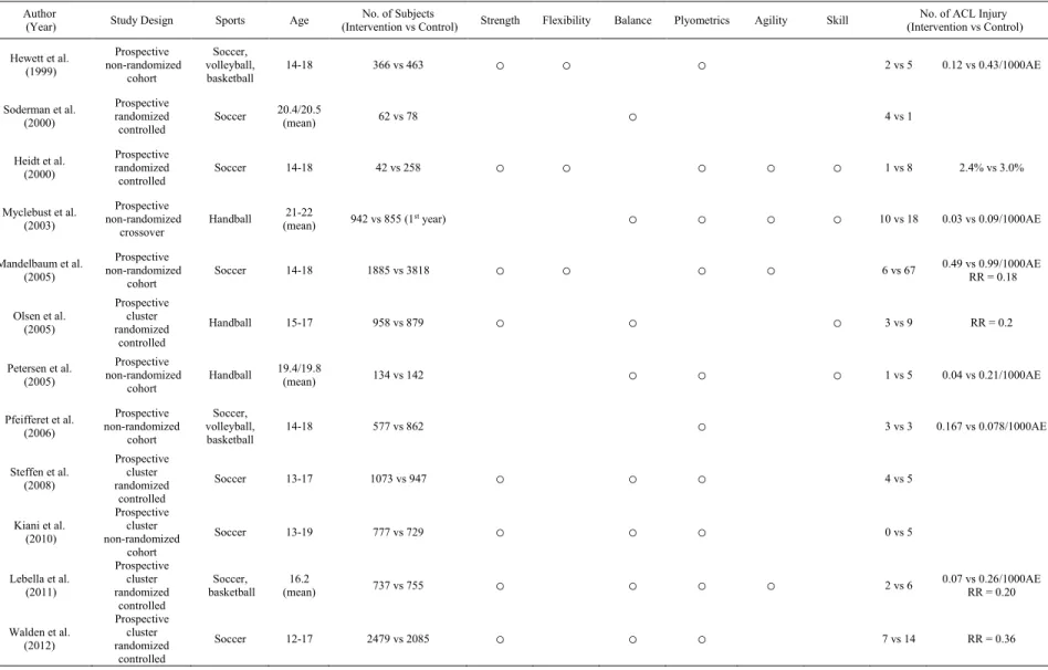
Several studies have investigated the effect of an injury prevention program on the incidence of ACL injury (Heidt Jr et al., 2000; Hewett et al., 1999; Kiani et al., 2010; LaBella et al., 2011; Mandelbaum et al., 2005; Myklebust, Engebretsen, et al., 2003; Olsen et al., 2005; Petersen et al., 2005; Pfeiffer, Shea, Roberts, Grandstrand, &
25
Bond, 2006; Söderman, Werner, Pietilä, Engström, & Alfredson, 2000; Steffen et al., 2008; Waldén et al., 2012). Table 2-2 presents a summary of these studies. Although some studies had methodological issues, an injury prevention training seems to be effective in reducing the number of ACL injuries.
26
Author
(Year) Study Design Sports Age No. of Subjects
(Intervention vs Control) Strength Flexibility Balance Plyometrics Agility Skill No. of ACL Injury (Intervention vs Control) Hewett et al.
(1999)
Prospective non-randomized
cohort
Soccer, volleyball,
basketball 14-18 366 vs 463 ○ ○ ○ 2 vs 5 0.12 vs 0.43/1000AE
Soderman et al.
(2000)
Prospective randomized
controlled Soccer 20.4/20.5
(mean) 62 vs 78 ○ 4 vs 1
Heidt et al.
(2000)
Prospective randomized
controlled Soccer 14-18 42 vs 258 ○ ○ ○ ○ ○ 1 vs 8 2.4% vs 3.0%
Myclebust et al.
(2003)
Prospective non-randomized
crossover
Handball 21-22
(mean) 942 vs 855 (1st year) ○ ○ ○ ○ 10 vs 18 0.03 vs 0.09/1000AE
Mandelbaum et al.
(2005)
Prospective non-randomized
cohort Soccer 14-18 1885 vs 3818 ○ ○ ○ ○ 6 vs 67 0.49 vs 0.99/1000AE
RR = 0.18
Olsen et al.
(2005)
Prospective cluster randomized
controlled
Handball 15-17 958 vs 879 ○ ○ ○ 3 vs 9 RR = 0.2
Petersen et al.
(2005)
Prospective non-randomized
cohort Handball 19.4/19.8
(mean) 134 vs 142 ○ ○ ○ 1 vs 5 0.04 vs 0.21/1000AE
Pfeifferet et al.
(2006)
Prospective non-randomized
cohort
Soccer, volleyball,
basketball 14-18 577 vs 862 ○ 3 vs 3 0.167 vs 0.078/1000AE
Steffen et al.
(2008)
Prospective cluster randomized
controlled
Soccer 13-17 1073 vs 947 ○ ○ ○ 4 vs 5
Kiani et al.
(2010)
Prospective cluster non-randomized
cohort
Soccer 13-19 777 vs 729 ○ ○ ○ 0 vs 5
Lebella et al.
(2011)
Prospective cluster randomized
controlled
Soccer,
basketball 16.2
(mean) 737 vs 755 ○ ○ ○ ○ 2 vs 6 0.07 vs 0.26/1000AE
RR = 0.20
Walden et al.
(2012)
Prospective cluster randomized
controlled
Soccer 12-17 2479 vs 2085 ○ ○ ○ 7 vs 14 RR = 0.36
Table 2-2 Summary of ACL injury prevention intervention studies
27 CHAPTER 3
Study 1: ACL Injury Risk in Female Athletes during Maturation
3.1 INTRODUCTION
The incidence of ACL injury starts to increase after puberty in female athletes (Granan et al., 2008). It is important to understand why this trend occurs during this period. As physical growth greatly influences movement strategy, this study
investigated the changes in biomechanical risk factors during puberty. Identifying when the risk for ACL injury increase would help determine when to initiate an ACL injury prevention training. The purpose of this study is to evaluate whether the biomechanical risk factors of ACL injury increase during puberty in female basketball players.
3.2 METHODS Subjects
Ninety-two female basketball players were recruited from the local elementary school, and junior and senior high schools. Subjects were included if they were members of school basketball teams and practiced more than three times per week.
Subjects were excluded from the study if they had a history of ACL injury, a lower extremity injury within 6 weeks that prevented full participation in basketball games, any medical or neurological pathology, or had previously participated in an injury prevention program. The maturational stage was evaluated by using the self-
administered rating scale for pubertal development (Carskadon & Acebo, 1993). The development of body structure and muscular function depends more on maturational stages rather than chronological ages; thus, the ACL injury risk was compared among
28
the different maturational stages. Subjects were categorized into four maturational stages: early pubertal (n = 14, age 11.1 ±1.2 years), middle pubertal (n = 13, age 13.1 ± 1.0 years), late pubertal (n = 43, age 13.1 ± 0.7 years), and post-pubertal (n = 22, age 15.5 ± 1.1 years). The early-, middle-, late-, and post-pubertal stages were equivalent to the Tannar stages 2 to 5, respectively (Carskadon & Acebo, 1993). Once it was
determined that the subjects met the inclusion and were not excluded by the exclusion criteria, the subjects and their guardians were informed about the nature of the study, and were asked if they would like to participate. All subjects and their guardians were required to provide written consent to participate in the study. This study was approved by the Academic Research Ethical Review Committee of Waseda University.
Data collection
An ACL injury prediction algorithm developed by Myer et al. (2010) was used to evaluate the knee mechanics and ACL injury risk. This algorithm was reported to have high sensitivity and specificity, and was able to identify female athletes who
demonstrate a high knee abduction moment, which increases their risk of sustaining an ACL injury (Myer et al., 2010).
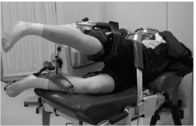
The height, weight, and tibia length were measured. Tibia length was measured the distance between the lateral knee joint line and the prominence of the lateral malleolus with the subjects standing with their knees extended. On the basis of the method described by Myer et al. (2010), the quadriceps-to-hamstrings (QH) strength ratio was obtained through surrogate calculations by multiplying the female athlete’s body mass by 0.01 and adding the resultant value to 1.10.
29
Two-dimensional lower extremity kinematics measurements were conducted.
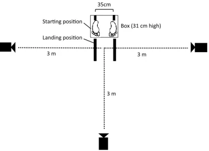
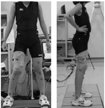
Eight bilateral markers were placed on each subject in the following locations: the greater trochanter, lateral knee joint line, patella, and lateral malleolus. Frontal and sagittal plane images were simultaneously captured with three video cameras (30 Hz;
CASIO EXILIM, Tokyo, Japan) (Figure 3-1). A basketball goal was used as an overhead target. The subjects performed a drop vertical jump (DVJ), as described previously (Myer et al., 2010). The subjects stood on a box (31 cm high) with their feet positioned 35 cm apart. They were instructed to jump off the box and then immediately perform a maximum vertical jump, raising both arms toward the target. Before testing, the subjects were allowed to perform one to three practice trials to familiarize
themselves with the test maneuver. Once they were able to perform the test maneuver, each subject performed three DVJ trials. No feedback was provided between the trials.
Figure 3-1 Experiment setup
30 Data analysis
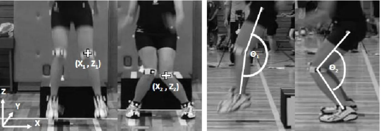
Frontal and sagittal images of the first DVJ landing were analyzed. The video files were first de-interlaced by using VirtualDub software (Avery Lee, GNU general public license). Then, the data were imported into ImageJ software (National Institute of Health, USA) to measure the knee valgus motion and knee flexion range of motion. The knee valgus motion was defined as the displacement between the patellar markers at the frame before initial contact and at the frame with a maximum medial position (Figure 3- 2) (Myer et al., 2010) . The displacement measurements were calibrated by using a known distance. The knee flexion angle was measured with the angle made by the greater trochanter, lateral knee joint line, and lateral malleolus. The knee flexion range of motion was defined as the difference in the knee flexion angles at the frame before initial contact and maximum knee flexion (Figure 3-2) (Myer et al., 2010). With the tibia length, body mass, QH ratio, knee valgus motion, and knee flexion range of motion as variables, the ACL injury prediction algorithm was used to obtain the probability of a high knee abduction moment (pKAM), which was defined as the measure of ACL injury risk in this study (Figure 3-3). Only the left leg was analyzed, as previous studies have found that female athletes were more likely to tear their left ACL more frequently than the right (Brophy, Silvers, Gonzales, & Mandelbaum, 2010; Negrete, Schick, &
Cooper, 2007; Ruedl et al., 2012).
31
Figure 3-3 Example of use of the ACL injury prediction algorithm (modified from Myer, Ford, Khoury, Succop, and Hewett (2010))
With 36.0 cm in tibia length, 6.5 cm in knee valgus motion, 70.0° in knee flexion range of motion, 50.0 kg in weight, and 1.6 in QH ratio, this athlete would have a 66% chance of demonstrating high knee abduction moment (> 21.74 Nm).
Figure 3-2 Calculations of knee valgus motion and knee flexion range of motion Left: The displacement measure between the two marked knee alignments (X2 – X1) is representative of the amount of knee valgus motion. Right: The displacement measure between the two knee flexion angles (Θ1 – Θ 2) is representative of the amount of knee flexion range of motion.
32 Statistical analysis
The mean and standard deviation of the knee valgus motion, knee flexion range of motion, and pKAM were calculated for each group. A one-way analysis of variance was used to compare the means among the four groups for each dependent variable. When there were significant differences, a post hoc test was performed with a Bonferroni least significant test. The alpha level was set at 0.05. SPSS ver. 21 (SPSS Inc., Chicago, IL, USA) was used to perform the statistical analysis.
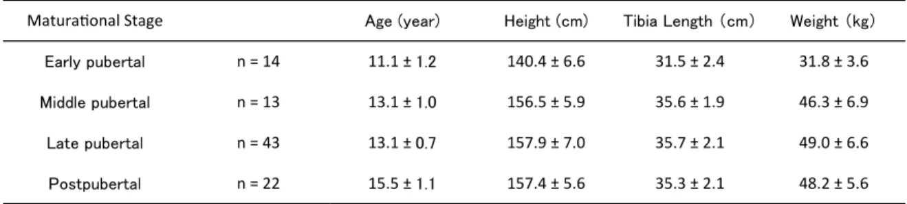
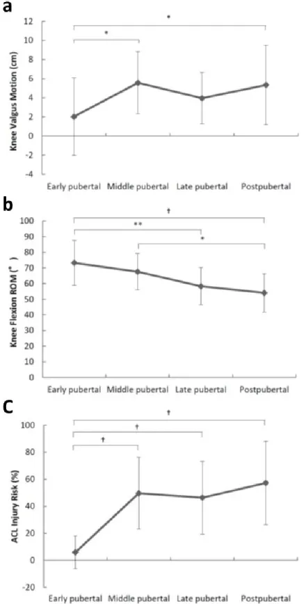
3.3 RESULTS
Table 3-1 presents the mean and standard deviation of age, height, tibia length, and weight by group. The height, weight, and tibia length were significantly larger in the middle-, late-, and post-pubertal stages than in the early-pubertal stage (p < 0.001).
However, there were no significant differences among the middle-, late-, and post- pubertal stages.
The knee valgus motion was significantly different among the groups (F(3,88) = 3.57, p = 0.017). The middle- and post-pubertal groups demonstrated significantly larger knee valgus motion than the early pubertal group (early = 2.0 ± 4.1cm vs. middle
= 5.6 ± 3.2 cm, p = 0.046; early = 2.0 ± 4.1 cm vs. post = 5.3 ± 4.1 cm, p = 0.030) (Figure 3-4a). There were no significant differences in knee valgus motion among the other groups. The knee flexion range of motion was also significantly different among groups (F(3,88) = 8.91, p < 0.001). The knee flexion range of motion was significantly smaller in the late- and post-pubertal groups compared with the early-pubertal group (early = 73.2 ± 14.3° vs. late = 58.3 ± 11.8°, p = 0.001; early = 73.2 ± 14.3° vs. post = 54.1 ± 12.2°, p < 0.001) (Figure 3-4b). Also, the knee flexion range of motion was
33
significantly smaller in the post-pubertal group than in the middle-pubertal group (middle = 67.6 ± 11.6° vs. post = 54.1 ± 12.2°, p = 0.013) (Figure 3-4b). There were also significant differences in the pKAM among groups (F(3,88) = 12.03, p < 0.001).
The pKAM was significantly higher in the middle-, late-, and post-pubertal groups when compared to the early-pubertal group (early = 5.9 ± 12.2% vs. middle = 49.6 ± 26.5%, p < 0.001; early =5.9 ± 12.2% vs. late = 46.3 ± 26.8%, p < 0.001; early = 5.9 ± 12.2% vs. post = 57.2 ± 30.8%, p < 0.001) (Figure 3-4c). No other significant
differences were observed in pKAM among the groups.
Table 3-1 Subject demographics. Data are shown as mean ± standard deviation.
34
Figure 3-4 Comparison of knee valgus motion (a), knee flexion range of motion (b), and pKAM (c) among maturational stages *:p<0.05, **:p<0.01, †:p<0.001
35 3.4 DISCUSSION
Knee valgus motion is one of the risk factors of ACL injury. It was identified, through analysis of videos of ACL injury situations, that the main mechanisms of ACL injury involved increased knee valgus motion combined with internal tibial rotation (Koga et al., 2010). Biomechanical studies with cadavers have also identified that the knee abduction moment is an essential factor that contributes to ACL injury (Kiapour et al., 2014; Levine et al., 2013). Furthermore, a large prospective study demonstrated that increased knee valgus angle and moment are significant predictors of future ACL injuries (Hewett et al., 2005). In this study, it was therefore investigated when an increase in this knee valgus motion is manifested during female maturation. The results of this study showed that the knee valgus motion significantly increased after the early- pubertal stage. Previous studies also reported that an increase in knee valgus angle was observed after girls reached puberty (Ford, Myer, & Hewett, 2010; Hewett et al., 2004;
Schmitz et al., 2009).
Another risk factor of ACL injury, a decrease in knee flexion, was also observed in this study. The knee flexion range of motion was significantly reduced as girls mature. This result was in agreement with a previous study by Yu et al. (2005) that found that the knee flexion angle during landing was significantly decreased as age increased, especially after 14 years of age in female athletes. A decrease in knee flexion angle increases the anterior tibial shear force generated by the quadriceps, thereby straining the ACL. Reduced knee flexion angle was commonly observed in injury situations (Koga et al., 2010; Krosshaug et al., 2007).
These biomechanical changes seemed to occur between the early- and middle- pubertal stages. As shown in Table 3-1, the height, weight, and tibia length were
36
significantly increased between the early- and middle-pubertal stages. During this period, the height, weight, and tibia length increased by 16.0 cm, 14.5 kg, 4.2 cm, respectively. The peak height velocity in Japanese girls was 10.2 ± 1.9 cm/year, and the mean age at peak height velocity was 11.5 ± 1.4 years (Satake, Kikuta, & Ozaki, 1993).
The peak weight velocity was 8.0 ± 1.8 kg/year, and the mean age at peak weight velocity was 12.6 ± 1.4 years (Satake et al., 1993). The mean ages in the early- and middle-pubertal stages were 11.1 and 13.1 years; therefore, the peak height and weight velocity might have occurred between the early- and middle-pubertal stages. These significant changes in structure might affect their movement patterns. A rapid increase in height and weight was reported to increase the torque around the knee joint
(Quatman, Ford, Myer, & Hewett, 2006); therefore, female adolescents might have demonstrated changes in movement patterns. Moreover, it was reported that female adolescents do not have a significant “neuromuscular development” during puberty compared with male adolescents (Hewett et al., 2004). Male adolescents increased their quadriceps and hamstrings peak torque with increasing maturity, whereas female adolescents did not (Hewett et al., 2004). Therefore, female adolescents might have reduced neuromuscular control of the knee joint during puberty. With a combination of structural and neuromuscular changes, girls develop high-risk movement patterns as they mature.
These changes in structure and movement patterns increased the overall risk of ACL injury, as measured by pKAM. The pKAM was significantly increased between the early- and middle-pubertal stages. Okuwaki (2012) reported that the number of ACL injury starts to increase at around the age of 13-14 years, and peaks at around the age of
37
16-17 years. This period seems to be in agreement with the period of pKAM increase observed in this study.
The difference in knee valgus motion between the early- and middle-pubertal stages was about 4 cm. Also, the difference in the knee flexion range of motion between the early- and late-pubertal stages was about 15°. It was indicated that a decrease in the knee flexion angle by about 15° might result in increased activation of the quadriceps (Malinzak, Colby, Kirkendall, Yu, & Garrett, 2001). Moreover, a decrease in the knee flexion angle was reported to increase the force in the ACL (Mesfar & Shirazi-Adl, 2005). Therefore, the difference of 15° in the knee flexion angle might be clinically meaningful. Although these differences in the knee valgus motion and knee flexion range of motion seem small, the pKAM, which accounts for both biomechanical and structural factors, was increased by 44% between the early- and middle-pubertal stages.
This change in pKAM seems clinically important. When evaluating maturing subjects, it might be necessary to take into account both the changes in structure and the
biomechanics.
One of the limitations of this study was its cross sectional nature which did not allow determining changes over time. Another limitation was that the knee kinematics were evaluated by using two-dimensional measurements in this study. Although a good correlation between two-dimensional and three-dimensional analysis was reported in previous studies (Myer et al., 2010), two-dimensional measures might not represent the complex movements of the knee. In addition, the use of the pKAM might not be as accurate as the laboratory-based assessment in evaluating the risk of ACL injury.
However, this assessment tool is less costly and easier to apply; thus, it is more
38
clinically relevant. Also, the number of subjects in each group was not balanced;
therefore, it might have affected the results of this study.
3.5 CONCLUSION
The knee valgus motion, knee flexion range of motion, and pKAM were
significantly changed during pubertal growth in female athletes; the period between the early- and middle-pubertal stages seemed especially critical. Therefore, intervention for ACL injury prevention should be initiated during this period to reduce the risk of ACL injury.