Pemafibrate Dramatically Ameliorated the Values of Liver Function Tests and
Fibrosis Marker in Patients with Non-Alcoholic Fatty Liver Disease
Suguru Ikeda,* Takaaki Sugihara,* Yoshiki Hoshino,* Yukako Matsuki,* Takakazu Nagahara,* Jun-ichi Okano,* Sonoko Kitao,† Youhei Fujioka,† Kazuhiro Yamamoto† and Hajime Isomoto*
*Division of Medicine and Clinical Science, Department of Gastroenterology and Nephrology, School of Medicine, Faculty of Medicine, Tottori University, Yonago 683-8504, Japan and †Division of Medicine and Clinical Science, Department of Cardiovascular Medicine and Endocrinology and Metabolism, School of Medicine, Faculty of Medicine, Tottori University, Yonago 683-8504, Japan
ABSTRACT
Background Non-alcoholic fatty liver disease (NAFLD) is a chronic liver disease related to metabolic syndrome, which can progress to liver cir-rhosis. Standard medication has not been established. Pemafibrate is a selective peroxisome proliferator-activated receptor (PPAR) α modulator. We retrospec-tively evaluated the efficacy of pemafibrate in patients with NAFLD.
Methods We retrospectively enrolled 17 patients (ten men, seven women; median age, 63 years; range, 27–81 years). They were all proven to have fatty liver through imaging and had little or no history of drinking (ethanol consumption of < 20 g/day for women and < 30 g/day for men). They were administered pemafibrate from October 2018 to June 2020.
Results After administration, serum triglyceride (TG) tended to be decreased (300.5 ± 22.5 to 239.5 ± 34.3 mg/dL, P = 0.06). Serum high-density lipoprotein (HDL) cholesterol and low-density lipoprotein (LDL) cholesterol levels did not change. ALT was significantly decreased (–47.4%) for six months (57.5 ± 8.8 to 30.3 ± 5.8 U/L, P < 0.01). The values of serum GGT signifi-cantly decreased (–48.7%) for sixth months (63.9 ± 10.3 to 32.8 ± 6.6 U/L, P < 0.01). Aspartate aminotransferase (AST) to platelet ratio (APRI), a fibrosis marker, also was significantly decreased in the sixth month (0.7 ± 0.1 to 0.4 ± 0.1, P < 0.05). Body mass index (BMI) and hemoglobin A1c (HbA1c) showed no significant change.
Conclusion Pemafibrate dramatically ameliorated the values of liver function tests and APRI in patients with NAFLD.
Key words non-alcoholic fatty liver disease; non- alcoholic steatohepatitis; pemafibrate; liver function; liver fibrosis
Non-alcoholic fatty liver disease (NAFLD), including steatohepatitis (NASH), has emerged as a common public health problem in recent decades. The overall global prevalence of NAFLD diagnosed by imaging is estimated at around 25%.1 It is also increasing in the
Asia-Pacific region.2 The treatment for NAFLD consists
of a variety of strategies, including lifestyle modification and several drugs such as metabolism ameliorator, anti-inflammatory agents, and antifibrotic agents. However, the recommended pharmacotherapies are extremely limited, and vitamin E or pioglitazone are indicated only for a certain limited number of patients according to the guidelines.3 The standard medication for NAFLD
has not yet been established.
Peroxisome proliferator-activated receptors (PPARs) form a subfamily of the nuclear receptor superfamily.4 There are three different types of PPAR
(α, δ, and γ). Among them, PPARα (nuclear receptor subfamily 1, group C, member 1, NR1C1) is a ligand-activated nuclear receptor highly expressed in the liver. PPARα was initially identified as the molecular target of xenobiotics inducing peroxisome proliferation in rodents.5 PPARα has been well-established as a
critical modulator of lipid transport and metabolism, notably mitochondrial and peroxisomal fatty acid beta-oxidation.6 It negatively regulates pro-inflammatory and
acute phase response signaling pathways.7 Modulating
PPARα has been considered as the key to treating meta-bolic disease, including NAFLD.8
Pemafibrate (K-877, Parmodia® tablet, Kowa Company, Ltd., Nagoya, Japan), a selective PPARα modulator (SPPARMα), was approved in July 2017 and released in June 2018 in Japan. In Japan, phase 2 and 3 trials demonstrated that pemafibrate significantly ame-liorated lipid abnormalities in patients with dyslipidemia without increasing adverse effects.9, 10 A large-scale
trial, PROMINENT, for dyslipidemic patients with type 2 diabetes (T2DM) is now ongoing.11
Corresponding author: Takaaki Sugihara, MD, PhD sugitaka@tottori-u.ac.jp
Received 2020 July 17 Accepted 2020 July 30
Online published 2020 August 7
Abbreviations: ALT, alanine aminotransferase; APRI, aspartate aminotransferase to platelet ratio; AST, aspartate aminotransfer-ase; BMI, body mass index; GGT, gamma-glutamyl transferaminotransfer-ase; LFTs, liver function tests; NAFLD, non-alcoholic fatty liver disease; NASH, non-alcoholic steatohepatitis; PPAR, peroxisome proliferator-activated receptor; TG, triglycerides
Pemafibrate, which improves liver pathology in the diet-induced rodent model of NASH,12 is expected
to become a promising therapeutic agent for human NASH. However, the efficacy of pemafibrate in patients with NAFLD has not yet been reported.
Serum aspartate aminotransferase (AST) and alanine aminotransferase (ALT) values have long been used as surrogate markers for liver injury.13, 14 ALT is
also a marker for hepatocyte death, and predicts the presence of liver fibrosis and risk for fibrosis progression in NASH.15 It has also been reported that
normaliza-tion of ALT levels after intervennormaliza-tion is associated with fibrosis improvement in NASH.16, 17 On the other hand,
gamma-glutamyl transpeptidase (GGT) levels indirectly reflect the oxidative stress related to the glutathione levels in the liver.18 Oxidative stress is the core
patho-genesis of NAFLD.19 Therefore, reducing the values
of serum liver function tests (LFTs) (mainly ALT and GGT) is considered as a predictive factor of long-term
treatment success in NAFLD.
In this study, we aimed to evaluate the efficacy of pemafibrate on LFTs in patients with NAFLD retrospectively.
SUBJECTS AND METHODS
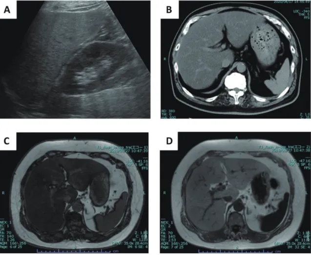
This was a single-center retrospective observational study. Between October 2018 and June 2020, a total of 77 patients in our hospital that were administered pemafibrate were enrolled. Then, only patients with fatty liver proven by imaging (urtrasonography; US, computed tomography; CT, or magnetic resonance of imaging; MRI) were selected (Figs. 1A, B, C and D) for the study. Patients who had other causes for chronic hepatitis (hepatitis B virus, hepatitis C virus, autoim-mune hepatitis, primary biliary cholangitis) were not enrolled.
Table 1 demonstrates the definition of fatty liver by imaging.20 Patients who discontinued pemafibrate for
Fig. 1. Representative images of fatty liver. (A) Ultrasonography demonstrated higher echogenicity of the liver than the renal cortex, (B) Unenhanced computed tomography demonstrated lower density than the spleen, Magnetic resonance of imaging demonstrated the signal loss on (C) out-of-phase compared with (D) in-phase imaging.
any reason, had a history of drinking (ethanol consump-tion of > 20 g/day for women and > 30 g/day for men), and short duration of using pemafibrate (< 3 months) were excluded from the study. Finally, we selected 17 patients (ten men, seven women) (Fig. 2). We obtained each patient’s information concerning gender, age, height, and body weight, and then calculated body mass index (BMI), both pre-treatment and most recent. We also collected the information about concomitant medi-cations, which are considered useful for NAFLD/NASH treatment as follows; dipeptidyl peptidase-4 (DPP4) inhibitor, metformin, sodium-glucose cotransporter 2 (SGLT2) inhibitor, ursodeoxycholic acid (UDCA), eicosapentaenoic acid (EPA), statin, and ezetimibe. Fasting laboratory data [triglyceride (TG), high-density lipoprotein (HDL) cholesterol, low-density lipoprotein
(LDL) cholesterol, AST, ALT, GGT, fibrosis index based on the four factors (FIB-4) index using age, AST, ALT and platelet values,21–23 AST to platelet ratio index
(APRI)24, 25] were collected and calculated as well. Six
months were selected for the median duration of treat-ment. Missing values were inputted using the median of pre- and post values.
Statistical analysis
The Friedman test and Dunn’s test were applied for multiple paired comparisons. The Wilcoxon test was applied for comparing two paired groups. The Mann-Whitney test was applied for comparing two independent groups. One-way ANOVA was applied for comparing three independent groups. All statistical tests were performed using StatFlex (Windows ver. 6.0;
Table 1. The definition of fatty liver on imaging
Modalities Definition
US (1) Increased echogenicity, hepatomegaly, and intra-hepatic vascular blurring. (2) Higher echogenicity than renal cortex
(1) and/ or (2)
CT An unenhanced CTL-S (ratio of liver to splenic attenuation values) of less than 1 HU.
MRI The signal loss on out-of-phase (OP) compared with in-phase (IP) imaging. CT, computed tomography; MRI, magnetic resonance imaging; HU, Hounsfield Unit; US, ultrasonography.
Fig. 2. Flow-chart of patient selection. Initially, there were seventy-seven eligible patients. Forty-one patients were not enrolled because they had not been proven to have fatty liver through imaging. Patients with other causes of chronic hepatitis were also not enrolled. Patients who stopped pemafibrate for any reason, and/or with a history of drinking and short duration of using pemafibrate were ex-cluded from the study. Seventeen patients were ultimately selected for this study.
Artech, Osaka, Japan). Values are expressed as median (range) or mean with a standard error of the mean (SEM). Statistical significance was set at P < 0.05.
Ethical Considerations
The study protocol was approved by the Institutional Review Board of our institute (No. 20A070) under the guidelines of the 1975 Declaration of Helsinki.
RESULTS
Baseline characteristics of the patients
The baseline characteristics of the patients are presented in Table 2. The median age of patients was 63 years; range, 27–81 years. Ten patients had T2DM (58.8%). Ten patients (58.8%), six patients (35.3%), and one patient (5.9%) were proven to have fatty liver by US, CT, and MRI, respectively. Three patients had been previously proven to have NASH by liver biopsy. Pre-treatment laboratory values as follows; TG 300.5 ± 22.5 mg/dL, HDL-cholesterol 46.5 ± 2.4 mg/dL, LDL-cholesterol 109.5 ± 10.6 mg/dL, AST 43.8 ± 5.4 U/L, ALT 57.4 ± 8.8 U/L, GGT 63.9 ± 10.3 U/L. DPP4 inhibi-tor, metformin, SGLT2 inhibiinhibi-tor, EPA, statin, ezetimibe, and UDCA had been already prescribed in six (35.3%), three (17.6%), two (11.8%), three (17.6%), five (29.4%), seven (41.2%) and four (23.5%) patients, respectively. Three patients (17.6%) were given 0.1 mg bid (twice-daily), twelve (70.6%), 0.2 mg bid, and two (11.8%), 0.4 mg bid of pemafibrate, respectively. The dosage of pemafibrate was selected according to pre-treatment serum TG levels (0.1 mg: 0.2 mg: 0.4 mg = 275.3 ± 41.0: 298.0 ± 29.2: 353.5 ± 10.3 mg/dL). The median dura-tion of administradura-tion was 6.1 months; range, 3.0–19.1 months.
Serum TG tended to be decreased (–12.4%) (300.5 ± 22.5 to 239.5 ± 34.3 mg/dL, P = 0.06) for six months (Fig. 3A). The serum HDL cholesterol levels and LDL cholesterol did not change (46.5 ± 2.4 to 44.6 ± 1.5 mg/dL; 109.5 ± 10.6 to 124.7 ± 19.0 mg/dL) (Figs. 3B and C). The values of serum AST decreased (–34.2%); however, there is no statisti-cal significance (43.8 ± 5.4 to 28.5 ± 2.7 U/L, P = 0.23) (Fig. 3D). ALT was significantly decreased (–47.4%) for six months (57.5 ± 8.8 to 30.3 ± 5.8 U/L,
P < 0.01) (Fig. 3E). The values of serum GGT were
significantly decreased (–48.7%) for six months (63.9 ± 10.3 to 32.8 ± 6.6 U/L, P < 0.01) (Fig. 2F). BMI (26.8 ± 1.0 to 26.1 ± 1.0 kg/m2) and HbA1c (7.7 ± 0.5 to 7.0
± 0.2 %) did not change for six months (Figs. 4A and B). There was no difference in BMI between T2DM and non-T2DM patients (25.2 ± 1.3 vs. 25.0 ± 1.4 kg/ m2). There was also no significant change in BMI in
both groups in the sixth month (25.6 ± 1.2 and 24.4 ± 1.5 kg/m2). Gender made no difference in the efficacy of
pemafibrate.
FIB-4 index also did not change in the sixth month (1.7 ± 0.2 to 1.4 ± 0.2); on the other hand, APRI was significantly decreased in the sixth month (0.7 ± 0.1 to 0.4 ± 0.1, P < 0.05).
Three biopsy-proven NASH patients were admin-istered 0.2 mg bid of pemafibrate. The median duration was 5.6 months; range, 3.7–14.5 months. Serum TG and values of LFTs were decreased (AST –41.7%, ALT –53.8%, GGT –22.2%) in six months (Figs. 5A, B, C and D). APRI was also decreased (1.0 ± 0.2 to 0.5 ± 0.1) in three months.
DISCUSSION
In this study, we demonstrated that pemafibrate dramat-ically ameliorated the values of ALT, GGT, and APRI in NAFLD. To our knowledge, this is the first report demonstrating the biological effects of pemafibrate in humans with NAFLD.
Several trials reported the efficacy of fibrates on NAFLD patients. Miranda et al. reported that fenofi-brate for 48 weeks demonstrated a significant decrease in abnormal LFTs (AST; 88 ± 39 to 64 ± 31 U/L, ALT; 47 ± 23 to 38 ± 12 U/L, GGT; 90 ± 79 to 55 ± 41 U/L) in 16 patients, although liver histology did not change significantly.26 Gandhi N. et al. also reported that
fenofi-brate treatment for median 0.3 (0.02–2.1) years led to an improvement in LFTs (ALT; 43.1 ± 33.5 to 37.5 ± 24.5 U/L, GGT; 79.5 ± 59.8 to 57.1 ± 43.6 U/L) in 116 pa-tients.27 The reduction rate (around –20%) of fenofibrate
on ALT and GGT is lower than our data.
The difference between previously released fibrates and pemafibrate is the high selectivity of PPARα. The PPARα activation by pemafibrate was reported to be > 2,500 times stronger than fenofibric acid because of the Y-shaped structure, unlike conventional fibrates.28
The high selectivity enables pemafibrate to be used at a reduced dosage. Fenofibrate administration needs to be at least 106.6 mg to 160 mg qd (once-daily); on the other hand, only 0.2 to 0.4 mg bid of pemafibrate is needed. Although the major excretion route is the liver, the high selectivity enables pemafibrate to be used in any under-lying liver disease except for decompensated cirrhosis (Child-Pugh B or C). Moreover, pemafibrate is the first fibrate that can be used safely in combination with a statin. In this study, five patients had been prescribed a statin. There were no differences in LDL cholesterol and LFTs between combination and monotherapy groups for six months.
liver were administered pemafibrate (among these, 14 subjects were administered the clinical dose).6 In phase
3, 58 (53.2%) subjects with fatty liver were administered 0.2 mg or 0.4 mg bid of pemafibrate; however, there were 68 (62.4%) patients with a drinking habit. Since these trials did not exclude patients with alcoholic liver disease, the effects of pemafibrate on NAFLD could not be evaluated.
In phases 2 and 3, subjects with AST or ALT levels more than 2- or 3-fold higher than the upper limit of the reference range were excluded.6, 7 So our study reports
the first demonstration of the efficacy of clinical doses of pemafibrate for LFTs in patients with NAFLD.
In this study, BMI and HbA1c did not change for six months, which indicated that the improvement of ALT and GGT was achieved only by PPARα activation. We also evaluated the scoring system of fibrosis, and only APRI significantly decreased in sixth months after treatment with pemafibrate. The mean of ROC for APRI was reportedly 0.85 with an optimal cut-off of 0.98, giving a sensitivity of 75% and a specificity of 86%.25 In our study, the mean of APRI was 0.65, and
only two patients were over 0.98. APRI can reportedly distinguish patients with lobular inflammation and ballooning from those without.29 Decrease of AST is
directly considered as an improvement of liver injury associated with inflammation. Platelets, another factor of APRI, are reported as an active participant in the process of liver inflammation.30 Platelets exert potent
inflammatory effects and play a central part in the progression from simple steatosis to NASH.31 Excessive
platelet consumption is one of the mechanisms of thrombocytopenia in chronic liver disease.32 Platelets
were also reported to improve liver fibrosis and ac-celerate liver regeneration.33 In patients with chronic
hepatitis B with significant fibrosis receiving anti-viral therapy, the increase of platelets was associated with the
Table 2. Characteristics of 17 patients treated with pemafibrate
Male/Female 10:7
Age (years) 63 (27–81)
Height (m) 1.7 (1.5–1.8)
Pre-treatment body weight (kg) 70.6 (48.6–101.7) Pre-treatment BMI (kg/m2)* 26.8 (19.2–33.8) Patients of BMI > 25 kg/m2 11 (64.7%) Comorbidities T2DM 10 (58.8) Chronic hepatitis B† 1 (5.9) CAD 1 (5.9) IBD 1 (5.9) GERD 1 (5.9) Other‡ 1 (5.9) Imaging modalities US 10 (58.8) CT 6 (35.3) MRI 1 (5.9)
Biopsy proven NASH 3 (17.6)
Pre-treatment laboratory values
TG (mg/dL) 300.5 ± 22.5 HDL-cholesterol (mg/dL) 46.5 ± 2.4 LDL-cholesterol (mg/dL) 109.5 ± 10.6 AST (U/L) 43.8 ± 5.4 ALT (U/L) 57.5 ± 8.8 GGT (U/L) 63.9 ± 10.3 FIB-4 index 1.7 ± 0.2 APRI 0.7 ± 0.1 HbA1c (%) 7.7 ± 0.5 Concomitant medications DPP4 inhibitor 6 (35.3) Metformin 3 (17.6) SGLT2 inhibitor 2 (11.8) EPA 3 (17.6) Statin 5 (29.4) Ezetimibe 7 (41.2) UDCA 4 (23.5) Dosage of pemafibrate 0.1 mg 3 (17.6) 0.2 mg 12 (70.6) 0.4 mg 2 (11.8) Duration of pemafibrate administration (months) 6.1 (3.0–19.1)
APRI, aspartate aminotransferase to platelet ratio index; AST, aspartate aminotransferase; ALT, alanine aminotransferase; BMI, body mass index; CAD, coronary artery disease; CT, computed tomography; DPP4, dipeptidyl peptidase-4; EPA, eicosapentaenoic acid; GERD, gastroesophageal reflux disease; GGT, gamma-glutamyl transpeptidase; HDL, high-density lipoprotein; IBD, inflammatory bowel disease; LDL, low-density lipoprotein; MRI, the magnetic resonance of imaging; SGLT2, sodium-glucose cotransporter 2; T2DM, type 2 diabetes mellitus; TG, triglyceride; UDCA, ursodeoxycholic acid; US, ultrasonography. *BMI cannot be calculated in two patients because of a lack of body hight. †HBV DNA is controlled under the detective level by nucleotide analog treatment. ‡Ovarian insufficiency. Data are expressed as median (range) or mean ± SEM.
Fig. 3. Laboratory data during pemafibrate treatment for six months. (A) Triglyceride, (B) High-density lipoprotein cholesterol, (C) Low-density lipoprotein cholesterol, (D) Aspartate aminotransferase, (E) Alanine aminotransferase, (F) Gamma-glutamyl transferase. Data are expressed as mean with a standard error of the mean (SEM). *P < 0.05, **P < 0.01
improvement of liver fibrosis.34 In our study,
pemafi-brate decreased the AST level (–35%) and increased platelet count (+10%). AST and platelet counts could not demonstrate significant changes independently; however, APRI was significantly decreased in the sixth month after treatment with pemafibrate. Decreasing APRI can be considered as an improvement in hepatic inflammation and liver fibrosis. This significant change was not found by other medications.
It has already known that metformin can improve biochemical and metabolic features in NAFLD.35 In
our study, three patients had already taken metformin. There was no significant difference between patients with or without metformin. Metformin also has an effect of improving BMI via improvement in insulin resistance. Although BMI did not decrease in our three patients for six months (29.5 ± 0.3 to 29.1 ± 0.3 kg/m2),
the synergic effect of metformin and pemafibrate might be achieved by more prolonged treatment.
SGLT2 inhibitors have also been reported to have favorable effects on NAFLD in T2DM patients.36 In our
study, there were only two patients who had taken an
Fig. 4. The differences in BMI and HbA1c between pre-treatment and the sixth month.
(A) Body mass index did not change between pre-treatment and the sixth month. (B) HbA1c did not change between pre-treatment and the sixth month. Data are expressed as mean with a standard error of the mean (SEM). *P < 0.05. n.s., not significant.
SGLT2 inhibitor. There were no significant differences in the data obtained in this study. The other medications also did not demonstrate any differences in the data between patients with or without it.
Dose dependency of pemafibrate was also evalu-ated; however, there were no statistically significant differences in LFTs among three dosages because of the small sample size.
Among the excluded patients, four discontinued pemafibrate. One patient stopped it because of suc-cessful weight reduction, another patient stopped it for unknown reasons, and two were due to the elevation
of LFTs. One patient experienced AST 59 U/L to 119 U/L and ALT 55 U/L to 113 U/L after 12 weeks from administration. Another patient experienced AST 69 U/L to 201 U/L, ALT56 U/L to 181 U/L, and GGT 198 U/L to 677 U/L after 19 weeks from administration. Both were administered 0.2 mg bid. The adverse events on LFTs reported in the phase 3 trial were in only two patients (3.7%) complicated with GGT > three times the upper normal limit. There is no appropriate explanation for this discrepancy for now. Because of these results, we should pay much more attention to LFTs for at least four months after administration in order to gather more
Fig. 5. Laboratory data during pemafibrate treatment in biopsy-proven NASH patients.
(A) Triglyceride, (B) Aspartate aminotransferase, (C) Alanine aminotransferase, (D) Gamma-glutamyl transferase. Data are expressed as mean with a standard error of the mean (SEM).
information and experience.
Our study had some limitations. It was a small size retrospective observational study of only six months’ duration, and there was missing data that needed to be compensated by using median values. Selection bias could not be avoided because this study enrolled only imaging-proven NAFLD patients retrospectively. However, these data are consistent with other experi-mental and trial data.9, 12 For this reason, we believe
that we could demonstrate the efficacy of pemafibrate for NAFLD. Liver biopsy is undoubtedly the gold standard for diagnosing NASH at present. In this study, only three patients had a biopsy performed on them. Although it was difficult to obtain statistical differences, pemafibrate could also decrease LFTs in biopsy-proven NASH in a similar fashion to the patients that had NAFLD. Whether pemafibrate can reduce fibrosis or not was not evaluated by biopsy in this study. In Japan, clinical trials for the antifibrotic effect of pemafibrate on NAFLD are ongoing. The results are expected to eluci-date the further efficacy of pemafibrate for NAFLD.
In conclusion, we demonstrated that pemafibrate, the new SPPARMα, dramatically ameliorated the values of LFTs in patients with NAFLD. Pemafibrate has the potential to be the first standard medication of NAFLD.
The authors declare no conflict of interest. REFERENCES
1 Chalasani N, Younossi Z, Lavine JE, Charlton M, Cusi K, Rinella M, et al. The diagnosis and management of nonalco-holic fatty liver disease: Practice guidance from the American Association for the Study of Liver Diseases. Hepatology. 2018;67:328-57. DOI: 10.1002/hep.29367, PMID: 28714183 2 Liu CJ. Prevalence and risk factors for non-alcoholic
fatty liver disease in Asian people who are not obese. J Gastroenterol Hepatol. 2012;27:1555-60. DOI: 10.1111/j.1440-1746.2012.07222.x, PMID: 22741595
3 Sumida Y, Yoneda M. Current and future pharmacological therapies for NAFLD/NASH. J Gastroenterol. 2018;53:362-76. DOI: 10.1007/s00535-017-1415-1, PMID: 29247356 4 Berger J, Moller DE. The mechanisms of action of PPARs.
Annu Rev Med. 2002;53:409-35. DOI: 10.1146/annurev. med.53.082901.104018, PMID: 11818483
5 Issemann I, Green S. Activation of a member of the steroid hormone receptor superfamily by peroxisome proliferators. Nature. 1990;347:645-50. DOI: 10.1038/347645a0, PMID: 2129546
6 Abdelmegeed MA, Moon KH, Hardwick JP, Gonzalez FJ, Song BJ. Role of peroxisome proliferator-activated receptor-α in fasting-mediated oxidative stress. Free Radic Biol Med. 2009;47:767-78. DOI: 10.1016/j.freeradbiomed.2009.06.017, PMID: 19539749
7 Gervois P, Kleemann R, Pilon A, Percevault F, Koenig W, Staels B, et al. Global suppression of IL-6-induced acute phase response gene expression after chronic in vivo treat-ment with the peroxisome proliferator-activated receptor-α activator fenofibrate. J Biol Chem. 2004;279:16154-60. DOI: 10.1074/jbc.M400346200, PMID: 14764586
8 Pawlak M, Lefebvre P, Staels B. Molecular mechanism of PPARα action and its impact on lipid metabolism, inflamma-tion and fibrosis in non-alcoholic fatty liver disease. J Hepatol. 2015;62:720-33. DOI: 10.1016/j.jhep.2014.10.039, PMID: 25450203
9 Ishibashi S, Yamashita S, Arai H, Araki E, Yokote K, Suganami H, et al.; K-877-04 Study Group. Effects of K-877, a novel selective PPARα modulator (SPPARMα), in dyslipidaemic patients: A randomized, double blind, active- and placebo-controlled, phase 2 trial. Atherosclerosis. 2016;249:36-43. DOI: 10.1016/j.atherosclerosis.2016.02.029, PMID: 27062408
10 Araki E, Yamashita S, Arai H, Yokote K, Satoh J, Inoguchi T, et al. Effects of pemafibrate, a novel selective PPARa modula-tor, on lipid and glucose metabolism in patients with type 2 diabetes and hypertriglyceridemia: A randomized, double-blind, placebo-controlled, phase 3 trial. Diabetes Care. 2018;41:538-46. DOI: 10.2337/dc17-1589, PMID: 29298800 11 Yamashita S, Masuda D, Matsuzawa Y. Pemafibrate, a new
selective PPARα modulator: drug concept and its clinical applications for dyslipidemia and metabolic diseases. Curr Atheroscler Rep. 2020;22:5. DOI: 10.1007/s11883-020-0823-5, PMID: 31974794
12 Honda Y, Kessoku T, Ogawa Y, Tomeno W, Imajo K, Fujita K, et al. Pemafibrate, a novel selective peroxisome proliferator-activated receptor alpha modulator, improves the pathogenesis in a rodent model of nonalcoholic steatohepatitis. Sci Rep. 2017;7:42477. DOI: 10.1038/srep42477, PMID: 28195199 13 Kallai L, Hahn A, Röder V, Županić V. Correlation between
histological findings and serum transaminase values in chronic diseases of the liver. Acta Med Scand. 1964;175:49-56. DOI: 10.1111/j.0954-6820.1964.tb00549.x, PMID: 14110637 14 Gowda S, Desai PB, Hull VV, Math AA, Vernekar SN,
Kulkarni SS. A review on laboratory liver function tests. Pan Afr Med J. 2009;3:17. PMID: 21532726
15 Schwabe RF, Tabas I, Pajvani UB. Mechanisms of Fibrosis Development in Nonalcoholic Steatohepatitis. Gastroenterol-ogy. 2020;158:1913-28. DOI: 10.1053/j.gastro.2019.11.311, PMID: 32044315
16 Vilar-Gomez E, Calzadilla-Bertot L, Friedman SL, Gra-Oramas B, Gonzalez-Fabian L, Lazo-del Vallin S, et al. Serum biomarkers can predict a change in liver fibrosis 1 year after lifestyle intervention for biopsy-proven NASH. Liver Int. 2017;37:1887-96. DOI: 10.1111/liv.13480, PMID: 28544769 17 Kleiner DE, Brunt EM, Wilson LA, Behling C, Guy C,
Contos M, et al.; Nonalcoholic Steatohepatitis Clinical Research Network. Association of Histologic Disease Activ-ity With Progression of Nonalcoholic Fatty Liver Disease. JAMA Netw Open. 2019;2:e1912565. DOI: 10.1001/jamanet-workopen.2019.12565, PMID: 31584681
18 Irie M, Sohda T, Anan A, Fukunaga A, Takata K, Tanaka T. Reduced Glutathione suppresses Oxidative Stress in Nonal-coholic Fatty Liver Disease. Euroasian J Hepatogastroenterol. 2016;6:13-8. DOI: 10.5005/jp-journals-10018-1159, PMID: 29201718
19 Chen Z, Tian R, She Z, Cai J, Li H. Role of oxidative stress in the pathogenesis of nonalcoholic fatty liver disease. Free Radic Biol Med. 2020;152:116-41. DOI: 10.1016/ j.freeradbiomed.2020.02.025, PMID: 32156524
20 Li Q, Dhyani M, Grajo JR, Sirlin C, Samir AE. Current status of imaging in nonalcoholic fatty liver disease. World J Hepatol. 2018;10:530-42. DOI: 10.4254/wjh.v10.i8.530, PMID: 30190781
21 Vallet-Pichard A, Mallet V, Nalpas B, Verkarre V, Nalpas A, Dhalluin-Venier V, et al. FIB-4: an inexpensive and accurate marker of fibrosis in HCV infection. comparison with liver biopsy and fibrotest. Hepatology. 2007;46:32-6. DOI: 10.1002/ hep.21669, PMID: 17567829
22 McPherson S, Stewart SF, Henderson E, Burt AD, Day CP. Simple non-invasive fibrosis scoring systems can reliably exclude advanced fibrosis in patients with non-alcoholic fatty liver disease. Gut. 2010;59:1265-9. DOI: 10.1136/ gut.2010.216077, PMID: 20801772
23 Shah AG, Lydecker A, Murray K, Tetri BN, Contos MJ, Sanyal AJ; Nash Clinical Research Network. Comparison of noninvasive markers of fibrosis in patients with nonalcoholic fatty liver disease. Clin Gastroenterol Hepatol. 2009;7:1104-12. DOI: 10.1016/j.cgh.2009.05.033, PMID: 19523535 24 Wai C, Greenson JK, Fontana RJ, Kalbfleisch JD, Marrero
JA, Conjeevaram HS, et al. A simple noninvasive index can predict both significant fibrosis and cirrhosis in patients with chronic hepatitis C. Hepatology. 2003;38:518-26. DOI: 10.1053/jhep.2003.50346, PMID: 12883497
25 Kruger FC, Daniels CR, Kidd M, Swart G, Brundyn K, van Rensburg C, et al. APRI: a simple bedside marker for advanced fibrosis that can avoid liver biopsy in patients with NAFLD/NASH. S Afr Med J. 2011;101:477-80. PMID: 21920102
26 Fernández-Miranda C, Pérez-Carreras M, Colina F, López-Alonso G, Vargas C, Solís-Herruzo JA. A pilot trial of fenofibrate for the treatment of non-alcoholic fatty liver disease. Dig Liver Dis. 2008;40:200-5. DOI: 10.1016/ j.dld.2007.10.002, PMID: 18261709
27 Gandhi N, Lenton R, Bhartia M, Abbas A, Raju J, Ramachandran S. Effect of fibrate treatment on liver function tests in patients with the metabolic syndrome. Springerplus. 2014;3:14. DOI: 10.1186/2193-1801-3-14, PMID: 24455467 28 Yamashita S, Masuda D, Matsuzawa Y. Clinical
applica-tions of a novel selective PPARα modulator, pemafibrate, in dyslipidemia and metabolic diseases. J Atheroscler Thromb. 2019;26:389-402. DOI: 10.5551/jat.48918, PMID: 30930344 29 Polyzos S, Slavakis A, Koumerkeridis G, Katsinelos P,
Kountouras J. Noninvasive Liver Fibrosis Tests in Patients with Nonalcoholic Fatty Liver Disease: An External Valida-tion Cohort. Horm Metab Res. 2019;51:134-40. DOI: 10.1055/ a-0713-1330, PMID: 30273934
30 Luyendyk J, Lisman T. Platelets as Modulators of Liver Dis-eases. Semin Thromb Hemost. 2018;44:114-25. DOI: 10.1055/ s-0037-1604091, PMID: 28898899
31 Diggs LP, Greten TF. The effects of platelet accumulation in fatty liver disease. Nat Rev Gastroenterol Hepatol. 2019;16:393-4. DOI: 10.1038/s41575-019-0160-8, PMID: 31114057
32 Mitchell O, Feldman DM, Diakow M, Sigal SH. The patho-physiology of thrombocytopenia in chronic liver disease. Hepat Med. 2016;8:39-50. PMID: 27186144
33 Kurokawa T, Ohkohchi N. Platelets in liver disease, cancer and regeneration. World J Gastroenterol. 2017;23:3228-39. DOI: 10.3748/wjg.v23.i18.3228, PMID: 28566882
34 Wang L, Wang B, You H, Wu X, Zhou J, Ou X, et al. Platelets’ increase is associated with improvement of liver fibrosis in entecavir-treated chronic hepatitis B patients with significant liver fibrosis. Hepatol Int. 2018;12:237-43. DOI: 10.1007/s12072-018-9864-z, PMID: 29700765
35 Li Y, Liu L, Wang B, Wang J, Chen D. Metformin in non-alcoholic fatty liver disease: A systematic review and meta-analysis. Biomed Rep. 2013;1:57-64. DOI: 10.3892/br.2012.18, PMID: 24648894
36 Scheen AJ. Beneficial effects of SGLT2 inhibitors on fatty liver in type 2 diabetes: A common comorbidity associated with severe complications. Diabetes Metab. 2019;45:213-23. DOI: 10.1016/j.diabet.2019.01.008, PMID: 30708071