Accepted Manuscript
Title: Molecular dynamics study on conformational
differences between dGMP and 8-oxo-dGMP: Effects of metal ions
Author: Shin-ichi Fujiwara Kenichiro Sawada Takashi Amisaki
PII: S1093-3263(14)00080-1
DOI: http://dx.doi.org/doi:10.1016/j.jmgm.2014.05.007
Reference: JMG 6415
To appear in: Journal of Molecular Graphics and Modelling Received date: 11-11-2013
Revised date: 9-5-2014 Accepted date: 22-5-2014
Please cite this article as: S.-i. Fujiwara, K. Sawada, T. Amisaki, Molecular dynamics study on conformational differences between dGMP and 8-oxo-dGMP: effects of metal ions, Journal of Molecular Graphics and Modelling (2014), http://dx.doi.org/10.1016/j.jmgm.2014.05.007
This is a PDF file of an unedited manuscript that has been accepted for publication. As a service to our customers we are providing this early version of the manuscript. The manuscript will undergo copyediting, typesetting, and review of the resulting proof before it is published in its final form. Please note that during the production process errors may be discovered which could affect the content, and all legal disclaimers that apply to the journal pertain.
Accepted Manuscript
Molecular dynamics study on conformational differences between dGMP and 8-oxo-dGMP: effects of metal ions
Shin-ichi Fujiwara*, Kenichiro Sawada, and Takashi Amisaki
Department of Biological Regulation, Faculty of Medicine, Tottori University, 86 Nishi-cho, Yonago 683-8503, Japan
*To whom correspondence should be addressed. Tel: +81-858-38-6356, Fax: +81-859-38-6350, E-mail: [email protected]
Accepted Manuscript
Highlights
• Molecular dynamics simulations of dGMP and 8-oxo-dGMP were performed. • 8-oxo-dGMP adopted both the syn and anti conformations.
• Metal ions increased the proportion of the anti conformation of 8-oxo-dGMP. • Ionic radius and charge influenced the stable anti conformations of 8-oxo-dGMP. • Metal ions may promote the binding of 8-oxo-G to nucleotide-sanitizing enzymes.
Abstract
The modified nucleotide base 7,8-dihydro-8-oxo-guanine (8-oxo-G) is one of the major sources of spontaneous mutagenesis. Nucleotide-sanitizing enzymes, such as the MutT homolog-1 (MTH1) and nudix-type motif 5 (NUDT5), selectively remove 8-oxo-G from the cellular pool of nucleotides. Previous studies showed that, although the syn conformation generally predominates in purine nucleotides with a bulky substituent at the 8-position, 8-oxo-dGMP binds to both MTH1 and NUDT5 in the anti conformation. This study was initiated to investigate the possibility that 8-oxo-dGMP itself may adopt the anti conformation. Molecular dynamics simulations of mononucleotides (dGMP, 8-oxo-dGMP) in aqueous solution were performed. 8-oxo-dGMP adopted the anti conformation as well as the syn conformation, and the proportion of adopting the anti conformation increased in the presence of metal ions. When 8-oxo-dGMP was in the anti conformation, a metal ion was located between the oxygen atom of phosphate and the oxygen atom at the 8-position of 8-oxo-G. The types of stable anti conformations of 8-oxo-dGMP differed, depending on the ionic radii and charges of coexisting ions. These data suggested a role for metal ions, other than as cofactors for the hydrolysis of the di- and tri-phosphate forms of mononucleotides; that the metal ions help retain the anti conformation of the N-glycosidic torsion angle of 8-oxo-dGMP to
Accepted Manuscript
promote the binding between the 8-oxo-G deoxynucleotide and the nucleotide-sanitizing enzymes.
Keywords: mononucleotide; molecular dynamics simulations; 8-oxo-dGTP; nucleotide pool;
Accepted Manuscript
Introduction
Nucleic acid bases in cells are easily modified by reactive oxygen species. Among these modified bases, 7,8-dihydro-8-oxo-guanine (8-oxo-G) is a major source of spontaneous mutagenesis. Cells have evolved different mechanisms to reduce the mutagenic effect of 8-oxo-G [1]. One mechanism is nucleotide-sanitization. This involves selective removal of modified bases from the nucleotide pool by nucleotide hydrolases so that the modified nucleotides cannot be incorporated into DNA by DNA polymerases. For example, 8-oxo-dGTPases such as MutT and the MutT homolog-1 (MTH1) hydrolyze 8-oxo-dGTP into 8-oxo-dGMP [1, 2]. 8-oxo-dGDPases such as nudix (nucleoside diphosphate linked moiety X)-type motif 5 (NUDT5) hydrolyze 8-oxo-dGDP into 8-oxo-dGMP [3]. The enzymes MutT, MTH1, and NUDT5 can discriminate among the slight structural differences between normal and modified nucleotides.
Structural studies have been performed to investigate the specific recognition of 8-oxo-G by MutT [4], MTH1 [5], and NUDT5 [6]. Crystal structures indicated that 8-oxo-dGMP bound to MutT adopted the syn conformation about the N-glycosidic bond that links the base to the sugar. Hydrogen bonds were observed between the enzyme and the oxygen atom at the 8-position (O8 atom) of 8-oxo-dGMP [4]. On the other hand, 8-oxo-dGMP was bound to MTH1 and NUDT5 in the anti and the high-anti conformations, respectively [5, 6]. However, the syn conformation should generally predominate in purine nucleotides with a bulky substituent at the 8-position [7]. No direct interaction, such as hydrogen bonding between MTH1 and NUDT5 and the O8 atom of 8-oxo-dGMP, was observed in the crystal structures of either enzyme [5, 6].
Conformations of mononucleotides have been analyzed experimentally [7], and recently, by computational analyses. Quantum mechanics (QM) calculations have been widely performed to compare potential energies of the anti and syn conformations of
Accepted Manuscript
mononucleotides [8-18]. Although these QM results gave insights into preferable conformations of mononucleotides in terms of the potential energies, water molecules and ions were either excluded or included only implicitly in many of these studies. The N-glycosidic conformation of 8-oxo-dGMP can be affected by water molecules and ions. However, it is generally very difficult to perform QM calculations that include water molecules or ions explicitly. On the other hand, although molecular dynamics (MD) simulations are less rigorous, they can generate a collection of conformations of a small ligand in explicit water molecules and ions [19]. The preferable conformation of mononucleotides can be found by comparing the numbers of the anti or syn conformations obtained by MD simulations. Numerous MD simulations of mononucleotides complexed with G protein-coupled receptors [20], and gas-phase conformations of mononucleotides [8], have been reported. However, to our knowledge, no previously published works apply MD simulations to 8-oxo-G mononucleotides in explicit solvents.
In this study, we performed MD simulations of dGMP and 8-oxo-dGMP in explicit solvents. Under the assumption that the proportion of the anti or syn conformations of mononucleotides might be influenced by water molecules, metal ions, or the state of protonation of the phosphate groups of mononucleotides, we examined different protonation states of phosphate, and the effects of different kinds of metal ions in MD simulations. We also tested different starting structures and two different parameters related to the N-glycosidic torsion angle. A series of simulations indicated that 8-oxo-dGMP can adopt the
anti conformation, and that exposure to metal ions increases the proportion of adopting the anti conformation.
Methods
Accepted Manuscript
The four kinds of mononucleotides studied were phosphate-deprotonated dGMP (dGMPdep), phosphate-protonated dGMP (dGMPH), phosphate-deprotonated 8-oxo-dGMP
(8OGdep), and phosphate-protonated 8-oxo-dGMP (8OGH) (Fig. 1). Given that the phosphate
group of nucleotides has pKa = 5.9−7.0 [21], both deprotonated (dGMPdep, 8OGdep) and
protonated (dGMPH, 8OGH) forms of mononucleotides were considered. A series of model constructions and MD simulations were performed using AMBER11 and AmberTools1.5 [22]. The force field parameters of mononucleotides were generated by the antechamber module, based on the ff99SB force field [23]. Two parameters were examined for the dihedral angle of the N-glycosidic bond. One was the standard parameter generated by the antechamber module (denoted by χstd). The other was the modified parameter (denoted by χmod), which was based
on the literature [24]. The force field parameters of 8-oxo-G [25] and protonated phosphate [26] were based on published values. QM calculations of the mononucleotides with the anti conformation were performed at the HF/6-31G**//B3LYP/cc-pVTZ level of theory in implicit diethylether (energy minimization) and water (point-charge calculation) using Gaussian 03 [27]. Restrained electrostatic potential was used as the charge method. Both the
anti and syn conformations were used as the initial structures to examine the effects on the
MD simulations. The syn conformation of each mononucleotide was created by rotating the N-glycosidic bond manually from the energy-minimized anti conformation using Discovery Studio Visualizer (version 3.5). The force field parameters used in this study are summarized in Doc. S1 of Supporting information.
The LEaP module was used to construct a model of each mononucleotide in explicit solvents. A truncated octahedral box of TIP3P water [28] was added around the anti or syn conformation of each mononucleotide, with the buffering distance set to at least 15 Å. An ion was placed at the 1.0-Å grid point of the lowest electrostatic potential energy around each starting conformation (Fig. S1). Either no ion, one divalent (Mg2+), or two monovalent (Na+)
Accepted Manuscript
ions were placed in the phosphate-deprotonated systems (dGMPdep and 8OGdep). Either no ion
or one ion (Mg2+ or Na+) was placed in the phosphate-protonated systems (dGMPH and 8OGH). In addition, one Ca2+ ion or two Li+ ions were placed in the 8OGdep system, and one
ion (Ca2+ or Li+) was placed in the 8OGH system. Positions of the metal ions placed for each
starting conformation are shown in Fig. S1. The force field parameters of the ions were taken from the literature [29]. The total number of mononucleotide, water molecules, and ions was set to 2130 in all the systems. In summary, 12 systems for dGMPdep and dGMPH, and 16
systems for 8OGdep and 8OGH were constructed, differing in the N-glycosidic bond
parameters (χstd or χmod), ions (no ion, Na+, Mg2+, Li+, Ca2+), and the initial conformation (anti
or syn) of the mononucleotides.
MD simulations of mononucleotides
The pmemd module was used for energy minimization and MD calculations. We performed 500 steps of energy minimization constraining the mononucleotide and ions, followed by 500 steps of energy minimization with no constraints. For each system, constant-volume MD calculations were carried out for 80 ps, during which the temperature was raised from 10 to 310 K, followed by a total of 200 ps constant-pressure MD calculations for equilibration. Production runs, each lasting 5.2 ns, were conducted for each system. Temperature and pressure were maintained using the weak-coupling algorithm [30] with coupling constants τT and τP of 1.0 ps and 0.2 ps, respectively (310 K, 1 atm). The
non-bonded list was generated using an atom-based cutoff of 10 Å. The long-range electrostatic interactions were handled by the particle mesh Ewald algorithm [31]. In non-neutralized systems (no-ion systems, and dGMPH and 8OGH systems with divalent ions), a uniform neutralizing plasma was used to prevent the energy in the net-charge system from diverging [32]. The time step was set to 2.0 fs, and the SHAKE algorithm [33] was used. For
Accepted Manuscript
each system, MD simulations were performed twice by changing the initial velocity of each atom, which was assigned from a Maxwell-Boltzmann distribution at 10 K. Snapshots were saved every 1 ps. For analyses, 20,000 snapshots, each between 201 ps and 5.2 ns, were used. Published values were used for torsion angles of the sugar-phosphate backbone (β, γ), and the N-glycosidic torsion angle (χ) that determines the syn, anti, and high-anti conformations [34]. The torsion angles χ, β, and γ were defined by O4′−C1′−N9−C4, P−Ο5′−C5′−C4′, and Ο5′−C5′−C4′−C3′, respectively.
Results and Discussion
Conformation of dGMP in explicit solvent
Figure 2A shows the probability density of the χ angle (N-glycosidic torsion angle) of dGMP with a standard N-glycosidic torsion parameter (χstd). In both dGMPdep and dGMPH
systems, dGMP adopted the syn (0−90°, 330−360°), anti (90−270°) and high-anti (270−330°) conformations. It is generally recognized that steric constraints restrict bases in purine nucleotides to two stable conformations (anti and syn) with respect to deoxyribose [35]. Given that QM conformational studies involving implicit water with a Na+ counterion [17] indicated that the potential energy of dGMPH was lower in the syn conformation than in the
anti conformation by only 1.5 kJ/mol, it is reasonable to consider that dGMPH can adopt both
conformations in water. Accordingly, the MD simulation of dGMPH also indicated that dGMPH, in the presence of Na+, adopted a conformational ratio of about 54 (anti and
high-anti) : 46 (syn).
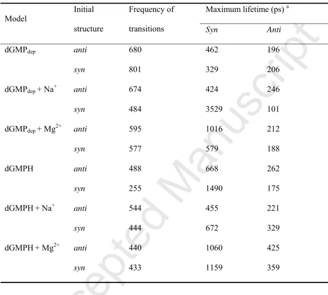
In the dGMPdep systems, when the syn conformation was the initial structure, the
proportion of the syn conformation was larger in the presence than in the absence of metal ions (Fig. 2A). This resulted from the longer maximum lifetime, which is defined as the time from the moment of the original syn/anti conformation until the moment at which it is
Accepted Manuscript
changed into the opposite anti/syn conformation (Table 1). In examining a series of the syn conformations with the maximum lifetime, characteristic syn conformations of dGMP were observed in the presence of metal ions (Figs. 2B and 2C). In dGMP that adopted the syn conformation, a hydrogen bond was formed between the hydrogen atom of the guanine 2-amino group and the oxygen atom of phosphate. This interaction probably resulted from a longer maximum lifetime in the syn conformation than in the anti conformation (Table 1). Furthermore, in the syn conformation of dGMP, electrostatically favorable interactions were formed between Na+ or Mg2+ and the nitrogen and oxygen atoms of dGMP (Figs. 2B and 2C).
In the dGMPH system without ions, the proportion of the syn conformation was larger than that in the dGMPdep system (Fig. 2A), owing to a longer maximum lifetime of the syn
conformation in the dGMPH system than in the dGMPdep system (Table 1). In the syn
conformation with maximum lifetime, a hydrogen bond was formed between the nitrogen atoms of guanine and the phosphate hydrogen atom (Fig. 2D). These interactions should also occur in the 8OGdep and 8OGH systems, because the only differences between dGMP and
8-oxo-dGMP are the kind of atoms found at the 8-position of guanine, and the protonation of the nitrogen atom at the 7-position of guanine (Fig. 1).
Conformation of 8-oxo-dGMP in explicit solvent
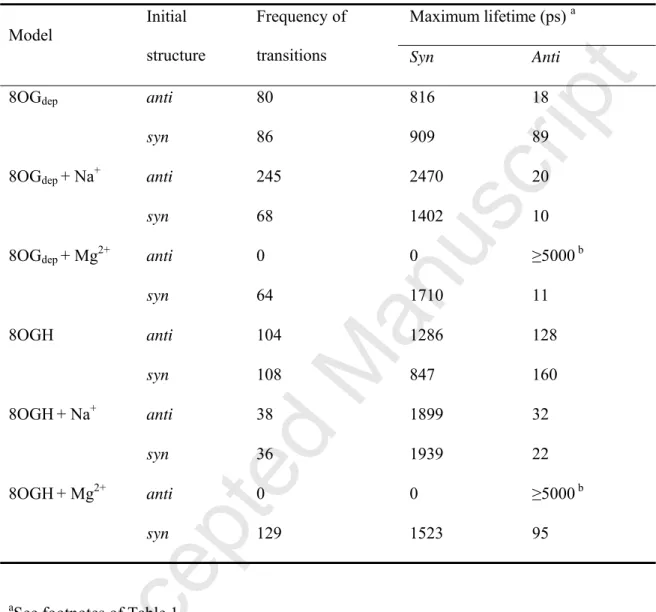
Figure 3A shows the probability density of the χ angle of 8-oxo-dGMP under the χstd
parameter. In both 8OGdep and 8OGH systems, 8-oxo-dGMP mainly adopted the syn
conformation. This result was consistent with an earlier experimental analysis [7]. As were the cases with the dGMPdep and dGMPH systems, the maximum lifetime of the syn
conformation was longer in the 8OGdep and 8OGH systems with metal ions than in those
systems without ions (Table 2). On the other hand, a large proportion for the anti conformation was observed in the 8OGdep and 8OGH systems with Mg2+ (Fig. 3A). When the
Accepted Manuscript
anti conformation was the initial structure, no anti-to-syn transition occurred during the MD
simulations (Table 2). This suggested that 8-oxo-dGMP retained the anti conformation through a strong interaction between 8-oxo-dGMP and Mg2+. In the anti conformations observed in the 8OGdep and 8OGH systems, Mg2+ was, in most cases, located between the
oxygen atom of phosphate and the O8 atom (Fig. S2), thus forming electrostatic interactions with the O8 atom and the phosphate oxygen atom of 8-oxo-dGMP to retain the anti conformation (Figs. 3B and 3C). Divalent cations such as Mg2+ or Mn2+ serve as cofactors of
MTH1 [36-38] and NUDT5 [6] and play an important role in the hydrolysis of the di- and tri-phosphate forms of the mononucleotide. These MD results implied another role of Mg2+; Mg2+ retains the stable anti conformations of 8-oxo-dGMP for the binding of the mononucleotide to the enzymes.
The syn-to-anti transition was also observed in 8-oxo-dGMP, as shown by the frequency of the observed transitions (Table 2). When the syn conformation was the initial structure, the maximum lifetime during which 8-oxo-dGMP retained the anti conformation was approximately 100 ps. This indicated that the syn conformation of 8-oxo-dGMP shifted to the
anti conformation, and that the anti conformation was retained for about 100 ps (Table 2).
Examination of the anti conformation at the maximum lifetime indicated hydrogen bonding occurred between the O8 atoms and the hydrogen atom of phosphate in the 8OGH system without ions (Fig. 3D). In the 8OGdep system with Mg2+, Mg2+ formed electrostatic
interactions with the O8 atom and the phosphate oxygen atom of 8-oxo-dGMP to retain the
anti conformation (Fig. 3E). Despite that, the interaction lasted for only 100 ps. This
suggested that the anti conformations observed as Figs. 3C and 3E differed from each other. To examine differences in the anti conformations of 8-oxo-dGMP in detail, we analyzed other torsion angles of the mononucleotides, pseudorotation of the deoxyribose ring, and the torsion angles of the sugar-phosphate backbone (β, γ). These angles are associated with the
Accepted Manuscript
interactions between 8-oxo-dGMP and Mg2+. Although no remarkable difference in the pseudorotation was observed between the no-ion systems and the Mg2+ systems (Figs. S3−S10), the distribution pattern of the β and γ angles was quite different between the systems (Fig. 4). The β and γ angles observed in the stable anti conformations were within the range of (120°−160°/210°−250°, 40°−100°) and (60°−210°, 40°−70°) in the 8OGdep−Mg2+and
the 8OGH−Mg2+ systems, respectively. However, some of the other anti conformations
observed through the syn-to-anti transitions were within the range of the β and γ angles in which stable anti conformations were observed. This suggested that 8-oxo-dGMP may shift to the stable anti conformation after the syn-to-anti transition in the presence of Mg2+, although the stable anti conformation was not observed after the syn-to-anti transition in the present MD simulations.
In the crystal structures of MTH1 (PDB: 3ZR0) [5] and NUDT5 (PDB: 3AC9 and PDB: 3L85) [6], the respective β and γ angles of 8-oxo-dGMP and 8-oxo-dGDP were 262.04° and 291.69° (chain A of MTH1), 237.21° and 302.21° (chain B of MTH1), 105.22° and 273.03° (PDB: 3AC9, in the presence of Mn2+), and 140.81° and 211.71° (PDB: 3L85, in the absence of ions). Comparison of these β and γ angles with those obtained from the MD simulations (Fig. 4) indicated that 8-oxo-dGMP does not bind to these enzymes in the anti conformation observed through MD simulations.
There are several points to note for the crystal structure of NUDT5 (PDB: 3AC9) concerning the selective recognition of 8-oxo-dGDP that has adopted the anti conformation [6]. First, the guanine base is located between two tryptophan side chains. The difference in electron distribution between guanine and 8-oxo-G may result in different strengths of interaction with the tryptophan residues. This is somewhat similar to the recognition of methylated bases by 3-methyladenine glycosylase from Escherichia coli [39]. Second, a water-mediated hydrogen bond is formed between N7(H) of 8-oxo-G and a carbonyl oxygen
Accepted Manuscript
of tyrosine. This bond would not be formed between a normal guanine and NUDT5. Third, the anti conformation, with a much more extended conformation compared to that observed in the MD simulations, is stabilized by interactions between the phosphate groups of 8-oxo-dGDP and Mn2+. Based on these observations, the selective recognition of NUDT5 and
the anti conformation of 8-oxo-dGDP can be explained. Some bias for an anti conformation in 8-oxo-dGMP mediated by metal ions, which was observed in the MD simulations, may assist in this mechanism. The anti conformations observed in the 8OGdep and 8OGH systems
may thus play a role in retaining the anti conformation until binding of 8-oxo-G mononucleotides to the enzyme occurs.
Conformational analyses of dGMP and 8-oxo-dGMP using the modified N-glycosidic torsion angle parameter
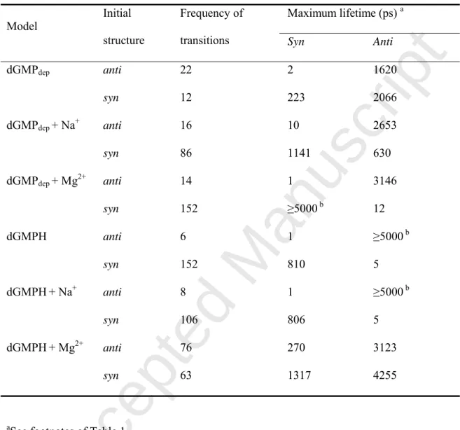
Recent works have proposed various force field modifications of the χ torsional potential for DNA and RNA based on QM calculations of small molecular models that represent mononucleotides [24, 40, 41]. In this study, the modified parameter (χmod) was taken from the
literature [24]. This was proven to be suitable for MD simulations of DNA duplexes [24]. The difference between the χstd and χmod parameters lies in an energy barrier between the anti and syn conformations of mononucleotides. The energy barrier was higher in the χmod parameter
than in the χstd parameter [24]. Put differently, the anti-to-syn or syn-to-anti conformational
transition can occur less frequently in MD simulations that use the χmod parameter than those
that use the χstd parameter. In MD simulations of the dGMPdep and dGMPH systems using the
χmod parameter, the frequency of the transition was much less than that of the χstd parameter
(Table 3). Together with the higher energy barrier in the χmod parameter, the maximum
lifetime of the anti conformation was longer with the χmod parameter than with the χstd
Accepted Manuscript
Figure 5 shows the probability density of the χ angle of mononucleotides using the χmod
parameter. In the dGMPdep systems, the proportion of the anti conformation was much larger
than that of the syn conformation, compared with the case of the χstd parameter. In the
dGMPdep no-ion system, dGMP adopted mainly the anti conformation. When the initial
structure was the syn conformation, the proportion of the syn conformation was larger in the dGMPdep systems with Na+ or Mg2+ than those without ions. In these systems, electrostatic
interactions between the syn conformer of dGMP and metal ions were observed, as well as in the case of the MD simulations using the χstd parameter (Figs. 2B and 2C). In dGMPH
systems that used the syn conformation as the initial structure, the proportion of the syn conformation was larger than systems that used the anti conformation. Hydrogen bonding between the nitrogen atoms of guanine and the hydrogen atom of phosphate was observed in the syn conformation (Figs. 2B and 2C). The effect of protonation of phosphate, and the kinds of metal ions, on the anti/syn conformation of dGMP was observed more clearly in the χmod
parameter than in the χstd parameter.
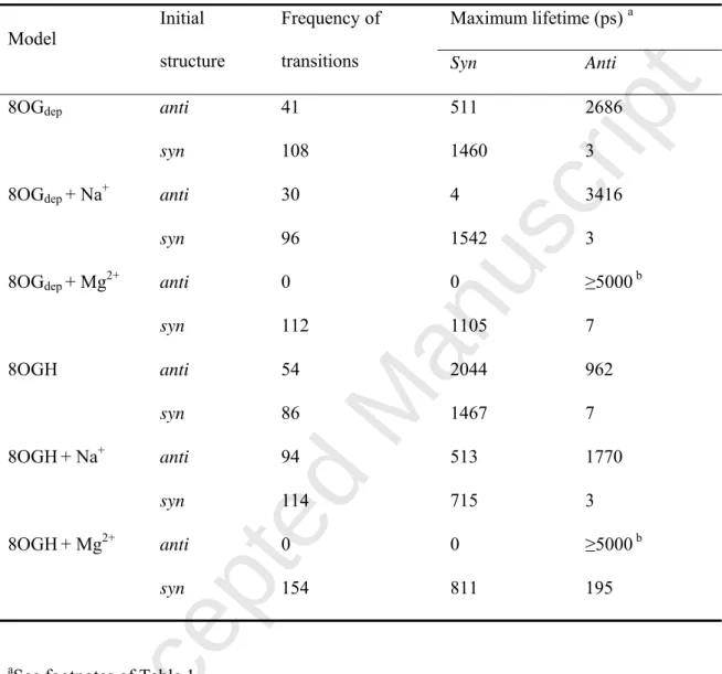
When the anti conformation of 8-oxo-dGMP was the initial structure, the anti conformation was retained in both 8OGdep and 8OGH systems without ions and with Na+, as
well as with Mg2+ (Fig. 5). This probably resulted from the higher energy barrier between the anti and syn conformations of mononucleotides in the χmod parameter. In the 8OGH no-ion
system, the mononucleotide adopted mainly the syn conformation because of the possible hydrogen bond between the nitrogen atoms of guanine and the hydrogen atom of phosphate (Fig. 2D). When the anti conformation was the initial structure, the proportion of the anti conformation was larger in the presence of Na+ than without ions. The maximum lifetime of the anti conformation was longer in the 8OGH−Na+ system than in the 8OGH no-ion system
(Table 4). In the anti conformations observed in the 8OGH systems, Na+ was located between the phosphate oxygen atom and the O8 atom in a manner similar to what was observed for
Accepted Manuscript
Mg2+ (Fig. 3C). This suggested that metal ions other than Mg2+ can also retain the anti conformation of 8-oxo-dGMP.
Effect of ion size and charge on the conformations of 8-oxo-dGMP
The effects of Li+ and Ca2+ were examined to elucidate whether ions other than Mg2+ can
influence the conformations of 8-oxo-dGMP in a similar manner. Both Li+ and Ca2+ differ
from Mg2+ in terms of their ionic radii and charges. Table S1 summarizes the force field
parameters of the metal ions used in this study. The ionic radius (corresponding to R*) of Li+ and the ionic radius of Ca2+ are similar with that of Mg2+.
In terms of both χstd and χmod parameters, 8-oxo-dGMP adopted both the anti and syn
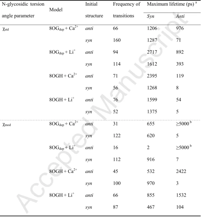
conformations in the presence of Li+ and Ca2+ (Fig. 6). The observation that the proportion of the anti conformation was smaller in the presence of Li+ and Ca2+ than with Mg2+ suggested that both ion size and charge can influence the proportion of the syn/anti conformations of 8-oxo-dGMP. The syn-to-anti transition was also observed in a series of MD simulations. In the 8OGdep−Li+ system, 8-oxo-dGMP shifted from the syn to anti conformation, and retained
the stable state for a maximum of 393 ps (Table 5). The β and γ angles observed in the anti conformations with maximum lifetime were within the range of 130°−200° and 150°−210° (Fig. 7), which differed from the stable anti conformations observed in the 8OGdep−Mg2+ and
8OGH−Mg2+ systems. In the anti conformations with maximum lifetime, Li+ was often
located between the phosphate oxygen atom and the O8 atom, similar to what was observed for Mg2+ (Fig. 3E). These results suggest that multiple types of stable anti conformers exist in
8-oxo-dGMP.
In this study, the force field parameter of Mg2+ was taken from the data by Åqvist [29]. This non-bonded model was adopted in early AMBER force fields. It is generally very difficult to model divalent ions for MD simulations because the non-bonded model of the
Accepted Manuscript
interaction between the ions and their surrounding residues is oversimplified, and a single point poorly represents the charge distribution of most ions [42]. To overcome these problems, improvements in the force field parameter of Mg2+ have been devised [42-46]. For example, Allnér et al. [46] focused on the exchange rate between Mg2+ and water. They found from
their MD simulations that the exchange rate between Mg2+ derived by Åqvist [29] and TIP3P
water was slower than the experimental data. They developed a new set of Mg2+ parameters
based on the Mg2+−water exchange rate. In comparison with the non-bonded Mg2+ force field
parameters recently developed [42-46], the ionic radius (R*) of Mg2+ derived by Allnér et al. [46] was close to that of Ca2+ derived by Åqvist (Table S1). Therefore, it is expected that the probability density profile of the χ angle in the 8OGdep and 8OGH systems with Allnér’s
estimate for Mg2+, using the anti conformation as the initial structure, may be close to that using Åqvist’s estimate for Ca2+. Although further improvements are needed to develop an accurate Mg2+ force field parameter, under the present MD simulations, the larger proportion
of the anti conformation will be observed in the 8OGdep and 8OGH systems with metal ions,
and the types of the stable anti conformations might differ depending upon the properties of metal ions, such as ionic radius and charge.
Conformational sampling of mononucleotide using MD simulations
MD simulations are typically performed at laboratory temperatures and often require cost-prohibitive time scales to produce results that are consistent with those obtained by conformational sampling [47]. In the present 10-ns MD simulations, the conformational sampling of mononucleotides was sometimes influenced by the initial anti or syn conformations. In the χmod parameter, the probability density profiles were quite different
between the initial anti and syn conformations (Fig. 5). In addition, when 8-oxo-dGMP was simulated in the presence of Mg2+ using the anti conformation as the initial structure, no
Accepted Manuscript
anti-to-syn transition occurred (Tables 2 and 4). The transition did not occur even during the
additional 95-ns MD simulations for each run (Fig. S11). Mg2+ was not moved away from the O8 atom of 8-oxo-dGMP (Fig. S12). The transitions were observed in the high-temperature (573 K) MD calculations. The simulations overcame the barriers between the anti and syn conformations in the 8OGdep and 8OGH systems (starting conformation: anti) with Mg2+ (Fig.
S13). The anti-to-syn transition occurred in either or both of the two MD runs of the 8OGdep
and 8OGH systems when Mg2+ was moved away from the O8 atom (Fig. S14). These findings
indicate that the results of conformational sampling had not converged after two runs of the 5-ns MD simulations and that the probability density profiles of χangle might be inaccurate. It is also possible that the force field parameter of Mg2+ derived by Åqvist [29] causes too strong an interaction between Mg2+ and the O8 atom of 8-oxo-dGMP. Enhanced sampling approaches, such as replica exchange MD [48], as well as improvements in the force field parameter of Mg2+ as discussed above, might be needed for converged sampling to obtain the accurate χ-angle probability density profiles of mononucleotides. However, a series of MD simulations of mononucleotides has clarified the effects of metal ions on the conformations of 8-oxo-dGMP.
Conclusions
A series of MD simulations of dGMP and 8-oxo-dGMP indicated that 8-oxo-dGMP can adopt both the syn and anti conformations. The proportion adopting the anti conformation increased in the presence of metal ions. When 8-oxo-dGMP adopted the anti conformation, metal ions were often located between the oxygen atom of phosphate and the O8 atom of 8-oxo-G. The types of stable anti conformations of 8-oxo-dGMP differed, depending on the ionic radii and charges of coexisting ions. This paper also described common issues associated with MD simulations, such as a lack of convergence, dependence on starting conformation, and
Accepted Manuscript
dependability of force field parameters, particularly for ions. Further exploration of the dependence on χ dihedral parameters would be useful, not only for comparisons between parameters but also for gaining further insights into the conformations of mononucleotides. Nevertheless, there does seem to be a general trend in the MD simulations that the anti conformation of 8-oxo-dGMP is favored in the presence of metal ions. The MD results suggest the possibility that the 8-oxo-G deoxynucleotide adopts the anti conformation and binds to MTH1 or NUDT5. This study also suggested a role for metal ions other than as cofactors for the hydrolysis of di- and tri-phosphate forms of mononucleotides. Specifically, these ions can retain the anti conformation to promote the binding of 8-oxo-G deoxynucleotides to nucleotide-sanitizing enzymes. This study contributes to our understanding of the effects of metal ions and the protonation of mononucleotides on their conformations, and describes limitations and areas for improvement in order to gain further insights into conformations of mononucleotides through MD simulations.
Supporting information
Additional supporting information may be found in the online version of this article at the publisher’s website.
Acknowledgements
This study was supported in part by Grants-in-Aid for Scientific Research from the Ministry of Education, Culture, Sports, Science and Technology of Japan.
Accepted Manuscript
References
[1] Van Loon, B., Markkanen, E., Hubscher, U. Oxygen as a friend and enemy: How to combat the mutational potential of 8-oxo-guanine. DNA Repair. 2010, 9, 604-616.
[2] Nakabeppu, Y., Kajitani, K., Sakamoto, K., Yamaguchi, H., Tsuchimoto, D. MTH1, an oxidized purine nucleoside triphosphatase, prevents the cytotoxicity and neurotoxicity of oxidized purine nucleotides. DNA Repair. 2006, 5, 761-772.
[3] Kamiya, H., Hori, M., Arimori, T., Sekiguchi, M., Yamagata, Y., Harashima, H. NUDT5 hydrolyzes oxidized deoxyribonucleoside diphosphates with broad substrate specificity. DNA Repair. 2009, 8, 1250-1254.
[4] Nakamura, T., Meshitsuka, S., Kitagawa, S., Abe, N., Yamada, J., Ishino, T., Nakano, H., Tsuzuki, T., Doi, T., Kobayashi, Y., Fujii, S., Sekiguchi, M., Yamagata, Y. Structural and dynamic features of the MutT protein in the recognition of nucleotides with the mutagenic 8-oxoguanine base. J. Biol. Chem. 2010, 285, 444-452.
[5] Svensson, L.M., Jemth, A.S., Desroses, M., Loseva, O., Helleday, T., Hogbom, M., Stenmark, P. Crystal structure of human MTH1 and the 8-oxo-dGMP product complex. FEBS Lett. 2011, 585, 2617-2621.
[6] Arimori, T., Tamaoki, H., Nakamura, T., Kamiya, H., Ikemizu, S., Takagi, Y., Ishibashi, T., Harashima, H., Sekiguchi, M., Yamagata, Y. Diverse substrate recognition and hydrolysis mechanisms of human NUDT5. Nucleic Acids Res. 2011, 39, 8972-8983.
[7] Uesugi, S., Ikehara, M. Carbon-13 magnetic resonance spectra of 8-substituted purine nucleosides. Characteristic shifts for the syn conformation. J. Am. Chem. Soc. 1977, 99, 3250-3253.
[8] Gidden, J., Bowers, M.T. Gas-phase conformations of deprotonated and protonated mononucleotides determined by ion mobility and theoretical modeling. J. Phys. Chem. B. 2003, 107, 12829-12837.
Accepted Manuscript
[9] Shishkin, O.V., Gorb, L., Zhikol, O.A., Leszczynski, J. Conformational analysis of canonical 2-deoxyribonucleotides. 1. Pyrimidine nucleotides. J. Biomol. Struct. Dyn. 2004, 21, 537-554.
[10] Shishkin, O.V., Gorb, L., Zhikol, O.A., Leszczynski, J. Conformational analysis of canonical 2-deoxyribonucleotides. 2. Purine nucleotides. J. Biomol. Struct. Dyn. 2004, 22, 227-244.
[11] Gorb, L., Shishkin, O., Leszczynski, J. Charges of phosphate groups. A role in stabilization of 2'-deoxyribonucleotides. A DFT investigation. J. Biomol. Struct. Dyn. 2005, 22, 441-454.
[12] Shishkin, O.V., Palamarchuk, G.V., Gorb, L., Leszczynski, J. Intramolecular hydrogen bonds in canonical 2'-deoxyribonucleotides: an atoms in molecules study. J. Phys. Chem. B. 2006, 110, 4413-4422.
[13] Liu, D., Wyttenbach, T., Bowers, M.T. Hydration of mononucleotides. J. Am. Chem. Soc. 2006, 128, 15155-15163.
[14] Kosenkov, D., Gorb, L., Shishkin, O.V., Sponer, J., Leszczynski, J. Tautomeric equilibrium, stability, and hydrogen bonding in 2'-deoxyguanosine monophosphate complexed with Mg2+. J. Phys. Chem. B. 2008, 112, 150-157.
[15] Close, D.M., Øhman, K.T. Ionization energies of the nucleotides. J. Phys. Chem. A. 2008, 112, 11207-11212.
[16] Kosenkov, D., Kholod, Y.A., Gorb, L., Shishkin, O.V., Kuramshina, G.M., Dovbeshko, G.I., Leszczynski, J. Effect of a pH change on the conformational stability of the modified nucleotide queuosine monophosphate. J. Phys. Chem. A. 2009, 113, 9386-9395.
[17] Millen, A.L., Manderville, R.A., Wetmore, S.D. Conformational flexibility of c8-phenoxyl-2'-deoxyguanosine nucleotide adducts. J. Phys. Chem. B. 2010, 114, 4373-4382. [18] Palamarchuk, G.V., Shishkin, O.V., Gorb, L., Leszczynski, J. Nucleic acid bases in
Accepted Manuscript
anionic 2'-deoxyribonucleotides: a DFT/B3LYP study of structures, relative stability, and proton affinities. J. Phys. Chem. B. 2013, 117, 2841-2849.
[19] Calvo, F., Douady, J. Stepwise hydration and evaporation of adenosine monophosphate nucleotide anions: a multiscale theoretical study. Phys. Chem. Chem. Phys. 2010, 12, 3404-3414.
[20] Grossfield, A. Recent progress in the study of G protein-coupled receptors with molecular dynamics computer simulations. Biochim. Biophys. Acta. 2011, 1808, 1868-1878. [21] Dawson, R.M.C., Elliot, D.C., Elliot, W.H., Jones, K.M. Data for Biochemical Research. 3rd ed. Oxford, Clarendon Press, 1986.
[22] AMBER11, 2010, University of California, San Francisco.
[23] Wang, J.M., Cieplak, P., Kollman, P.A. How well does a restrained electrostatic potential (RESP) model perform in calculating conformational energies of organic and biological molecules? J. Comput. Chem. 2000, 21, 1049-1074.
[24] Ode, H., Matsuo, Y., Neya, S., Hoshino, T. Force field parameters for rotation around chi torsion axis in nucleic acids. J. Comput. Chem. 2008, 29, 2531-2542.
[25] Miller, J.H., Fan-Chiang, C.C., Straatsma, T.P., Kennedy, M.A. 8-Oxoguanine enhances bending of DNA that favors binding to glycosylases. J. Am. Chem. Soc. 2003, 125, 6331-6336.
[26] Homeyer, N., Horn, A.H., Lanig, H., Sticht, H. AMBER force-field parameters for phosphorylated amino acids in different protonation states: phosphoserine, phosphothreonine, phosphotyrosine, and phosphohistidine. J. Mol. Model. 2006, 12, 281-289.
[27] Gaussian 03, 2003, Gaussian, Inc., Pittsburgh.
[28] Jorgensen, W.L., Chandrasekhar, J., Madura, J.D., Impey, R.W., Klein, M.L. Comparison of simple potential functions for simulating liquid water. J. Chem. Phys. 1983, 79, 926-935.
Accepted Manuscript
[29] Åqvist, J. Ion water interaction potentials derived from free-energy perturbation simulations. J. Phys. Chem. 1990, 94, 8021-8024.
[30] Berendsen, H.J.C., Postma, J.P.M., Van Gunsteren, W.F., DiNola, A., Haak, J.R. Molecular dynamics with coupling to an external bath. J. Chem. Phys. 1984, 81, 3684-3690. [31] Darden, T., York, D., Pedersen, L. Particle mesh Ewald - an Nlog(N) method for Ewald sums in large systems. J. Chem. Phys. 1993, 98, 10089-10092.
[32] Darden, T., Pearlman, D., Pedersen, L.G. Ionic charging free energies: Spherical versus periodic boundary conditions. J. Chem. Phys. 1998, 109, 10921-10935.
[33] Van Gunsteren, W.F., Berendsen, H.J.C. Algorithm for macromolecular dynamics and constraint dynamics. Mol. Phys. 1977, 34, 1311-1327.
[34] IUPAC-IUB Joint Commission on Biochemical Nomenclature. Abbreviations and symbols for the description of conformations of polynucleotide chains. Eur. J. Biochem. 1983, 131, 9-15.
[35] Nelson, D.L., Cox, M.M. Lehninger Principles of Biochemistry. 5 ed. New York, W.H. Freeman and Company, 2008.
[36] Fujikawa, K., Kamiya, H., Yakushiji, H., Fujii, Y., Nakabeppu, Y., Kasai, H. The oxidized forms of dATP are substrates for the human MutT homologue, the hMTH1 protein. J. Biol. Chem. 1999, 274, 18201-18205.
[37] Fujikawa, K., Kamiya, H., Yakushiji, H., Nakabeppu, Y., Kasai, H. Human MTH1 protein hydrolyzes the oxidized ribonucleotide, 2-hydroxy-ATP. Nucleic Acids Res. 2001, 29, 449-454.
[38] Mishima, M., Sakai, Y., Itoh, N., Kamiya, H., Furuichi, M., Takahashi, M., Yamagata, Y., Iwai, S., Nakabeppu, Y., Shirakawa, M. Structure of human MTH1, a Nudix family hydrolase that selectively degrades oxidized purine nucleoside triphosphates. J. Biol. Chem. 2004, 279, 33806-33815.
Accepted Manuscript
[39] Hollis, T., Ichikawa, Y., Ellenberger, T. DNA bending and a flip-out mechanism for base excision by the helix-hairpin-helix DNA glycosylase, Escherichia coli AlkA. EMBO J. 2000, 19, 758-766.
[40] Yildirim, I., Stern, H.A., Kennedy, S.D., Tubbs, J.D., Turner, D.H. Reparameterization of RNA chi torsion parameters for the AMBER force field and comparison to NMR spectra for cytidine and uridine. J. Chem. Theory Comput. 2010, 6, 1520-1531.
[41] Zgarbová, M., Otyepka, M., Šponer, J., Mládek, A., Banáš, P., Cheatham, T.E., 3rd, Jurečka, P. Refinement of the Cornell et al. nucleic acids force field based on reference quantum chemical calculations of glycosidic torsion profiles. J. Chem. Theory Comput. 2011, 7, 2886-2902.
[42] Li, F., Roberts, B.P., Chakravorty, D.K., Merz, K.M., Jr. Rational design of particle mesh Ewald compatible Lennard-Jones parameters for +2 metal cations in explicit solvent. J. Chem. Theory Comput. 2013, 9, 2733-2748.
[43] Florián, J., Goodman, M.F., Warshel, A. Computer simulation of the chemical catalysis of DNA polymerases: discriminating between alternative nucleotide insertion mechanisms for T7 DNA polymerase. J. Am. Chem. Soc. 2003, 125, 8163-8177.
[44] Babu, C.S., Lim, C. Empirical force fields for biologically active divalent metal cations in water. J. Phys. Chem. A. 2006, 110, 691-699.
[45] Oelschlaeger, P., Klahn, M., Beard, W.A., Wilson, S.H., Warshel, A. Magnesium-cationic dummy atom molecules enhance representation of DNA polymerase beta in molecular dynamics simulations: improved accuracy in studies of structural features and mutational effects. J. Mol. Biol. 2007, 366, 687-701.
[46] Allnér, O., Nilsson, L., Villa, A. Magnesium ion-water coordination and exchange in biomolecular simulations. J. Chem. Theory Comput. 2012, 8, 1493-1502.
Accepted Manuscript
dynamics simulations of protein structure and function. Curr. Opin. Struct. Biol. 2009, 19, 120-127.
[48] Sugita, Y., Okamoto, Y. Replica-exchange molecular dynamics method for protein folding. Chem. Phys. Lett. 1999, 314, 141-151.
[49] Humphrey, W., Dalke, A., Schulten, K. VMD: Visual molecular dynamics. J. Mol. Graphics. 1996, 14, 33-38.
Accepted Manuscript
Table 1. Frequency of the anti-to-syn and syn-to-anti transitions, and maximum lifetime of
the anti/syn conformations in MD simulations of dGMP using the χstd parameter.
Maximum lifetime (ps) a
Model Initial
structure
Frequency of
transitions Syn Anti
dGMPdep anti 680 462 196 syn 801 329 206 dGMPdep + Na+ anti 674 424 246 syn 484 3529 101 dGMPdep + Mg2+ anti 595 1016 212 syn 577 579 188 dGMPH anti 488 668 262 syn 255 1490 175 dGMPH+ Na+ anti 544 455 221 syn 444 672 329 dGMPH+ Mg2+ anti 440 1060 425 syn 433 1159 359
aThe syn and anti conformations are defined as χ angle of 0−90° and 330−360°, and 90−330°,
Accepted Manuscript
Table 2. Frequency of the anti-to-syn and syn-to-anti transitions, and maximum lifetime of
the anti/syn conformations in MD simulations of 8-oxo-dGMP using the χstd parameter.
Maximum lifetime (ps) a
Model Initial
structure
Frequency of
transitions Syn Anti
8OGdep anti 80 816 18
syn 86 909 89
8OGdep + Na+ anti 245 2470 20
syn 68 1402 10
8OGdep + Mg2+ anti 0 0 ≥5000 b
syn 64 1710 11 8OGH anti 104 1286 128 syn 108 847 160 8OGH+ Na+ anti 38 1899 32 syn 36 1939 22 8OGH+ Mg2+ anti 0 0 ≥5000 b syn 129 1523 95
aSee footnotes of Table 1.
bThe anti conformationis retained during 5 ns in both of the MD runs. The stability of the anti
conformation is discussed in Conformational sampling of mononucleotide using MD
Accepted Manuscript
Table 3. Frequency of the anti-to-syn and syn-to-anti transitions, and maximum lifetime of
the anti/syn conformations in MD simulations of dGMP using the χmod parameter.
Maximum lifetime (ps) a
Model Initial
structure
Frequency of
transitions Syn Anti
dGMPdep anti 22 2 1620 syn 12 223 2066 dGMPdep + Na+ anti 16 10 2653 syn 86 1141 630 dGMPdep + Mg2+ anti 14 1 3146 syn 152 ≥5000 b 12 dGMPH anti 6 1 ≥5000 b syn 152 810 5 dGMPH+ Na+ anti 8 1 ≥5000 b syn 106 806 5 dGMPH+ Mg2+ anti 76 270 3123 syn 63 1317 4255
aSee footnotes of Table 1.
Accepted Manuscript
Table 4. Frequency of the anti-to-syn and syn-to-anti transitions, and maximum lifetime of
the anti/syn conformations in MD simulations of 8-oxo-dGMP using the χmod force field.
Maximum lifetime (ps) a
Model Initial
structure
Frequency of
transitions Syn Anti
8OGdep anti 41 511 2686
syn 108 1460 3
8OGdep + Na+ anti 30 4 3416
syn 96 1542 3
8OGdep + Mg2+ anti 0 0 ≥5000 b
syn 112 1105 7 8OGH anti 54 2044 962 syn 86 1467 7 8OGH+ Na+ anti 94 513 1770 syn 114 715 3 8OGH+ Mg2+ anti 0 0 ≥5000 b syn 154 811 195
aSee footnotes of Table 1.
bThe anti conformationis retained during 5 ns in both of the MD runs. The stability of the anti
conformation is discussed in Conformational sampling of mononucleotide using MD
Accepted Manuscript
Table 5. Frequency of the anti-to-syn and syn-to-anti transitions, and maximum lifetime of
the anti/syn conformations in MD simulations of 8-oxo-dGMP in the presence of Ca2+ or Li+. Maximum lifetime (ps) a N-glycosidic torsion angle parameter Model Initial structure Frequency of
transitions Syn Anti
χstd 8OGdep + Ca2+ anti 66 1206 976
syn 160 1287 71
8OGdep + Li+ anti 94 2717 892
syn 114 1612 393
8OGH+ Ca2+ anti 71 2395 119
syn 56 1268 8
8OGH+ Li+ anti 76 1599 54
syn 52 1375 5
χmod 8OGdep + Ca2+ anti 31 655 ≥5000 b
syn 122 620 5
8OGdep + Li+ anti 16 2 ≥5000 b
syn 112 916 7
8OGH+ Ca2+ anti 45 532 2422
syn 100 970 3
8OGH+ Li+ anti 66 855 1532
syn 87 467 104
aSee footnotes of Table 1.
Accepted Manuscript
Figure captions
Fig. 1. Mononucleotide models analyzed in this study. Atom numbering is shown for
dGMPdep.
Fig. 2. (A) Probability density of the χ angle of dGMP in the dGMPdep and dGMPH systems
using the standard N-glycosidic bond parameter (χstd). Initial structures (anti/syn) are
indicated in parentheses. (B) One of the syn conformations observed in the dGMPdep system
with Na+. (C) One of the syn conformations observed in the dGMP
dep system with Mg2+. (D)
One of the syn conformations observed in the dGMPH system. These molecular diagrams are generated with VMD (version 1.9.1) [49].
Fig. 3. (A) Probability density of the χ angle of 8-oxo-dGMP in the 8OGdep and 8OGH
systems using the χstd parameter. Initial structures (anti/syn) are indicated in parentheses. (B)
One of the anti conformations observed in the 8OGdep system with Mg2+. χ, β, and γ angles
are 218.6°, 134.9°, and 51.9°, respectively. (C) One of the anti conformations observed in the 8OGH system with Mg2+. The angles of χ, β, and γ are 240.3°, 82.3°, and 40.5°, respectively. (D) One of the anti conformations observed in the 8OGH system without ions during the
syn-to-anti transition. The angles of χ, β, and γ are 247.2°, 270.1°, and 190.3°, respectively.
(E) One of the anti conformations observed in the 8OGH system with Mg2+ during the syn-to-anti transition. The angles of χ, β, and γ are 261.7°, 180.5°, and 65.3°, respectively.
These molecular diagrams are generated with VMD (version 1.9.1) [49].
Fig. 4. Scatter plot of torsion angles (β, γ) of the sugar-phosphate backbone in the 8OGdep and
8OGHsystems using the χstd parameter. In the systems without ions, green and red points
indicate the syn and anti conformations, respectively. In the systems with Mg2+, green, red, and blue points indicate the syn conformations, anti conformations obtained from MD simulations using the anti conformation as initial structures, and anti conformations obtained from MD simulations using the syn conformation as initial structures, respectively. β and γ
Accepted Manuscript
angles of 8-oxo-dGMP or 8-oxo-dGDP in the crystal structures of MTH1 [5] and NUDT5 [6] are also shown. These figures are modified from Figs. S6 and S8 in Supporting information.
Fig. 5. Probability density of the χ angle of dGMP and 8-oxo-dGMP using the modified
N-glycosidic bond parameter (χmod). Initial structures (anti/syn) are indicated in parentheses. Fig. 6. Probability density of the χ angle of 8-oxo-dGMP in the 8OGdep and 8OGH systems
with Mg2+, Li+ or Ca2+. Initial structures (anti/syn) are indicated in parentheses.
Fig. 7. Scatter plot of torsion angles (β, γ) of the sugar-phosphate backbone in the 8OGdep–Li+
system using the χstd parameter. Green, red, and blue points indicate the syn conformations, anti conformations, and anti conformations with maximum lifetime obtained from MD