73
Yonago Acta medica 2008;51:73–76
Abbreviation: VATS, video–assisted thoracic surgery
Video-Assisted Thoracic Surgery (VATS) for Stabbing
Thoracic Injury
Yuji Taniguchi, Hiroshige Nakamura*, Tomohiro Haruki*, Shinji Fujioka*, Yoshin Adachi*, Ken Miwa*, Satoshi Kamihira†, Kengo Nishimura† and Motonobu Nishimura†
Operating Room Division, *Division of Thoracic Surgery, Tottori University Hospital and †Divi-sion of Organ Regeneration Surgery, Department of Surgery, School of Medicine, Tottori Univer-sity Faculty of Medicine, Yonago, Yonago 683-8504 Japan
The patient was a 30-year-old male who was found after he had stabbed himself in the left side of the chest and collapsed. He was brought to our hospital to undergo immediate drainage of the thoracic cavity. The chest X-ray and chest computed tomography findings showed that the knife was situated from the left cardiac border toward the proximity of the diaphragm, thus resulting in hemopneumothorax. The patient’s vital signs were sta-ble, and we believed that it was important to avoid secondary injury when removing the knife from the thoracic cavity. Therefore, we performed video-assisted thoracic surgery (VATS). We resected the considerably crushed lung and sutured the lacerated myocar-dium by VATS. The postoperative course was good. We believe that VATS for thoracic injury can be an effective surgical option if a patient’s vital signs are stable.
Key words: stab wound; thoracic injury; video-assisted thoracic surgery
Recently video-assisted thoracic surger y (VATS) has become widely used for treatment in thoracic surgery. VATS is now performed in approximately 50% of all respiratory surgical procedures in Japan (Kazui et al., 2006). In this report, we show that VATS is performed for approximately 88% of all cases of pneumothorax, which is a commonly treated quasi-emergency operation. On the other hand, due to the low rate of VATS for thoracic injury, which is treated with emergency surgery, the frequency of VATS for thoracic injury is not described. However, in some hospitals, VATS has been performed for chest trauma (Manlulu et al., 2004; Maitani et al., 2006). We performed emergency VATS to treat a stab wound in the chest incurred during a suicide attempt.
Patient Report
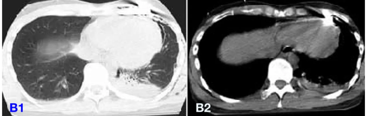
The patient was a 30-year-old male who had been receiving inpatient care at a local hospital for schizophrenia. During a short leave from the hospital, he was found lying on the floor after he had stabbed himself in the left side of the chest with a knife. He was brought to our emergency room at Tottori University Hospital. His blood pressure and pulse were 95/52 mmHg and 110 bpm, respectively, and he had a clear sensorium. The knife was inserted from the 4th left intercostal midclavicular line and was directed slightly laterally and downward (Fig. 1). According to the chest X-ray and chest computed tomography findings, the knife appeared to be lodged from the left cardiac border toward the proximity of the diaphragm, thus resulting in
74
Y. Taniguchi et al.
hemopneumothorax (Fig. 2). At the Division of Emergency Medicine, the patient was immediately inserted into a thoracostomy tube from the 5th left intercostal posterior axillary line. Immediately after insertion, approximately 500 mL of blood was drained, but subsequent hemorrhaging was only in small amounts, and the hemodynamics were stable. The patient was diagnosed as having left hemopneumothorax, which was caused by the stab wound to the chest. He was then referred to the Division of Thoracic Surgery for emergency operation approximately 2 h after arriving at our hospital and taken to the operating room to undergo VATS within 3 h after his arrival.
The operation was performed in the left half lateral decubitus position with care to maintain the position of the knife. A trocar was inserted from the 8th intercostal anterior axillary line, and the thoracostomy tube that had been inserted from the 5th intercostal posterior axillary line
Fig. 1. The wound before surgery. Fig. 2A. Chest X-ray at the time of admission. A1:
Posteroante-rior view. A2: Lateral view.
Fig. 2B. Chest computed tomography at the time of admission. B1: Lung window image. B2: Mediastinal window image.
Fig. 3. The knife has not reached the heart or lungs.
was removed for insertion of a trocar. Rough adhesion and moderate hematoma were observed in the thoracic cavity, but there was almost no continuous bleeding in the thoracic cavity or from the thoracic wall. The knife was located near the anterior mediastinum in the proximity of the pericardium without penetrating the heart or lungs (Fig. 3), and it was removed without
A1
B1
A2
75
VATS for stabbing thoracic injury
causing any secondary injury to the heart or lungs under thorascopic vision. The 4th left intercostal wound caused by the knife was extended to approximately 10 cm for a minithoracotomy and was supplemented with a Multi-Flap Gate (Akita Sumitomo Bake, Akita, Japan). There
Fig. 5B. The wound after being sutured and reinforced
with fleece-coated fibrin glue.
Fig. 4. Excised specimen. The perforating wound
caused by the knife (arrow).
Fig. 5A. The lacerated wound in the myocardium
caused by the knife (arrow).
was a severely crushed perforating wound caused by the knife in the lingular segment, which was resected with an automatic suturing instrument (Fig. 4). In addition, the pericardium was gaped in the length of about 2 cm, and the surface of the heart was lacerated about 1 cm (Fig. 5A). After the epicardium had been sutured under direct vision without extending the minithoracotomy, the suture line was reinforced with fleece-coated fibrin glue (Fig. 5B). The wound was sutured and temporarily closed. The surgery time was 2 h and 8 min, and the amount of intraoperative bleeding was 200 mL. The postoperative course was good. The patient was transferred to the Division of Neuropsychiatry on day 5 after the surgery and transferred to the hospital where he had originally been 6 weeks after the surgery.
Discussion
It appears as though few hospitals use VATS as their primary method in emergency surgery for stab wounds of the chest. We believe the reason for this is that injury to the heart and great vessels is often life threatening and is believed to be difficult to treat by VATS. In fact, 70 to 80% of all patients with injury to the heart die before arriving at the hospital (Demetriades and van der Veen, 1983). Even among patients who survive and arrive at the hospital, 80% have unstable hemodynamics and thus undergo emergency thoracotomy. However, it is believed that the remaining 20% have stable hemodynamics (Demetriades and van der Veen, 1983; Ordog et al., 1994). Based on this, Pons et al. (2002) have reported that when videothoracoscopy was implemented for a case of a penetrating injury with stable hemodynamics, it was safe and effective in determining the degree of damage. Moreover, Manlulu et al. (2004) implemented VATS for chest trauma and defined the following conditions for application: i) bleeding that persists even after drainage of the thoracic cavity, ii) stable hemodynamics and iii) no findings
76
Y. Taniguchi et al.
thereby allowing for safe suture treatment.
VATS for stab wounds in the chest can be a very effective surgical procedure if the patient’s vital signs are stable and some concern exists for secondary injury caused by a knife or other object that is lodged in the thoracic cavity. However, we believe that there should be no hesitation in converting the treatment to a conventional thoracotomy if there is more serious injury than initially predicted preoperatively.
References
1 Demetriades D, van der Veen BW. Penetrating inju-ries of the heart: experience over two years in South Africa. J Trauma 1983;23:1034–1041.
2 Kazui T, Osada H, Fujita H. Thoracic and cardiovas-cular surgery in Japan during 2004: annual report by the Japanese Association for Thoracic Surgery. Jpn J Thorac Cardiovasc Surg 2006;54:363–386. 3 Maitani F, Nakagawa T, Masuda R, Inoue Y, Nishiumi
N, Iwazaki M, et al. Video-assisted thoracoscopic surgery in treatment of stabbing chest injuries. Tokai J Exp Clin Med 2006; 31:157–159.
4 Manlulu AV, Lee TW, Thung KH, Wong R, Yim APC. Current indications and results of VATS in the evaluation and management of hemodynamically stable thoracic injuries. Eur J Cardiothorac Surg 2004; 25: 1048–1053.
5 Nippon Gaisho Gakkai Shin-Daikekkan Sonsyo Bunrui Iinkai. [Classification for cardiovascular in-jury.] Nippon Gaisho Gakkai Zasshi 1999;13:327–33 (in Japanese).
6 Nippon Gaisho Gakkai Shin-Daikekkan Sonsyo Bunrui Iinkai. [Classification for chest wall and lung injury.] Nippon Gaisho Gakkai Zasshi 2000;14:299– 306 (in Japanese).
7 Ordog GJ, Wasserberger J, Balasubramanium S, Shoemaker W. Asymptomatic stab wounds of the chest. J Trauma 1994;36:680–684.
8 Pons F, Lang-Lazdunski L, de Kerangal X, Chapuis O, Bonnet PM, Jancovici R. The role of videotho-racoscopy in management of precordial thoracic penetrating injuries. Eur J Cardiothorac Surg. 2002;22:7–12.
Received June 23, 2008; accepted August 5, 2008 Corresponding author: Yuji Taniguchi, MD
in the diagnostic images that would indicate injury to the great vessels. In their report, 45% of all patients with chest trauma met the above conditions, and therefore VATS was performed, and no patients had to be converted from VATS to a conventional thoracotomy during the surgery or there were no cases of intraoperative mortality or reoperation. In the present case, the amount of bleeding after drainage of the thoracic cavity was relatively small, and the patient’s hemodynamics were stable, thus matching the conditions that have been defined by Manlulu et al. (2004). Moreover, the knife was lodged from the left cardiac border toward the proximity of the diaphragm, and therefore it was important to avoid injury to the heart, pulmonary artery and pulmonary vein when removing the knife during surgery. Therefore, we selected VATS rather than conventional thoracotomy as a method in which the position of the knife would not be easily displaced during the approach to the thoracic cavity. In addition, the laceration wound in the myocardium and the penetrating wound in the lung, which constituted the injuries in this case, corresponded to the category of non-full-thickness-type myocardium lacerated wound in the Cardiovascular Classification of the Japanese Association for the Surgery of Trauma (1999) and a deep-seated lacerated wound such as lung damage in the Thoracic and Lung Damage Classification by the same association (2000). Many believe that these types of damage do not require emergency surgery but may require thoracotomy in some cases. Accordingly, it appears as though our decision to use VATS in this case was appropriate. In addition, the Multi-Flap Gate that we used is a tool that has been developed for spreading wounds for small incision surgery that is also used frequently for VATS lobectomies and enables treatment of the pulmonary artery and pulmonary vein. Furthermore, in this case, the damaged area of the myocardium was identified under direct vision,