IRUCAA@TDC : Cyclic AMP-dependent protein kinase-independent angiotensin II-induced inhibition of calcium current in hamster submandibular ganglion neurons
6
0
0
全文
(2) 95. Bull. Tokyo dent. Coll., Vol. 43, No. 2, pp. 95⬃99, May, 2002. Short Communication. CYCLIC AMP-DEPENDENT PROTEIN KINASE-INDEPENDENT ANGIOTENSIN II-INDUCED INHIBITION OF CALCIUM CURRENT IN HAMSTER SUBMANDIBULAR GANGLION NEURONS TAKAYUKI ENDOH, MITSUHIRO ABE and TAKASHI SUZUKI Department of Physiology, Tokyo Dental College, 1-2-2 Masago, Mihama-ku, Chiba 261-8502, Japan. Received 6 March, 2002/Accepted for Publication 25 March, 2002. Abstract In a previous study, we demonstrated that angiotensin II (Ang II) inhibited voltagedependent calcium channels (VDCCs) currents (I Ca ) in hamster submandibular ganglion (SMG) neurons. In sinoatrial node cells, it has been reported that Ang II inhibits I Ca by suppressing cyclic AMP production. In this study, to investigate the possible involvement of a cyclic AMP-cyclic AMP dependent protein kinase (PKA) pathway in the Ang IIinduced inhibition of I Ca , effects of Ang II were examined in SMG neurons after treatment with an activator and inhibitor of PKA. Neither pretreatment of neurons with membrane permeable cyclic AMP nor intracellular dialysis of PKA blocker attenuated the Ang II-induced inhibition of I Ca . These results indicate that Ang II inhibited I Ca via a cyclic AMP-PKA-independent mechanism in SMG neurons. Key words:. Hamster submandibular ganglion neurons— Voltage-dependent calcium channels—Angiotensin II— Cyclic AMP—Protein kinase A. INTRODUCTION. channel currents and neuronal activity. It is well known that cyclic AMP-cyclic AMP dependent protein kinase (PKA) pathways are the most important second messenger pathways which mediate hormone-ion channel regulation. Intracellular Ca2Ⳮ, which acts as a second messenger, can interact with cyclic AMP, and diacylglycerol (DAG)-protein kinase C (PKC), which is another important second messenger, interacts with the cyclic AMP-PKA pathways. It has been reported that Ang II receptors cause inhibition of PKA through inhibition of. Angiotensin II (Ang II), one of the main active peptides of the renin-angiotensin system, exerts its multiple functions, including drinking, vasopressin and oxytocin release, natriuresis, cardiovascular, endocrine and neuronal targets. This peptide is one of the most important vasoconstrictive hormones but is also known to act as a neuromodulator and a neurotransmitter in the central and peripheral nervous systems. These actions of Ang II involve modulation of membrane ion 95.
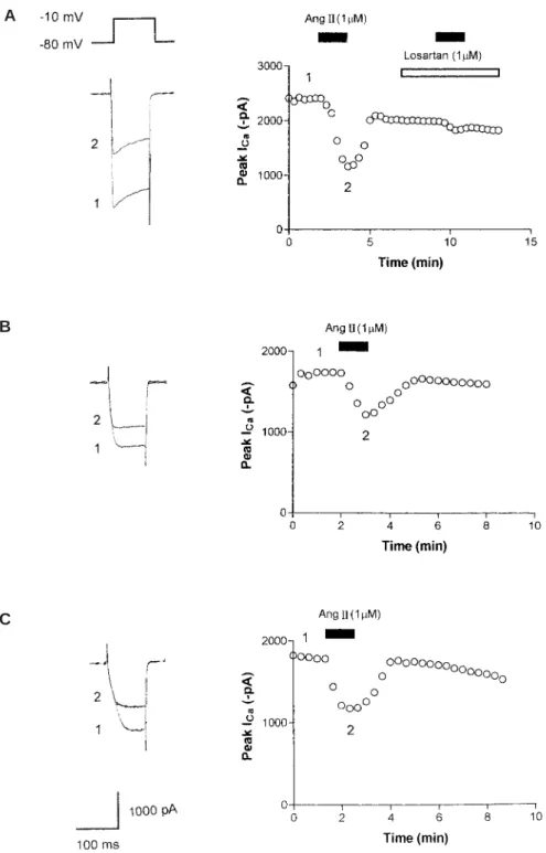
(3) 96. T. ENDOH et al.. adenylyl cyclase and reduction in cyclic AMP in various tissues1,2,10). Additionally, it has been demonstrated that Ang II inhibits I Ca by suppressing cyclic AMP production in sinoatrial node cells3). We previously reported that Ang II, mediated via Ang II type 1 (AT1) receptors, inhibits voltage-dependent calcium channels (VDCCs) currents (I Ca ) in submandibular ganglion (SMG) neurons7). It remains unclear, however, whether cyclic AMP-PKA pathways are mediated in the Ang II-induced inhibition of I Ca in this neuron. Thus, the purpose of the present study was to examine the contribution of cyclic AMP-PKA pathways in the Ang II-induced inhibition of I Ca in the SMG neuron.. MATERIALS AND METHODS Experiments were conducted according to the guidelines for the treatment of experimental animals at Tokyo Dental College. SMG neurons from hamsters were acutely dissociated with a modified version of the method described previously7). In brief, SMG neurons were isolated from 4–6 week-old hamsters and maintained in Ca2Ⳮ-free Krebs solution of the following composition (in mM): 136 NaCl, 5 KCl, 3 MgCl2 䡠 6H2O, 10.9 glucose, 11.9 NaHCO3 and 1.1 NaH2PO4 䡠 2H2O. The neurons were treated with collagenase type I (3 mg/ml in Ca2Ⳮ-free Krebs solution; Sigma, St. Louis, MO, U.S.A.) for 50 min at 37°C, followed by incubation in trypsin type I (1 mg/ml in Ca2Ⳮ-free Krebs solution; Sigma, St. Louis, MO, U.S.A.) for an additional 10 min. The supernatant was replaced with normal Krebs solution of the following composition (in mM): 136 NaCl, 5 KCl, 2.5 CaCl2, 0.5 MgCl2 䡠 6H2O, 10.9 glucose, 11.9 NaHCO3 and 1.1 NaH2PO4 䡠 2H2O. Neurons were then plated onto poly-l-lysine (Sigma)-coated glass coverslips. Voltage-clamp recordings were conducted using the whole-cell configuration of the patch clamp technique4). Fabricated recording pipettes (2–3M⍀) were filled with an. internal solution with the following composition (in mM): 100 CsCl, 1 MgCl2 , 10 HEPES, 10 BAPTA, 3.6 MgATP, 14 tris2phosphocreatine, 0.1 GTP, and 50 U/ml creatine phosphokinase. The pH was adjusted to 7.2 with CsOH. After the formation of a giga seal, in order to record I Ca , the external Krebs solution was replaced by solution containing the following (in mM): 67 cholinechloride, 100 tetraethylammonium chloride, 5.3 KCl, 5 CaCl2 and 10 HEPES. The pH was adjusted to 7.4 with Tris base. Command voltage protocols were generated with a computer software pCLAMP version 8 (Axon Instruments, Union City, CA, U.S.A.) and transformed to an analogue signal using a DigiData 1200 interface (Axon Instruments, Union City, CA, U.S.A.). The command pulses were applied to the cell through an L/M-EPC7 amplifier (HEKA Elektronik, Lambrecht, Germany). The currents were recorded with the amplifier and a computer software pCLAMP 8 acquisition system. All data analyses were performed using the pCLAMP 8 acquisition system. Statistical analyses were made by Student t -test. Probability (p) values of less than 0.05 were considered significant. Ang II was purchased from Peptide Institute. Losartan was a gift from Merck & Co., Inc. 8-(4-chlorophenylthio)-adenosine 3쎾: 5쎾cyclic monophosphate (CPT-cAMP) was purchased from Sigma, St. Louis, MO, U.S.A. The PKA inhibitor peptide, PKI (5–24), was purchased from BIOMOL Research Laboratories Inc. (Plymouth, PA, U.S.A.).. RESULTS An example of the effects of Ang II and AT1 receptor antagonist, losartan, on I Ca is shown in Fig. 1 (A). Application of 1M Ang II inhibited I Ca from ⳮ2,408 pA to ⳮ1,160 pA (51.8% inhibition) in this neuron. 1M losartan completely antagonized the inhibitory effect of Ang II. To evaluate the possible contribution of PKA to the Ang II-induced inhibition of I Ca ,.
(4) AT1 RECEPTOR PATHWAY IN GANGLION NEURON. A. B. C. Fig. 1 (A) Effects of Ang II and losartan on I Ca . Superimposed I Ca traces were recorded using a voltage protocol at the times indicated in the time course graph (right). Open points in the graph indicate I Ca . Ang II (1M) and losartan (1M) were bath applied during the times indicated by the filled and open bars, respectively. (B) Effects of Ang II on I Ca in a neuron pretreated with CPT-cAMP (10M for 10 min). (C) Effects of Ang II on I Ca in the presence of PKI (5–24) (20M for 10 min) contained in the recording pipette.. 97.
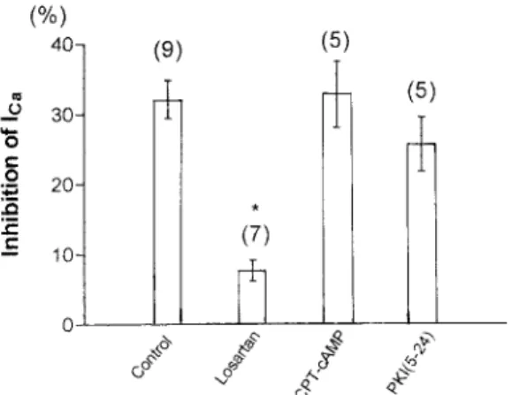
(5) 98. T. ENDOH et al.. the effects of Ang II on I Ca in neurons treated with CPT-cAMP (10M for 10 min), membrane permeable cyclic AMP, were investigated. We have previously demonstrated that this compound has a sufficient effect to cause the saturation of intracellular cyclic AMP levels11). As shown in Fig. 1 (B), Ang II-induced inhibition of I Ca was not affected by CPTcAMP treatment. In addition, intracellular dialysis of PKI (5–24) (20M for 10 min), PKA inhibitor, also did not attenuated the Ang II-induced inhibition of I Ca as shown in Fig. 1 (C). The data showing the effects of altering PKA activity and antagonizing AT1 receptors are summarized in Fig. 2. On average, the Ang II-induced inhibition of I Ca was 32.1 Ⳳ2.7% (n⳱9) for control, 7.6Ⳳ1.5% (n⳱7) for neurons treated with losartan, 32.7Ⳳ4.7% (n⳱5) for the neurons treated with CPTcAMP, and 25.6Ⳳ3.9% (n⳱5) when the recording pipette was filled with PKI (5–24).. DISCUSSION The present results show that Ang II inhibited I Ca by a cyclic AMP-PKA pathwayindependent mechanism. In contrast, there is considerable evidence that Ang II receptors cause inhibition of cyclic AMP-PKA through inhibition of adenylyl cyclase and reduction in cyclic AMP1–3,10). This evidence raises several questions: (1) which processes are absent in Ang II-induced inhibition of ICa in SMG?; (2) are cyclic AMP-PKA pathways between AT1 receptors and VDCCs possibly absent in SMG?; and (3) is there a possible lack of AT1 receptors that is selectively regulating cyclic AMP-PKA pathways in SMG? Various reports have demonstrated the distinct second messenger pathways in the Ang II-induced ion channel modulations: for instance, PKC pathways and calcium/ calmodulin-dependent protein kinase II (CaM K II) pathways in the central nervous system (CNS) neurons 9,12), Ras/Raf/mitogenactivated protein (MAP) kinase pathways and Janus kinase/signal transducers and. Fig. 2 Summary of Ang II-induced inhibition of I Ca determined when various conditions. Histogram demonstrating the degree of I Ca inhibition by 1M Ang II in control (untreated neurons), after losartan (AT1 receptor antagonist), after CPT-cAMP (membrane permeable cyclic AMP) and intracellular dialysis of PKI (5–24) (PKA inhibitor). Ordinates represent the Ang II-induced inhibition of I Ca . Number of neurons tested is indicated in parentheses. *Significant difference relative to control (p⬍0.05, by post hoc Dunnett’s test). activators of transcription ( JAK/STAT) pathways6,8), Fos-regulating kinase and c-Jun NH2terminal kinase5). The present data do not allow us to deduce the exact mechanism by which intracellular pathways may alter VDCCs function. It will be important in future experiments to determine what second messenger pathways contribute to the Ang II-induced inhibition of I Ca in SMG neurons.. ACKNOWLEDGEMENTS This work was supported by a Grant-in-Aid for Scientific Research from the Japanese Ministry of Education, Culture, Sports, Science and Technology (No. 12671816).. REFERENCES 1) Anand-Srivastava, M.B. (1983). Angiotensin II receptors negatively coupled to adenylate cyclase in rat aorta. Biochem Biophys Res.
(6) AT1 RECEPTOR PATHWAY IN GANGLION NEURON. Commun 117, 420–427. 2) Griendling, K.K., Rittenhouse, S.E., Brock, T.A., Ekstein, L.S., Gimbrone, M.A., Jr. and Alexander, R.H. (1986). Sustained diacylglycerol formation from inositol phospholipids in angiotensin II-stimulated vascular smooth muscle cells. J Biol Chem 261, 5901– 5906. 3) Habuchi, Y., Lu, L.-L., Morikawa, J. and Yoshimura, M. (1995). Angiotensin II inhibition of L-type Ca2Ⳮ current in sinoatrial node cells of rabbits. Am J Physiol 268, H1053– H1060. 4) Hamill, O.P., Marty, A., Neher, E., Sakmann, B. and Sigworth, F.J. (1981). Improved patchclamp techniques for high-resolution current recording from cells and cell-free membrane patches. Pflügers Archs 391, 85–100. 5) Huang, X.-C., Deng, T. and Sumners, C. (1998). Angiotensin stimulates activation of Fos-regulating kinase and c-Jun NH2-terminal kinase in neuronal cultures from rat brain. Endocrinology 139, 245–251. 6) Huang, X.-C., Richards, E.M. and Sumners, C. (1996). Mitogen-activated protein kinases in rat brain neuronal cultures are activated by angiotensin II type 1 receptors and inhibited by angiotensin II type 2 receptors. J Biol Chem 27, 15635–15641. 7) Ikegami, H., Endoh, T. and Suzuki, T. (2001). Angiotensin II-induced inhibition of calcium currents in hamster submandibular ganglion neurons. Neurosci Res 41, 227–232.. 99. 8) Marrero, M.B., Schieffer, B., Paxton, W.G., Heerdt, L., Berk, B.C., Delafontaine, P. and Bernstein, K.E. (1995). Direct stimulation of Jak/STAT pathway by the angiotensin II AT1 receptor. Nature 375, 247–250. 9) Sumners, C., Zhu, M., Gelband, C.H. and Posner, P. (1996). Angiotensin II type 1 receptor modulation of neuronal KⳭ and Ca2Ⳮ currents: Intracellular mechanisms. Am J Physiol 271, C154–C163. 10) Unger, T., Chung, O., Csikos, T., Culman, J., Gallinat, S., Gohlke, P., Hohle, S., Meffert, S., Stoll, M., Stroth, U. and Zhu, Y.Z. (1996). Angiotensin receptors. J Hypertens Suppl 14, S95–103. 11) Yamada, T., Endoh, T. and Suzuki, T. (1999). Inhibition of calcium channels by neurokinin receptor and signal transduction in hamster submandibular ganglion cells. J Auton Nerv Sys 76, 1–8. 12) Zhu, M., Gelband, C.H., Posner, P. and Sumners, C. (1999). Angiotensin II decreases neuronal delayed rectifier potassium current: Role of calcium/calmodulin-dependent protein kinase II. J Neurophysiol 82, 1560–1568. Reprint requests to: Dr. Takayuki Endoh Department of Physiology, Tokyo Dental College, 1-2-2 Masago, Mihama-ku, Chiba 261-8502, Japan.
(7)
図
関連したドキュメント
In addition, more than 50% of fluorescence positive cells exhibited shrinkage and rounding even in the absence of anti-Fas antibodies (about 56, 65, and 56% of PKR-, dN-,
[Journal Article] Two independent regions of human telomerase reverse transcriptase (hTERT) are important for their oligomerization and telomerase 2002.
Pim-3, a proto-oncogene with serine ⁄ threonine kinase activity, is aberrantly expressed in human pancreatic cancer and phosphorylates Bad to block Bad-mediated apoptosis in
These are several genetically modified animal models for cardiac hypertrophy, in which the cardiac RAS is targeted [26]. They are largely the mouse models that aimed at
Furthermore, administration of testosterone to female mice newly induces nuclear JunD/menin immunoreactivity in cells located in the proximal portions of the SD at 6-24 hrs,
GA or an aqueous extract of Schisandra chinensis induced not only endothelium- dependent but also endothelium-independent vascular relaxation in isolated thoracic aorta
7 The current density J z at the center of the channel is higher for a micropolar fluid than that for a Newtonian fluid, and it will decrease as the microrotation parameter
We then compute the cyclic spectrum of any finitely generated Boolean flow. We define when a sheaf of Boolean flows can be regarded as cyclic and find necessary conditions
