Report on the use of non-clinical studies in the
regulatory evaluation of oncology drugs
Yoshihiro Hayakawa,
1,2Manabu Kawada,
1,3Hiroyoshi Nishikawa,
1,4Takahiro Ochiya,
1,5Hideyuki Saya,
1,6Hiroyuki Seimiya,
1,7Ryoji Yao,
1,8Masahiro Hayashi,
1,9Chieko Kai,
1,10Akira Matsuda,
1,11Tomoki Naoe,
1,12Atsushi Ohtsu,
1,13Taku Okazaki,
1,14Hideo Saji,
1,15Masataka Sata,
1,16Haruhiko Sugimura,
1,17Yuichi Sugiyama,
1,18Masakazu Toi
1,19and Tatsuro Irimura
1,201Subcommittee on Non-clinical Studies, The Science Board to the Pharmaceuticals and Medical Devices Agency, Tokyo;2Division of Pathogenic Biochemistry, Department of Bioscience, Institute of Natural Medicine, University of Toyama, Toyama;3Institute of Microbial Chemistry, Microbial Chemistry Research Foundation, Numazu-shi;4Division of Cancer Immunology, Exploratory Oncology Research and Clinical Trial Center, National Cancer Center, Chiba;5Division of Molecular and Cellular Medicine, National Cancer Center Research Institute, Tokyo;6Division of Gene Regulation, Institute for Advanced Medical Research, School of Medicine, Keio University, Tokyo;7Division of Molecular Biotherapy, Cancer Chemotherapy Center, Japanese Foundation for Cancer Research, Tokyo;8Division of Cell Biology, Cancer Institute, Japanese Foundation for Cancer Research, Tokyo;9Department of Pharmacy, Toranomon Hospital, Tokyo;10Laboratory Animal Research Center, Institute of Medical Science, The University of Tokyo, Tokyo;11Department of Medicinal Chemistry, Faculty of Pharmaceutical Sciences, Hokkaido University, Sapporo;12National Hospital Organization Nagoya Medical Center, Nagoya; 13Exploratory Oncology Research and Clinical Trial Center, National Cancer Center, Chiba;14Division of Immune Regulation, Institute for Genome Research, Tokushima University, Tokushima;15Department of Patho-Functional Bioanalysis, Graduate School of Pharmaceutical Sciences, Kyoto University, Kyoto; 16Department of Cardiovascular Medicine, Institute of Biomedical Sciences, Tokushima University Graduate School, Tokushima;17Department of Tumor Pathology, Hamamatsu University School of Medicine, Shizuoka;18Sugiyama Laboratory, RIKEN Innovation Center, RIKEN Cluster for Industry Partnerships, Kanagawa;19Department of Breast Surgery, Graduate School of Medicine, Kyoto University, Kyoto;20Juntendo University School of Medicine, Tokyo, Japan
Key words
Animal model, cancer, drug development, oncology drug, regulatory science
Correspondence
Tatsuro Irimura, Pharmaceuticals and Medical Devises Agency, 3-3-2 Kasumigaseki, Chiyoda-ku, Tokyo 100-0013, Japan or Juntendo University School of Medicine, 2-1-1 Hongo, Bunkyo-ku, Tokyo 113-8421, Japan.
Tel: +81-3-3506-9407 (ext 3802) or +81-3-5802-1222; Fax: +81-3-3813-3307;
E-mail: t-irimura@juntendo.ac.jp Funding Information
The work was conducted as a part of activity of the Science Board of Pharmaceuticals and Medical Devices Agency.
Received October 21, 2015; Revised December 4, 2015; Accepted December 4, 2015
Cancer Sci 107 (2016) 189–202 doi: 10.1111/cas.12857
Non-clinical studies are necessary at each stage of the development of oncology drugs. Many experimental cancer models have been developed to investigate car-cinogenesis, cancer progression, metastasis, and other aspects in cancer biology and these models turned out to be useful in the efficacy evaluation and the safety prediction of oncology drugs. While the diversity and the degree of engagement in genetic changes in the initiation of cancer cell growth and pro-gression are widely accepted, it has become increasingly clear that the roles of host cells, tissue microenvironment, and the immune system also play important roles in cancer. Therefore, the methods used to develop oncology drugs should continuously be revised based on the advances in our understanding of cancer. In this review, we extensively summarize the effective use of those models, their advantages and disadvantages, ranges to be evaluated and limitations of the models currently used for the development and for the evaluation of oncology drugs.
Progress of Cancer Biology is Closely Linked to Oncology
Drug Development
T
he history of the development of oncology drugs, so-called
chemotherapeutic agents, is closely associated with the
progress of the biological understanding of cancer. Based on
the concept that cancer cells are capable of unlimited
prolifera-tion, substances that inhibit DNA replication or cell division
have been used as drugs for cancer treatment for a long period,
since the 1950s. Although the concept has remained unchanged
to the present day,
(1)the discovery of cancer cell-specific
metabolic pathways has led to the development of
antimetabo-lites.
(2)After the discovery of cancer cell-specific molecular and
cellular mechanisms that are essential for the survival and
growth of cancer cells, therapeutic drugs targeting these
mecha-nisms, so-called molecular targeted drugs, started to be
devel-oped.
(3)Research into viral oncogenesis, started in the 1960s,
led to the discovery of oncogenes,
(4)and research into the
genetic backgrounds of cancers led to the discovery of tumor
suppressor genes.
(5)In the course of such studies, it also became
apparent that cancer is caused by genetic abnormalities such as
mutations,
deletions,
duplications,
and
translocations.
(6–9)Molecular targeted cancer drugs appeared in the 1990s;
(10)can-© 2016 The Authors. Cancer Science published by John Wiley & Sons Australia, Ltd on behalf of Japanese Cancer Association.
This is an open access article under the terms of the Creative Commons Attribution-NonCommercial-NoDerivs License, which permits use and distribution
cer was considered a disease characterized by abnormal
differen-tiation, and the efficacy of differentiation-inducing agents was
demonstrated.
(11,12)Furthermore, it was shown that a solid tumor
tissue consists of cancer and host cells such as vascular cells,
fibroblasts, and cells in the immune system and that these host
cells are essential for tumor growth. Drugs targeting the function
of these host cells and their interactions with cancer cells were
proven to be effective.
(13)Based on these findings, it has been
thought that regulatory mechanisms for the entire organism are
involved in the action of oncology drugs that regulate the
immune system.
(14)Significance of Non-Clinical Studies in Efficacy Evaluation
and Safety Prediction
Non-clinical studies are necessary at each stage of the
develop-ment of oncology drugs. Particularly, the efficacy and the
safety of a drug must be examined and evaluated before
under-taking any clinical study of the drug. Types of non-clinical
studies and how critical they are vary depending on the types
and mechanisms of action of oncology drugs. Non-clinical
studies required to develop drugs targeting cancer
–host
interac-tions differ markedly from those on substances having direct
killing effects on cancer cells. Many experimental cancer
mod-els (animal modmod-els, ex vivo modmod-els, and in vitro modmod-els) have
been developed to investigate carcinogenesis, cancer
progres-sion, metastasis, and other aspects in cancer biology. These
models turned out to be useful in the efficacy evaluation and
the safety prediction of oncology drugs. The present review
summarizes the effective use of those models, their advantages
and disadvantages, ranges to be evaluated, and limitations of
the models used in non-clinical study.
Evaluation of Oncology Drugs Using Experimental Animal
Models
Two classes of experimental animal models for human
can-cers are currently used for the evaluation of oncology drugs:
transplantation models and autochthonous cancer models.
Transplantation models have been playing an important role
in the non-clinical evaluation of oncology drugs. They are
generally categorized into two types, namely xenograft
mod-els using human cancer cells and orthograft modmod-els using
murine cancer cells. There has been some debate that the
efficacy evaluation of oncology drugs in transplantation
mod-els might not be adequate for predicting the clinical efficacy
or the types of cancer for which the drug could be effective.
As
autochthonous
cancer
models,
chemical
carcinogen-induced models were first established and the subsequent
technological progress in gene manipulation allowed
research-ers to produce models harboring the genetic mutations of
human cancer. Although a number of technical issues
regard-ing the ability to maximize the utility of these models need
to be addressed, such as their usability, reproducibility, and
throughput compared with transplantation models,
autochtho-nous cancer models clearly show some promise. In Table 1,
we summarize the characteristics of those experimental
can-cer models used to evaluate the efficacy of oncology drugs in
non-clinical studies.
Transplantation cancer models.
In general, the s.c.
(hetero-topic) transplantation models with cancer cell lines have been
used, and the efficacies of oncology drug response are
evalu-ated based on tumor size. These models are particularly useful
when a drug has a marked antiproliferative effect on cancer
cells. It is also easy to access tumor tissue samples from these
models for subsequent pharmacodynamic evaluations. Despite
such clear advantages, these models may not reflect the actual
characteristics of the cancer microenvironment because the s.c.
tissue is “heterotopic” for most cancer cells. In this context,
orthotopic transplantation models may reproduce the cancer
microenvironment more faithfully, although their utility caused
by species differences should be considered. To analyze
metas-tasis dissemination of cancer cells, experimental metasmetas-tasis
models have been considered as useful for evaluating drug
efficacy in the process after the invasion of cancer cells from
the primary tumor into the nearby blood vessel. Although these
models have clear advantage in their usability and
repro-ducibility, they cannot reproduce the entire step before the
extravasation of cancer cells and may not accurately represent
actual metastases by injecting a substantial number of cancer
cells into the blood vessel. In this regard, spontaneous
metasta-sis models have been considered to reflect the process of the
metastasis of cancer cells more accurately than the heterotopic
or orthotopic transplantations. Despite the clear advantages of
these models, only a limited number of cancer cell lines are
available and the results of experiments often vary. In addition
to the above transplantation cancer models with cancer cell
lines, patient-derived xenograft models have been considered
as emerging animal models recapitulating the clinical condition
of individual cancer patients, and therefore attracted much
attention on precision treatment.
(15–17)Autochthonous cancer models.
There are two major types of
autochthonous cancer models, carcinogen-induced models and
gene-engineered mouse (GEM) models. Of these, GEM models
have been regarded as a better choice for testing drug efficacy,
because the drug effects can be evaluated on autochthonous
cancer cells induced by gene mutations resembling human
can-cer. As summarized in Table 2, there are several pros and cons
to using autochthonous cancer models for drug efficacy tests in
non-clinical studies. In particular, the timing of tumor
occur-rence and tissue specificity are often the major concerns of
carcinogen-induced models and conventional knockout
⁄
trans-genic mice. To overcome these issues, conditional gene
knock-out or gene expression technology provide us with the
opportunity to use GEM models that more closely represent
the pathology of human cancers. In addition to the above
tech-nical difficulties, the administrative challenges, such as
mainte-nance of mouse strains to acquire a sufficient number of mice
as well as the characters of each mouse model, including the
latency and incidence of tumor and other relevant issues, need
to be considered before undertaking efficacy studies testing
oncology drugs in GEM models. Nevertheless, new
technolo-gies, such as in vivo imaging methods for small animals, have
been introduced as powerful tools for quantitative evaluation
of cancer occurrence and subsequent growth in GEM models.
In Table 3, GEM models developing tumors induced by
genetic mutations found in corresponding human cancers are
summarized.
Spontaneous cancer models using companion animals.
Even in
companion animals, such as dogs and cats, the incidence of
cancer has been increasing, likely due to their life extension
together with genetic factors. In fact, cancer has become the
leading cause of death among those companion animals. In
particular, it has been known that the mortality from cancer is
reported to be 47% (based on the report by the Veterinary
Cancer Society, http://www.vetcancersociety.org/members/) in
large breed dogs aged 10 years or more. Therefore, the
estab-lishment of early diagnosis methods and the development of
Table 1. Characteristics of preclinical animal models for oncology drug development Model Outline Advantage Disadvantage Mouse cancer model Transplantation model Heterotopic model Models s.c. transplanted with tumor cell lines Easy to monitor the drug efficacy on tumor growth by examining visible size May not fully reproduce human cancer tissue because of poor stroma involvement Efficacy data in this model may not accurately correlate with clinical outcomes in some cases Orthotropic model Models transplanted tumor cell lines into tissue where they were originated or to which they metastasize Account for tissue microenvironment for cancer cells where originated or metastasized Requires relatively complicated methods for transplantation Difficult to monitor tumor growth over time Autochthonous model Carcinogen-induced model Models induced tumors by carcinogen such as chemicals or UV radiation Reproduce carcinogenesis-associated events such as host inflammation Requires complicated methods and expects potential variability among individual animals Difficulties in preparing a sufficient number of mice and relatively time-consuming GEM model Models induced tumors by modifying cancer-related genes Reproduce human tumor development in the genetic character and the originating tissue Difficult to maintain mouse with multiple mutant alleles May not accurately reproduce human cancer types Challenges for using drug efficacy evaluation (tumor latency, time for tumor formation etc.) Human cancer model Transplantation model Cell line Transplantation of human cancer cell lines or human tumor tissues into immune-compromise d mice Ability for testing human cell lines in relevant tumor types or with genetic backgrounds Accuracy of the model in its clinical relevance has been questioned in some cases PDX Direct transplantation of patient-derived cancer tissue into immune-compromise d mice Ability for testing clinical patient-derived tumor tissues Clear restriction in availability and utility Spontaneous dog cancer model Naturally occurring canine cancer Use of dogs who naturally develop cancers Conduct as veterinary clinical trial Share many characteristics with human malignancies Difficulties in preparing a sufficient number of dogs Summary of the characteristics of preclinical animal models and their potential advantages and disadvantages for use in oncology drug development. GEM, gene-engineered mouse; PDX, patient-derived xenograft.
therapeutic drugs for cancer in companion animals is being
actively pursued in the USA and Europe. Considering the
pathology of cancer in large breed dogs seems to be similar to
those in humans,
(68)the utility of spontaneous cancer in large
breed dogs for testing new oncology drugs has already been
initiated in the USA and Europe.
(69)In Japan, the leading
cause of death in dogs is also cancer with a mortality of 54%
(“The Ten Leading Causes of Death in Dogs and Cats”
reported by the Animal Insurance System Japan Animal Club),
which is much higher than the mortality rate of other diseases
such as heart disease (17%). Given these circumstances,
stud-ies for developing methods for the diagnosis and treatment of
cancer in dogs have been actively initiated. Based on the
results of these studies, the Japanese Society of Clinical
Veterinary Medicine have been discussing the significance of
cancer models using companion animals in non-clinical studies
for developing oncology drugs as well as preparing for the
establishment of relevant administrative and management
sys-tems for its application.
Evaluation of Oncology Drugs that Directly Target Cancer
Cells
The efforts of oncology drug development originally
concen-trated on the production of drugs that directly target the
prolif-eration or metabolic properties of cancer cells. Along with
discovery of oncogenic driver genes, development of
molecu-lar targeted drugs has been highlighted, which directly pinpoint
signal transduction pathways involving those driver genes, as
well as the protein degradation systems, epigenome, and
meta-bolic systems of cancer cells. As molecular targeted drugs,
tyr-osine kinase inhibitors (TKI), multi-targeted kinase inhibitors
(MTKI), and drugs that target molecular mechanisms for cell
cycle regulation and others have been successfully developed.
Although the classical anticancer chemotherapeutic drugs also
show cytotoxicity by attacking specific intracellular molecules,
the term “molecular targeted drug” in this report is defined as
a drug that has been developed through primary identification
of a molecule or a signaling pathway as a therapeutic target,
which is highly activated or deregulated in cancer cells.
Table 4 summarizes the pros and cons for evaluating
molecu-lar targeted drugs in non-clinical cancer models. The results
produced by the use of these models have been included in the
application of new drugs; the models believed to be essential.
Tyrosine kinase inhibitors and other kinase inhibitors.
Tyro-sine kinase inhibitors include epidermal growth factor receptor
inhibitors (gefitinib, erlotinib, lapatinib, and afatinib), human
epidermal growth factor receptor 2 inhibitors (lapatinib and
afatinib), anaplastic lymphoma kinase inhibitors (crizotinib,
ceritinib, and alectinib), BCR-ABL inhibitors (imatinib,
dasa-tinib, nilodasa-tinib, ponadasa-tinib, and bosutinib), a KIT inhibitor
(ima-tinib), SRC inhibitors (dasatinib and bosu(ima-tinib), a JAK
inhibitor (ruxolitinib), a Bruton’s tyrosine kinase inhibitor
(ibrutinib), and a dual kinase MEK inhibitor (trametinib).
There are several other kinase inhibitors, including BRAF
inhi-bitors (vemurafenib and dabrafenib), a phosphatidylinositol-3
kinase inhibitor (idelalisib), and mammalian target of
rapamy-cin inhibitors (temsirolimus and everolimus). In addition, drugs
that target p38, AKT, p70S6 kinase, insulin-like growth factor
1 receptor, platelet-derived growth factor receptor (PDGFR),
fibroblast growth factor receptor (FGFR), MET, ROS 1, and
RET are currently being developed. For evaluating the
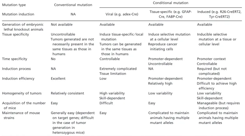
effica-Table 2. Characters of genetically engineered mouse modelsMutation type Conventional mutation Conditional mutation
Mutation induction NA Viral (e.g. adex-Cre) Tissue-specific (e.g. GFAP-Cre, FABP-Cre)
Induced (e.g. R26-CreERT2, Tyr-CreERT2) Generation of embryonic
lethal knockout animals
Not available Available Available Available
Tissue specificity Uncontrollable
Tumors generated are not necessarily present in the same tissues as those in humans
Induce tissue-specific⁄ local mutation
Tumors can be generated in the same tissues as those in humans
Induce selective mutation at a cellular level Reproduce cancer initiating cells Inducible selective mutation at a tissue or cellular level
Time specificity No Controllable Promoter-dependent
Uncontrollable
Promoter context Controllable Induction process NA Extremely complicated
Tissue limitation
NA Required (but not
complicated)
Induction efficiency Excellent Low Promoter-dependent
Relatively high
Promoter-dependent Difficult to achieve high
efficiency Homogeneity of tumors Relatively consistent High variability
Skill-dependent
Low variability Low variability Skill-dependent Acquisition of the number
of mice
Easy Difficult Easy Manageable (but requires
induction process) Maintenance of mouse
strains
Generally easy (dependent on target genes; difficult in the case of tumor generation in heterozygous mice)
Easy Complicated to maintain animals having multiple mutant alleles
Complicated to maintain animals having multiple mutant alleles
This table summarizes the advantages and potential problems in various types of genetically engineered mouse models for use in preclinical studies of oncology drugs. NA, not applicable.
Table 3. Mouse models corresponding to genetic mutations in human cancers
Human disease Mouse model
Cancer type Mutated
gene Mutated gene Mutation type
Mutation
induction Tumor produced Medulloblastoma RB1 Rb1/Tp53 Conditional KO⁄ conditional KO GFAP-Cre Medulloblastoma(18)
Rb1/Bmi1 Conditional KO⁄ conditional activation GFAP-Cre Medulloblastoma(19)
PTCH1 Ptch1 Conditional KO math1-cre⁄
GFAP-Cre
Medulloblastoma(20)
Gorlin syndrome PTCH1 Ptch1 Conventional Medulloblastoma,
rhabdomyosarcoma(21)
Pituitary gland tumor RB1 Rb1 Conventional KO Pituitary gland tumor(22,23)
Rb1 Conditional KO Pomc-Flp Pituitary gland tumor(24)
Lung cancer KRAS Kras Conventional KO (sporadic activation) Lung cancer(25)
BRAF Braf Conditional activation Adex-Cre Lung cancer(26,27)
RB1 Rb1/Tp53/Pten Conditional KO⁄ conditional KO ⁄ conditional KO
CGRP-CreER Lung cancer(28)
EML4-ALK EML4-ALK Conventional activation (SPC promoter) Lung cancer(29)
EML4-ALK Conditional activation Tet system Lung cancer(30)
KIF5B-RET KIF5B-RET Conventional activation (SPC promoter) Lung cancer(31)
EZR-ROS1 EZR-ROS1 Conventional activation (SPC promoter) Lung cancer(32)
Breast cancer PIK3CA Pik3ca Conditional activation MMTV-Cre Breast cancer(33)
TRP53 Pik3ca/Tp53 Conditional activation⁄ conditional KO MMTV-Cre Breast cancer, leukemia(34) PTEN Pten Conditional KO (stromal fibroblast) Fsp-Cre Breast cancer(35)
ERBB2 ErbB2 Conventional activation (MMTV promoter) Breast cancer(36,37)
ErbB2/Pten Conditional activation⁄ conventional KO MMTV-Cre Breast cancer(38)
RB1 Rb1/Tp53 Conditional KO⁄ conditional KO MMTV-Cre Breast cancer(39)
Hereditary breast cancer
BRCA1 Brca1/Tp53 Conditional KO⁄ conventional KO BLG-Cre Breast cancer(40)
Brca1/Chk2 Conditional KO⁄ conventional KO Wap-Cre Breast cancer(41)
BRCA2 Brca2/Tp53 Conditional KO⁄ conventional KO K14-Cre Breast cancer, skin tumor(42)
Colorectal cancer APC Apc/Kras Conditional KO⁄ conditional activation Adex-Cre Colorectal cancer(43)
KRAS Apc/Kras Conditional KO⁄ conditional activation Fapbl-Cre Colorectal cancer(44)
PTEN Apc/Pten Conditional KO⁄ conditional KO Cyp1a1-CreERT2
Tumor of the digestive tract(45)
Smad4 Apc/Smad4 Conventional KO⁄ conventional KO Tumor of the digestive tract(46) Familial adenomatous
polyposis
APC Apc Conventional KO Tumor of the digestive
tract(47–49)
Apc Conditional KO Adex-Cre Tumor of the digestive
tract,(50)liver cancer(51)
Hereditary non-polyposis colorectal cancer
MSH3 Msh3 Conventional KO Lymphoma(52)
MSH6 Msh6 Conventional KO Lymphoma,(52)tumor of the
digestive tract, skin cancer, uterine cancer(53)
Msh3/Msh6 Conventional KO Lymphoma,(52)tumor of the
digestive tract,(54)skin
tumor(53)
Cowden syndrome PTEN Pten Conventional KO Tumor of the digestive tract,
lymphoma, adrenal tumor, breast cancer, prostate cancer(55,56)
Pancreatic cancer KRAS Kras/Tp53 Conditional activation⁄ conditional KO pdx1-cre Pancreatic cancer(57)
Kras/Tgfbr2 Conditional activation⁄ conditional KO Ptf1a-cre Pancreatic cancer(58)
Kras/Pten Conditional activation⁄ conditional KO pdx1-cre Pancreatic cancer(59)
Endometrial cancer PTEN Pten/Mig6 Conditional KO⁄ conditional KO PR-Cre Endometrial cancer(60) Pten/Tp53 Conditional KO⁄ conditional KO PR-Cre Endometrial cancer(61)
Ovarian cancer KRAS Kras/Pten Conditional activation⁄ conditional KO Adex-Cre Ovarian cancer(62)
APC Apc Conditional KO Pgr-Cre Ovarian cancer(63)
BRCA2 Brca2/Tp53 Conditional KO⁄ conventional KO K18-Cre Ovarian cancer(64)
cies of those kinase inhibitors, transplantation models with
tar-get (mutant) gene-positive cancer cells or GEM models driven
by target (mutant) genes have been generally used. In general,
cancer cells that have potent driver gene mutations
(“gain-of-function” mutations) show a high degree of so-called oncogene
addiction, and therefore it would be relatively easy to predict
or
evaluate
the
drug
response
in
vivo.
These
non-clinical cancer models are also useful for evaluating
phar-macodynamics of the drugs by monitoring the phosphorylation
status of the target molecules, their downstream factors, or both.
Meanwhile, it should also be noted that established cancer cell
lines may have altered their phenotypes and characters compared
with the original cancers during in vitro culture, whereas
geneti-cally engineered cell lines may not be able to accurately
replicate the etiology of the relevant clinical cancer types.
Multitargeted kinase inhibitors.
Multitargeted kinase
inhibi-tors include a RAF
⁄ vascular endothelial growth factor
recep-tor-2 (VEGFR-2)
⁄ PDGFR-b inhibitor (sorafenib), a VEGFR2
⁄ PDGFR-b ⁄ KIT ⁄ FLT-3 inhibitor (sunitinib), a VEGFR ⁄ KIT
⁄ PDGFR inhibitor (pazopanib), a RET ⁄ VEGFR2 ⁄ EGFR
inhibi-tor
(vandetanib),
a
VEGF
⁄ PDGF inhibitor (axitinib), a
VEGFR
⁄ RET ⁄ KIT ⁄ PDGFR ⁄ RAF inhibitor (regorafenib), a
MET
⁄ RET ⁄ VEGFR ⁄ KIT ⁄ FLT-3 ⁄ TIE-2 ⁄ TRKB ⁄ AXL inhibitor
(cabozantinib),
and
a
VEGFR
⁄ FGFR ⁄ PDGFR ⁄ SRC ⁄ LCK
⁄ LYN ⁄ FLT-3 inhibitor (nintedanib). Similarly to TKIs, the
effi-cacy of MTKIs can be evaluated in non-clinical cancer
mod-els. However, MTKIs target multiple kinases and it is
generally difficult to prepare genetically engineered cell lines
that reproduce the pathology of the target cancers. In the case
of MTKIs that target angiogenic factors, such as VEGFR,
FGFR, and PDGFR, accurate prediction of in vitro efficacy
would be difficult: pazopanib, for example, does not
necessar-ily show a direct antiproliferative effect on many cancer cell
lines in vitro, but it significantly inhibits tumor growth in vivo
by blocking angiogenesis.
(74)Also, because MTKIs could have
multiple modes of action, establishment of the
proof-of-con-cept at the pharmacodynamic level in non-clinical cancer
mod-els might require a complex procedure.
Targeting cell cycle.
Palbociclib inhibits cyclin-dependent
kinases 4 and 6 (CDK4 and CDK6), which are involved in cell
cycle control. Furthermore, drugs targeting various cell cycle
regulators, such as WEE1, cell division cycle 7, checkpoint
kinase 1 and 2, ATR, Aurora, PLK, and mitotic kinesins, are
under clinical development. Efficacies of these drugs can be
evaluated using relevant cancer cell lines that have
abnormali-ties in the target molecules or their regulators (e.g. CCND1
⁄ CDK6 amplification or CDKN2 deletion ⁄ mutation) in
trans-plantation models.
Targeting protein degradation systems.
Protein degradation
systems have been recognized as an emerging therapeutic
target for particular types of cancer. While several target
mole-cules have been described in this category, proteasome
inhibi-tors, such as bortezomib and carfilzomib, have been developed
most extensively and approved as anticancer drugs.
Mean-while, other molecular targets include the NEDD8-activating
enzyme, the ubiquitin-activating enzyme, and stress proteins
that are involved in protein folding, such as heat shock protein
90 and glucose-regulated protein 78. Given that the
preferen-tial efficacies of proteasome inhibitors against multiple
mye-loma have been well established, transplantation models with
multiple myeloma cell lines could be applicable for evaluating
the efficacy of the drugs in this category. However, there are
several potential issues and limitations for predicting the
clini-cal efficacy of these drugs from non-cliniclini-cal cancer models:
detailed mechanisms for the action of the drugs and predictive
biomarkers for the drug responses are rather elusive, and
can-cer types that are susceptible to the anticancan-cer effects of the
drugs in non-clinical studies may not be consistent with those
in the clinical settings. Therefore, the latest knowledge from
basic research and clinical phase I studies on various cancer
types should be taken into consideration for additional
indica-tion of the drugs.
Targeting genomes and epigenomes.
The anticancer efficacies
of drugs that target cancer epigenomes, such as DNA
methyl-transferase inhibitors (azacytidine and decitabine) and histone
deacetylase (HDAC) inhibitors (vorinostat, panobinostat,
romi-depsin, and belinostat), have been shown in vivo, although the
cancer types against which the drugs are effective differ
between the non-clinical studies and clinical practice in some
cases.
(84)As these drugs affect many target sites in a
genome-wide manner, detailed mechanisms and predictive biomarkers
for the drug response often remain elusive. Drugs targeting the
genomic repair systems include poly(ADP-ribose) polymerase
(PARP) inhibitors, such as olaparib. Because there is a
syn-thetic lethal relationship between PARP and tumor suppressors,
BRCA1 and 2, It would be relatively easy to predict the
thera-peutic efficacy of PARP inhibitors by using transplant models
of cell lines with BRCA1 or 2 deficiency.
(85,86)Besides
BRCA1
⁄ 2, it has been also postulated that there are many
syn-thetic lethal factors with PARP inhibition. However, the
clini-cal validity of those candidates has not been fully established.
However, it should be also noted that synthetic lethality
con-firmed in the non-clinical studies (e.g. effect of a PARP
inhibi-tor on EWS-FLI1-positive Ewing’s sarcoma)
(87,89)could be
sometimes abolished by the formerly applied therapies in the
clinical settings.
Targeting cancer cell metabolisms.
Metabolic
enzymes
favored by cancer cells, such as isocitrate dehydrogenases 1
⁄ 2
(IDH1
⁄ 2) and fatty acid synthase, are potential targets for
can-cer therapy. For IDH1
⁄ 2 inhibitors, transplant models of IDH1
Table 3 (Continued)
Human disease Mouse model
Cancer type Mutated
gene Mutated gene Mutation type
Mutation
induction Tumor produced Skin tumor BRAF Braf Conditional activation Tyr-CreERT2 Malignant melanoma(66)
Braf/Pten Conditional activation⁄ conditional KO Tyr-CreERT2 Malignant melanoma(67)
PTCH1 Ptch1 Conditional KO R26-CreERT2 Basal cell tumor(20)
Mouse models reproducing generative tissues and mutations found in human caner. While many other scientifically excellent mouse models for human cancers have been generated, the table preferentially lists those harboring relatively simple mutant alleles suitable for preclinical studies. It should be noted some mouse models do not completely recapitulate pathologies of human cancer.
Table 4. Evaluation of drugs directly targeting cancer cells Classification (type of inhibitors) Target molecule Evaluation methods (drug efficacy study) Characteristics Problems Tyrosine kinases EGFR, HER2, ALK, BCR-ABL, KIT, SRC, JAK, BTK, IGF1R, PDGFR, FGFR, MET, ROS1, RET (i) Transplantation models of target (mutant) gene positive cancer cells Cancer cell lines with target (mutant) genes (70 ) Alternative cell lines into which target (mutant) genes are transfected (71) (e.g. Ba ⁄F3) (ii) GEM models (29) Can predict ⁄evaluate drug efficacy in the model with potent driver gene activities and oncogene addiction (72) Can generate resistant cells as negative control Can establish proof-of-concept pharmacodynamically by evaluating autophosphorylation of target kinases or phosphorylation of downstream factors (i) Cancer cell lines may change their phenotypes during the process of their establishment due to selective pressure and stresses (ii) Alternative cell lines may not accurately replicate the etiology of the relevant cancer types Kinases (multi-targeted) RAF, VEGFR-2, PDGFR-b , KIT, FLT-3, RET, EGFR, MET, RET, TIE-2, TRKB, AXL, SRC, LCK, LYN The same as (i) and (ii) above (31 ) For anti-angiogenic agents, Matrigel plug assay could be used (73) Can predict ⁄evaluate drug efficacy in the model with potent driver gene activities (31 ) In addition to (i) and (ii) above: It is difficult to generate alternative cell lines reproducing the pathology of target cancers by genetic engineering when the drug acts on multiple kinases in the target cancer cells In vitro cell growth assays do not reflect the antiangiogenic action in vivo (74 ) May require complicated pharmacodynamic analyses due to the presence of multiple targets MAPK pathway MEK, BRAF, p38 Cancer cell lines with mutations in the target pathway of interest (target molecule or upstream target) or transplantation animal models with alternative cell lines generated by genetic engineering (75 ,76) GEM models (27) Can predict ⁄evaluate drug efficacy in the model with potent driver gene activities (77 ) Can establish proof-of-concept pharmacodynamically by evaluating phosphorylation of downstream factors In addition to (i) and (ii) above: (iii) It is difficult to achieve sufficient drug response in some cancer types including colorectal cancer with less potent driver activities, in which other coexisting (i.e. not mutually exclusive) driver pathways contribute to tumor proliferation (77) PI3K ⁄mTOR pathway PI3K, mTOR, AKT, p70S6K Cancer cell lines with mutations in the target pathway of interest (target molecule or upstream target) or transplantation animal models with alternative cell lines generated by genetic engineering (78 ) GEM models (33) Can predict ⁄evaluate drug efficacy in the model with potent driver gene activities (79 ) Can establish proof-of-concept pharmacodynamically by evaluating phosphorylation of downstream factors The same as (i), (ii), and (iii) above Cell cycle CDK4 ⁄6, WEE1, CDC7, CHK1, CHK2, ATR, Aurora, PLK, mitotic kinesins Cancer cell lines with mutations in the target pathway of interest (target molecule or upstream target) or transplantation animal models with alternative cell lines generated by genetic engineering (80 ) Drug efficacy may be achieved in cancer cell lines with an abnormality as shown in the left-hand column The same as (i), (ii), and (iii) above
Table 4 (Continued) Classification (type of inhibitors) Target molecule Evaluation methods (drug efficacy study) Characteristics Problems
Protein degradation system
Proteasome, related target molecules (NEDD8-activating enzyme, ubiquitin-activating enzyme, HSP90, GRP78) Allograft ⁄xenograft models of multiple myeloma cell lines (81) Can predict ⁄evaluate drug efficacy with multiple myeloma cell lines used in the studies of previously developed drugs In addition to (i) above: (iv) Cancer types for which drugs are effective in preclinical studies may not be consistent with those in clinic Genome ⁄epigenome DNMT, related target molecules (histone methyltransferase, histone demethylase) Allograft ⁄xenograft models of MDS cell lines (82 ) MDS models generated by implanting MDS cell lines into genetically engineered NSG mice (83 ) MDS mouse models replicate the pathology more accurately than other transplantation animal models In addition to (i) and (iv) above: Due to a very small number of available cell lines, clinical relevance of the model may be limited (v) Due to the genome-wide distribution of target sites, detailed mechanisms of action and predictive biomarkers for the drug response remain unclear HDAC Allograft ⁄xenograft models of colorectal ⁄prostate ⁄lung cancer cell lines (84) Drug efficacy may be achieved in some cancer types in addition to those shown in the left-hand column The same as (i), (iv), and (v) above Cutaneous T-cell lymphoma and peripheral T-cell lymphoma are currently approved for HDAC inhibitors PARP1 ⁄PARP2, related target molecules (DNA-dependent protein kinase, telomerase) Allograft ⁄xenograft models of cancer cell lines with BRCA1 or BRCA2 (tumor suppressor gene) mutation or inactivation (85 ,86) Can predict ⁄evaluate drug efficacy by using cancer cell lines with BRCA1 ⁄2 deficiency: there is a synthetic lethal relationship between PARP1 ⁄2 and BRCA1 ⁄2 The same as (i) and (iv) above In addition to BRCA1 ⁄2, substantial numbers of synthetic lethal factors are reported, (however, most of them are described only at a basic research level and the clinical relevance has not been fully established) Synthetic lethality may be diminished by pretreatment in the clinical cases even if preclinically confirmed (87) Metabolic systems IDH1 ⁄IDH2 (mutant-type), Fatty acid synthase Xenograft models of IDH1 (R132) ⁄IDH2 (R172) mutant-positive AML or glioma cell lines (88) Can predict ⁄evaluate drug efficacy by examining the presence of mutation Pharmacodynamic study can be carried out by monitoring mutation-specific metabolites (oncometabolites) (88 ) Drugs targeting molecules that produce no oncometabolites may be effective to a wider range of cancer types If the target produces no oncometabolites, mechanisms of action or predictive biomarkers for the drug response may not be available and it may be difficult to design evidence-based studies to evaluate the drug response This table classifies the target molecules of approved ⁄investigational drugs used in Japan, overseas, or both and lists representative non-clinical evaluation methods of these drugs. Due to their usefulness and usability, evaluation results have been used for publication data of original papers and oncology drug application dossiers fo r approval. Meanwhile, it should be noted that these technologies have technical limitations and contain a number of limitations ⁄problems attributable to the properties or unclarified factors of target molecules and diseases. ALK, anaplastic lymphoma kinase; BTK, Bruton’s tyrosine kinase; CDC7, cell division cycle 7; CHK, checkpoint kinase; DMNT, DNA methyltransferase; EGFR , epidermal growth factor receptor; FGFR, fibroblast growth factor receptor; GRP, glucose-regulated protein; HDAC, histone deacetylase; HER2, human epidermal growth factor receptor 2; HSP , heat shock protein; IDH, isocitrate dehy-drogenase; IGF1R, insulin-like growth factor 1 receptor; MDS, myelodysplastic syndromes; mTOR, mammalian target of rapamycin; PARP, poly(ADP-ri bose) polymerase; PDGFR, platelet-derived growth factor receptor; PI3K, phosphatidylinositol-3 kinase; VEGFR, vascular endothelial growth factor receptor.
(R132) or IDH2(R172) mutation-positive AML and glioma cell
lines
are
useful
for
predicting
drug
efficacies.
(88)The
pharmacodynamics of these drugs can be evaluated by
moni-toring the mutation-specific metabolite (oncometabolite),
2-hydroxyglutaric acid. However, if the target molecule does not
produce a characteristic oncometabolite, one may expect a
broader spectrum of anticancer efficacies of the inhibitors. In
that case, however, it may be relatively difficult to evaluate
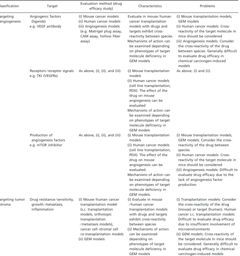
Table 5. Evaluations of drugs targeting angiogenesis and tumor stroma Classification Target Evaluation method (drug
efficacy study) Characteristics Problems Targeting
angiogenesis
Angiogenic factors (ligands) e.g. VEGF antibody
(i) Mouse cancer models (ii) Human cancer models (iii) Angiogenesis models (e.g. Matrigel plug assay, CAM assay, hollow fiber assay)
Evaluate in mouse⁄ human cancer transplantation models with drugs and targets exhibit cross-reactivity between species Mechanisms of action can
be examined depending on phenotypes of target molecule deficiency in GEM models
(i) Mouse transplantation models, GEM models
(ii) Human cancer models: Cross-reactivity of the target molecule in mice should be considered (iii) Angiogenesis models: Consider
the cross-reactivity of the drug between species. Generally difficult to evaluate drug efficacy in chemical carcinogen-induced models
Receptors⁄ receptor signals e.g. TKI (VEGFRs)
As above, (i), (ii), and (iii) (i) Mouse transplantation models
(ii) Human cancer models (cell line transplantation, PDX): The effect of the drug on mouse angiogenesis can be evaluated
Mechanisms of action can be examined depending on phenotypes of target molecule deficiency in GEM models
As above, (i) and (ii).
Production of angiogenesis factors e.g. mTOR inhibitor
As above, (i), (ii), and (iii) (i) Mouse transplantation models
(ii) Human cancer models (cell line transplantation, PDX): The effect of the drug on mouse angiogenesis can be evaluated.
Mechanisms of action can be examined depending on phenotypes of target molecule deficiency in GEM models.
(i) Mouse transplantation models, GEM models: Consider the cross-reactivity of the drug between species.
(ii) Human cancer models: Cross-reactivity of the target molecule in mice should be considered (iii) Angiogenesis models: Difficult to
evaluate drug efficacy due to the lack of angiogenesis factor production
Targeting tumor stroma
Drug resistance⁄ sensitivity, growth⁄ metastasis, inflammation
(i) Mouse⁄ human cancer transplantation model (s.c. transplantation models, orthotopic transplantation ⁄ metastasis models), cancer cell–stromal cell co-transplantation models (ii) GEM models
(i) Evaluate in mouse ⁄ human cancer transplantation models with drugs and targets exhibit cross-reactivity between species (ii) Mechanisms of action
can be examined depending on phenotypes of target molecule deficiency in GEM models
(i) Transplantation models: Consider the cross-reactivity of the drug (mouse) or target (human). Human cancer s.c. transplantation models: Difficult to evaluate drug efficacy due to insufficient involvement of microenvironments
(ii) GEM models: Cross-reactivity of the target molecule in mice should be considered. Generally difficult to evaluate drug efficacy in chemical carcinogen-induced models Animal (mainly mouse) models used for the evaluation of oncology drugs targeting angiogenesis and tumor stroma are classified in this table. As the efficacy of these drugs depends on cancer–host interactions or host factors, consideration should be given to the cross-reactivity of thera-peutic drugs and⁄ or their target molecules between species (mainly between humans and mice). CAM, chick chorioallantoic membrane; GEM, gene-engineered mouse; mTOR, mammalian target of rapamycin; PDX, patient-derived xenograft; TKI, tyrosine kinase inhibitor; VEGF, vascular endothelial growth factor; VEGFR, VEGF receptor.
the efficacy of the drugs because the mechanism of action and
predictive biomarkers would remain unclear.
Targeting Cancer Cell
–Host Interactions
The importance of microenvironments on the growth,
progres-sion, and therapeutic resistance of cancer cells has been drawn
much attention. Such tumor microenvironments have been
known to support cancer cell proliferation directly or indirectly
through interactions between surrounding stroma cells. In
gen-eral, it is relatively difficult to carry out an appropriate in vivo
efficacy test for drugs targeting interactions between cancer
cell and host microenvironment in non-clinical cancer models.
Targeting angiogenesis.
It has been widely recognized that
generation of new blood vessels into tumor (angiogenesis) is a
critical step for cancer cells to be adequately supplied nutrition
and oxygen, therefore, it is assumed that tumors are unable to
grow progressively without angiogenesis. There are also
sev-eral relevant studies suggesting that angiogenesis is involved
in not only cancer cell proliferation but also cancer cell
pro-gression, including metastases to distant organs. As represented
by VEGF inhibitors (bevacizumab), drugs targeting
angiogene-sis may not exert direct antitumor effects on cancer cells,
how-ever, should inhibit the activity of various angiogenic factors
that mainly affect vascular endothelial cells for generating new
blood vessels. Consequently, non-clinical evaluation of the
efficacy of drugs targeting angiogenesis can be greatly affected
by host factors in experimental animals; therefore, it is critical
to use appropriate models for drug evaluation, as summarized
in Table 5.
For carrying out appropriate in vivo tests for drugs targeting
angiogenesis, it is very important to consider whether cancer
cell lines or patient-derived samples produce angiogenic
factors for targeting and, moreover, their cross-reactivity in
non-clinical cancer models. It is also relevant for other
angio-genesis models such as the Matrigel plug assay, chick
chorioallantoic membrane assay, or hollow fiber assay.
Targeting cancer stroma.
Diverse cellular components of
tumor stroma (e.g. fibroblasts, mesenchymal cells, and
inflam-matory cells) and extracellular matrices (e.g. fibronectin,
colla-gen, laminin, and proteoglycan) have been shown to be
involved in cancer cell proliferation and progression. Although
tumor stroma is expected to be an attractive therapeutic target,
the development of drugs targeting cancer stroma is still in the
early stages.
Similar to those targeting angiogenesis, non-clinical
evalua-tion of drugs targeting tumor stroma should be greatly affected
by host factors. In immune-compromised mice (e.g. nude,
SCID, NOD
⁄ SCID, and NOG) often used for transplantation
models of human cancer cells display a range of different
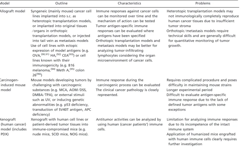
Table 6. Evaluations of drugs targeting host immune response
Model Outline Characteristics Problems
Allograft model Syngeneic (mainly mouse) cancer cell lines implanted into s.c. as heterotopic transplantation models, or implanted into original tissues ⁄ organs in orthotopic
transplantation models, or injected into tail vein as metastasis models Use of cell lines with ectopic
expression of model antigens (e.g. OVA,(90,91)HA,(92)CEA(93)) or cell
lines known with their immunogenicity (e.g. B16 melanoma,(94)Meth A,(95)colon
26(96))
Immune responses against cancer cells can be monitored over time and the mechanism of action can be tested Tumor antigen-specific immune
responses can be evaluated where antigens have been specified Orthotopic transplantation models and
metastasis models may be better for analyzing tumor-infiltrating lymphocytes considering the organ microenvironment of cancer cells.
Heterotopic transplantation models may not immunologically completely reproduce human cancer tissues due to insufficient tumor stroma
Orthotopic⁄ metastasis models require technical skills and are generally difficult for quantitative monitoring of tumor growth.
Carcinogen-induced mouse model
Mouse models developing tumors by challenging with carcinogenic substances (e.g. MCA, AOM⁄ DSS, DMBA⁄ TPA), or external stimuli such as UV, or inducing genetic abnormalities (e.g. p53 deficiency, transduction of SV40T antigen, APC deficiency)
Immune response during the
carcinogenic process can be evaluated The clinical cancer pathology is closely
represented.
Requires complicated procedure and poses difficulty in maintaining mouse strains Longer experimental period
Difficult to evaluate antigen-specific immune response due to the lack of defined tumor antigens with some exceptions
Xenograft (human cancer) model (includes PDX)
Xenograft with human cell lines or patient-derived tumor tissues into immune-compromised mice (e.g. nude mice, SCID mice, NOG mice).
Antitumor activities can be analyzed by using human (cancer patients’) immune cells.
Limitation for analyzing immune responses due to its incompetence of the intact immune system
Application of humanized mice engrafted with human immune cells clearly requires further investigation
Animal (mainly mouse) models used for evaluating drugs targeting host immune response are classified in this table. As the efficacy of cancer immunotherapy depends on the host’s immune system, concurrent use of multiple models should also be considered. In such a case, it is neces-sary to devise optimal combinations of models to be used, taking into account the potential limitations⁄ problems of each model presented in the table as advantages or disadvantages. AOM, azoxymethane; APC, Adenomatous polyposis coli; CEA, carcinoembryonic antigen; DMBA, 7,12-dimethylbenz(a)anthracene; DSS, Dextran sulfate sodium; HA, hemagglutinin; MCA, 3-Methylcholanthrene; OVA, ovalbumin; PDX, patient-derived xenograft; TPA, 12-O-TetradecanoyI-phorbol-13-acetate.
immunological environments. Even in these
immune-compro-mised animals, myeloid compartment and mesenchymal cells
are known as relatively normal, therefore the efficacy of drugs
targeting those stromal cells may be evaluated even in animal
models if the target shows cross-reactivity between species.
Targeting host immune responses.
The immune system has
been regarded as an important constituent of the tumor
microenvironment. Many series of studies have been
under-taken to understand the regulatory mechanisms by which
can-cer cells control, either positively or negatively, hosts’ immune
responses. Recent clinical successes of immune checkpoint
inhibitors, such as anti-CTLA-4 mAbs (ipilimumab and
treme-limumab)
and
anti-PD-1
mAbs
(nivolumab
and
pem-brolizumab) highlight targeting hosts’ immune responses
against cancer cells as a promising target for drug
develop-ment.
Obviously, drugs targeting hosts’ immune responses should
be tested in the appropriate non-clinical cancer models in
which the targets are involved in the immune responses against
cancer cells, for elucidating the mechanisms of action and
pre-dicting potential side-effects. In general, it is ideal to test the
importance of drug targets or potential drug candidates in
dif-ferent experimental models (multiple cell lines, difdif-ferent
mouse strains). Considering there should be a limitation for
predicting cancer types to which the drug shows clinical
bene-fit by testing only in non-clinical models, the results of phase I
clinical studies need to be carefully considered. For testing
drug candidates in which certain HLA haplotypes are required
to show antitumor effects (e.g. cancer vaccine therapy), an
application of humanized mice may be worth considering as
non-clinical models. In Table 6, we summarize pros and cons
of non-clinical models for testing drugs targeting hosts’
immune responses.
Evaluation of Oncology Drugs Based on New Concepts
Along with gaining our knowledge with the biological
charac-teristics of cancer, there are several new approaches to develop
oncology drugs, such as targeting cancer stem cells.
Targeting cancer stem cells.
The concept of cancer stem cells
was originally introduced in hematological malignancies and
further extended to solid cancers such as breast cancer and
brain tumors.
(97)Cancer stem cells have been characterized by
their self-renewal potential, multidirectional differentiation
potential, and niche dependence, similar to other stem cells, in
addition to their highly tumorigenic potential. Furthermore,
cancer stem cells have been known for their resistance to
con-ventional chemotherapy or radiotherapy; therefore, they may
be an emerging target for drug development. In Table 7, we
summarize the current methods for testing drugs targeting
can-cer stem cells in non-clinical evaluations.
Targeting other novel concepts or methods.
In Table 8, we
summarize the current status of oncology drug development
targeting new concepts other than cancer stem cells, or novel
methods for developing new oncology drugs. Non-clinical
evaluation of some of those oncology drugs targeting novel
Table 7. Evaluation of drugs targeting cancer stem cells
Evaluation method Outline Characteristics Problems
Spheroid formation potential
Culture a single non-adherent cell in the presence of specific growth factors (without serum) to test the capability of forming spheroids
Evaluation can be made using cultured cells, and the dose- and time-dependence can be quantitatively measured
General cytotoxicity of drugs mislead as positive without testing on normal tissue stem cells Cell surface marker Measuring the frequency of CD44
high⁄ CD24 low fraction, known as cancer stem cells in breast cancer by flow cytometry
Cytotoxic drugs can be tested by comparing effect on cancer stem cell fraction and others
Surface markers for cancer stem cell fractions differ depending on cancer types
ALDH ALDH activities positively correlate to chemoresistance and stemness in breast cancer, gastrointestinal tract cancer, and hematological tumors
Established methods for measuring activity by flow cytometry
Not all ALDH-positive cells are cancer stem cells
Xenograft models with human cancer stem cells in immune-compromised mouse
Human cancer stem cells transplanted into immune-compromised mice for testing drug efficacy on tumor formation ⁄ growth
Evaluating the inhibitory effect of drugs on tumor formation or growth and cancer stem cell frequency within tumor tissue (assessed based on surface markers, ALDH, and spheroid formation potential)
Not applicable for testing drugs targeting immune responses or
microenvironments Syngeneic mouse models
with mouse cancer stem cells
Mouse cancer stem cells transplanted into syngeneic mice for testing drug efficacy on tumor formation ⁄ growth
Evaluating the inhibitory effect of drugs on tumor formation or growth and cancer stem cell frequency within tumor tissue (assessed based on surface markers, ALDH, and spheroid formation potential)
Applicable for testing drugs targeting immune responses or microenvironments
Efficacy may need to be confirmed in models using human cancer stem cells
Genetically engineered animal models
Testing drugs targeting cancer stem cells using genetically engineered mice, rats, or zebrafish to develop tumors
Ideal models closely resembles an autochthonous tumor
Evaluation requires a prolonged time period because of late onset of cancer compared with transplantation models This table lists commonly used methods to evaluate cancer stem cell functions. ALDH, aldehyde dehydrogenase.
concepts may require approaches that are different from those
used for the evaluation of conventional oncology drugs.
A deeper understanding of the biological characteristics of
cancer is leading to the development of novel oncology drugs
based on new concepts such as “cancer stem cells” in addition
to the developmental targets presented in earlier sections.
Concluding Remarks
This review summarizes present non-clinical investigations by
listing the common methods currently used for the
develop-ment of oncology drugs as extensively as possible. Their types,
profiles, and problems are briefly described. Characteristics of
a variety of animal models, which provide indispensable
infor-mation to formulate clinical research and clinical trials, are
summarized according to each category of oncology drug.
Experimental models obtain the proof of evidence at the
molecular, cellular, and tissue levels, and unique oncology
drugs are also covered. It is hoped that this review provides
information to undertake regulatory science relevant to the
development of oncology drugs.
Studies with cancer models, including animal experiments,
ex vivo studies, and in vitro studies, are essential technology in
cancer biology and have contributed to the development and
evaluation of oncology drugs. Particularly, cancer cell lines
derived from humans and experimental animals have been
used for decades as indispensable tools for the biological
understanding of cancer and for the development of oncology
drugs. Properties of cancer cells represented by a cell have
been changing cell line, it was discovered that the
accumula-tion of multiple abnormalities in genes causes cancer and that
the properties of individual cancer cell lines depend not only
on their organ origins but also on the types of abnormal genes.
Growing knowledge on cancer as a disease has led to the
understanding that interactions between cancer and host cells
and the regulatory molecules play critical roles. The growth of
tumors strongly depends on tissue microenvironments and
immunological milieu that are difficult to reproduce in vitro.
As shown in this review, a substantial number of models
reflecting these various aspects of cancer
–host interactions
have been developed in the past decade. These models have
significantly contributed to the expansion of the range of
non-clinical studies and their role, in the exploration, development,
and clinical investigation of oncology drugs have become
indispensable.
The diversity and the degree of engagement in genetic
changes in the initiation of cancer cell growth and progression
are widely accepted. The roles of host cells, tissue, and the
immune system also vary depending on the type, properties,
and the stage of individual tumors are also becoming clear
than before. Therefore, the methods used to select and use
oncology drugs should continuously be revised based on the
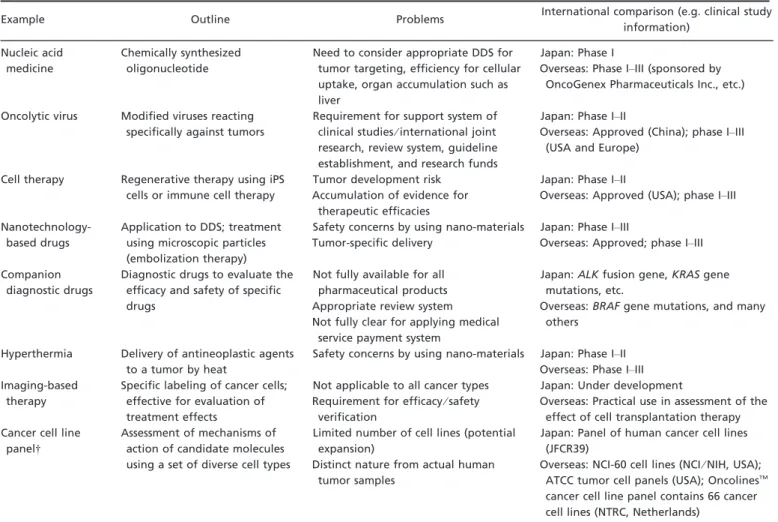
Table 8. Emerging new concepts in oncology drug developmentExample Outline Problems International comparison (e.g. clinical study
information) Nucleic acid
medicine
Chemically synthesized oligonucleotide
Need to consider appropriate DDS for tumor targeting, efficiency for cellular uptake, organ accumulation such as liver
Japan: Phase I
Overseas: Phase I–III (sponsored by OncoGenex Pharmaceuticals Inc., etc.) Oncolytic virus Modified viruses reacting
specifically against tumors
Requirement for support system of clinical studies⁄ international joint research, review system, guideline establishment, and research funds
Japan: Phase I–II
Overseas: Approved (China); phase I–III (USA and Europe)
Cell therapy Regenerative therapy using iPS cells or immune cell therapy
Tumor development risk Accumulation of evidence for
therapeutic efficacies
Japan: Phase I–II
Overseas: Approved (USA); phase I–III
Nanotechnology-based drugs
Application to DDS; treatment using microscopic particles (embolization therapy)
Safety concerns by using nano-materials Tumor-specific delivery
Japan: Phase I–III
Overseas: Approved; phase I–III Companion
diagnostic drugs
Diagnostic drugs to evaluate the efficacy and safety of specific drugs
Not fully available for all pharmaceutical products Appropriate review system Not fully clear for applying medical
service payment system
Japan: ALK fusion gene, KRAS gene mutations, etc.
Overseas: BRAF gene mutations, and many others
Hyperthermia Delivery of antineoplastic agents to a tumor by heat
Safety concerns by using nano-materials Japan: Phase I–II Overseas: Phase I–III Imaging-based
therapy
Specific labeling of cancer cells; effective for evaluation of treatment effects
Not applicable to all cancer types Requirement for efficacy⁄ safety
verification
Japan: Under development
Overseas: Practical use in assessment of the effect of cell transplantation therapy Cancer cell line
panel†
Assessment of mechanisms of action of candidate molecules using a set of diverse cell types
Limited number of cell lines (potential expansion)
Distinct nature from actual human tumor samples
Japan: Panel of human cancer cell lines (JFCR39)
Overseas: NCI-60 cell lines (NCI⁄ NIH, USA); ATCC tumor cell panels (USA); OncolinesTM
cancer cell line panel contains 66 cancer cell lines (NTRC, Netherlands)
This table exclusively presents oncology drugs that are being or about to be investigated in Japan and overseas based on new concepts. †Although “Cancer cell line panel” cannot be classified as a therapeutic drug, it is presented here as an assay that is extensively used in the development of new therapeutic drugs. DDS, drug delivery system; iPS, induced pluripotent stem cells.
advance in understanding of cancer. As stated earlier in this
review, models established for the biological understanding of
cancer have proven to be useful as tools for non-clinical
inves-tigations. When developing a new drug that is in the same
class as those for which efficacy and safety information was
already acquired from clinical studies, it is also useful to select
non-clinical models based on the clinical information.
Collec-tively, it will become increasingly important to design, to
select, and to use appropriate non-clinical models in order to
design clinical research and trials. Investigations with these
models should be effective in interpreting the results of such
investigations and to re-evaluate the effects of oncology drugs
used in clinical practice. It is strongly hoped that non-clinical
investigation will continuously be successfully used for the
development, approval, and proper use of oncology drugs,
which accelerate drug development.
Acknowledgments
This article was prepared as the summary statement of a subcommittee for non-clinical studies of the Science Board of the Pharmaceuticals and Medical Devices Agency. We are grateful to Takao Yamori, Tet-suo Nagano, Eiji Saito, and all other members in the Regulatory Science Division, Scientific Committee of the Pharmaceuticals and Medical Devices Agency for their assistance and discussion.
Disclosure Statement
The authors have no conflict of interest.
References
1 Hurley LH. DNA and its associated processes as targets for cancer therapy. Nat Rev Cancer 2002;2: 188–200.
2 DeVita VT Jr, Chu E. A history of cancer chemotherapy. Cancer Res 2008; 68: 8643–53.
3 Dobbelstein M, Moll U. Targeting tumour-supportive cellular machineries in anticancer drug development. Nat Rev Drug Discov 2014;13: 179–96. 4 Land H, Parada LF, Weinberg RA. Cellular oncogenes and multistep
car-cinogenesis. Science 1983;222: 771–8.
5 Hollingsworth RE, Lee WH. Tumor suppressor genes: new prospects for cancer research. J Natl Cancer Inst 1991;83: 91–6.
6 Croce CM. Genetic approaches to the study of the molecular basis of human cancer. Cancer Res 1991;51(18 Suppl): 5015s–8s.
7 Barrett JC, Thomassen DG, Hesterberg TW. Role of gene and chromosomal mutations in cell transformation. Ann N Y Acad Sci 1983;407: 291–300. 8 Cowell JK. Double minutes and homogeneously staining regions: gene
amplification in mammalian cells. Annu Rev Genet 1982;16: 21–59. 9 Bloomfield CD, Lindquist LL, Arthur D et al. Chromosomal abnormalities
in acute lymphoblastic leukemia. Cancer Res 1981;41(11 Pt 2): 4838–43. 10 Tsuruo T, Naito M, Tomida A et al. Molecular targeting therapy of cancer:
drug resistance, apoptosis and survival signal. Cancer Sci 2003;94(1): 15–21. 11 Pierce GB, Speers WC. Tumors as caricatures of the process of tissue
renewal: prospects for therapy by directing differentiation. Cancer Res 1988; 48: 1996–2004.
12 Hoffman SJ, Robinson WA. Use of differentiation-inducing agents in the myelodysplastic syndrome and acute non-lymphocytic leukemia. Am J Hematol 1988;28: 124–7.
13 McMillin DW, Negri JM, Mitsiades CS. The role of tumour-stromal interac-tions in modifying drug response: challenges and opportunities. Nat Rev Drug Discov 2013;12: 217–28.
14 Miller JF, Sadelain M. The journey from discoveries in fundamental immunology to cancer immunotherapy. Cancer Cell 2015;27: 439–49. 15 Siolas D, Hannon GJ. Patient-derived tumor xenografts: transforming clinical
samples into mouse models. Cancer Res 2013;73: 5315–9.
16 Marangoni E, Poupon MF. Patient-derived tumour xenografts as models for breast cancer drug development. Curr Opin Oncol 2014;26: 556–61. 17 Hidalgo M, Amant F, Biankin AV et al. Patient-derived xenograft models:
an emerging platform for translational cancer research. Cancer Discov 2014; 4: 998–1013.
18 Marino S, Vooijs M, van Der Gulden H, Jonkers J, Berns A. Induction of medulloblastomas in p53-null mutant mice by somatic inactivation of Rb in the external granular layer cells of the cerebellum. Genes Dev 2000;14: 994–1004.
19 Westerman BA, Blom M, Tanger E et al. GFAP-Cre-mediated transgenic activation of Bmi1 results in pituitary tumors. PLoS ONE 2012;7: e35943. 20 Yang ZJ, Ellis T, Markant SL et al. Medulloblastoma can be initiated by
deletion of Patched in lineage-restricted progenitors or stem cells. Cancer Cell 2008;14: 135–45.
21 Zibat A, Uhmann A, Nitzki F et al. Time-point and dosage of gene inactiva-tion determine the tumor spectrum in condiinactiva-tional Ptch knockouts. Carcino-genesis 2009;30: 918–26.
22 Tonks ID, Hacker E, Irwin N et al. Melanocytes in conditional Rb ⁄ mice are normal in vivo but exhibit proliferation and pigmentation defects in vitro. Pigment Cell Res 2005;18: 252–64.
23 Hu N, Gutsmann A, Herbert DC, Bradley A, Lee WH, Lee EY. Heterozy-gous Rb-1 delta 20⁄ +mice are predisposed to tumors of the pituitary gland with a nearly complete penetrance. Oncogene 1994;9: 1021–7.
24 Vooijs M, van der Valk M, te Riele H, Berns A. Flp-mediated tissue-specific inactivation of the retinoblastoma tumor suppressor gene in the mouse. Oncogene 1998;17(1): 1–12.
25 Shaw AT, Meissner A, Dowdle JA et al. Sprouty-2 regulates oncogenic K-ras in lung development and tumorigenesis. Genes Dev 2007;21: 694–707. 26 Andreadi C, Cheung LK, Giblett S et al. The intermediate-activity (L597V)
BRAF mutant acts as an epistatic modifier of oncogenic RAS by enhancing signaling through the RAF⁄ MEK ⁄ ERK pathway. Genes Dev 2012; 26: 1945–58.
27 Dankort D, Filenova E, Collado M, Serrano M, Jones K, McMahon M. A new mouse model to explore the initiation, progression, and therapy of BRAFV600E-induced lung tumors. Genes Dev 2007;21: 379–84.
28 Song H, Yao E, Lin C, Gacayan R, Chen MH, Chuang PT. Functional char-acterization of pulmonary neuroendocrine cells in lung development, injury, and tumorigenesis. Proc Natl Acad Sci USA 2012;109: 17531–6.
29 Soda M, Takada S, Takeuchi K et al. A mouse model for EML4-ALK-posi-tive lung cancer. Proc Natl Acad Sci USA 2008;105: 19893–7.
30 Chen Z, Sasaki T, Tan X et al. Inhibition of ALK, PI3K⁄ MEK, and HSP90 in murine lung adenocarcinoma induced by EML4-ALK fusion oncogene. Cancer Res 2010;70: 9827–36.
31 Saito M, Ishigame T, Tsuta K, Kumamoto K, Imai T, Kohno T. A mouse model of KIF5B-RET fusion-dependent lung tumorigenesis. Carcinogenesis 2014;35: 2452–6.
32 Arai Y, Totoki Y, Takahashi H et al. Mouse model for ROS1-rearranged lung cancer. PLoS ONE 2013;8: e56010.
33 Yuan W, Stawiski E, Janakiraman V et al. Conditional activation of Pik3ca (H1047R) in a knock-in mouse model promotes mammary tumorigenesis and emergence of mutations. Oncogene 2013;32: 318–26.
34 Adams JR, Xu K, Liu JC et al. Cooperation between Pik3ca and p53 muta-tions in mouse mammary tumor formation. Cancer Res 2011;71: 2706–17. 35 Trimboli AJ, Cantemir-Stone CZ, Li F et al. Pten in stromal fibroblasts
sup-presses mammary epithelial tumours. Nature 2009;461: 1084–91.
36 Finkle D, Quan ZR, Asghari V et al. HER2-targeted therapy reduces inci-dence and progression of midlife mammary tumors in female murine mam-mary tumor virus huHER2-transgenic mice. Clin Cancer Res 2004; 10: 2499–511.
37 Rao GN, Ney E, Herbert RA. Effect of melatonin and linolenic acid on mammary cancer in transgenic mice with c-neu breast cancer oncogene. Breast Cancer Res Treat 2000;64: 287–96.
38 Dourdin N, Schade B, Lesurf R et al. Phosphatase and tensin homologue deleted on chromosome 10 deficiency accelerates tumor induction in a mouse model of ErbB-2 mammary tumorigenesis. Cancer Res 2008; 68: 2122–31.
39 Cheng L, Zhou Z, Flesken-Nikitin A et al. Rb inactivation accelerates neo-plastic growth and substitutes for recurrent amplification of cIAP1, cIAP2 and Yap1 in sporadic mammary carcinoma associated with p53 deficiency. Oncogene 2010;29: 5700–11.
40 McCarthy A, Savage K, Gabriel A, Naceur C, Reis-Filho JS, Ashworth A. A mouse model of basal-like breast carcinoma with metaplastic elements. J Pathol 2007;211: 389–98.
41 McPherson JP, Lemmers B, Hirao A et al. Collaboration of Brca1 and Chk2 in tumorigenesis. Genes Dev 2004;18: 1144–53.
42 Jonkers J, Meuwissen R, van der Gulden H, Peterse H, van der Valk M, Berns A. Synergistic tumor suppressor activity of BRCA2 and p53 in a con-ditional mouse model for breast cancer. Nat Genet 2001;29: 418–25. 43 Hung KE, Maricevich MA, Richard LG et al. Development of a mouse
model for sporadic and metastatic colon tumors and its use in assessing drug treatment. Proc Natl Acad Sci USA 2010;107: 1565–70.