CASE REPORT
Successful Retrieval of Entrapped Guide Extension Catheter in the
Right Coronary Artery with Heavy Calcifications
Shinji Imura,
1MD, Takehiro Hashikata,
2MD, Motosu Ando,
1MD and Fumitaka Hosaka,
1MD
Summary
Usage of an optimal guide extension catheter often helps successful outcomes in complex percutaneous coronary intervention. Here, we report a case of successfully retrieving a guide extension catheter entrapped by a coronary stent in the middle RCA. The guide extension catheter was retrieved by anchoring with the stent de-livery balloon. Also, our in vitro experiment demonstrated that a “deep seating method” and an “anchoring guide extension catheter method” could be effective in bailing out guide extension catheter entrapment.
(Int Heart J 2021; 62: 682-686)
Key words: Bailout technique, Rotational atherectomy, Percutaneous coronary intervention
G
uide extension catheters were commonly used in percutaneous coronary intervention (PCI) to im-prove the device deliverability through deep seat-ing of the coronary artery. Guide extension catheters could help our procedures be safer and more efficient, especially in complex PCI, while on the other hand, some complica-tions related to them have been reported.1-3)Althoughen-trapment of a guide extension catheter is well known to be a major complication, reports on the bailout of this complication are limited. Here, we report the successful retrieval of a guide extension catheter entrapment in a heavily calcified lesion and present the following in vitro experiment.
Case Report
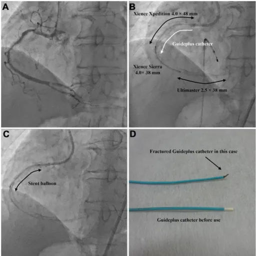
An 84-year-old male with stable angina was referred for PCI due to a heavily calcified lesion in the right coro-nary artery (RCA). One month before the current admis-sion, angiography showed a diffuse 75% lesion from the proximal to middle RCA segment and 90% lesion in the distal RCA segment with severe calcification (Figure 1A). A 7 Fr Launcher AL 0.75 (Medtronic, CA, USA) was used by the transfemoral approach. At first, rotational ath-erectomy (RA) was performed with a 1.25 mm and subse-quent a 2.0 mm burr over a Rota Wire floppy (Boston Scientific, MA, USA) in the distal and proximal RCA cal-cified lesion. Balloon pre-dilation was carried out with 2.5-4.0 mm Scoreflex NC (OrbusNeich, HK) balloons from the distal to proximal RCA lesion. After a Guideplus (Nipro, Japan) guide extension catheter was introduced, a 2.5 × 38 mm Ultimaster DES was successfully deployed in the distal RCA. Since a 3.5 × 48 mm Xience Xpedition 48 (Abbott, CA, USA) DES could not be passed into the
Guideplus, the DES was deployed in the proximal RCA. Subsequently, a 4.0 × 38 mm Xience Sierra DES was ad-vanced into the middle RCA with the Guideplus (Figure 1 B). After the Xience Sierra 4.0 × 38 mm DES was de-ployed in the middle RCA, we noted that the Guideplus was entrapped by the deployed DES. Although we at-tempted to intubate the 7 Fr guide catheter deeply into the RCA following the method of a previous report,4) it was
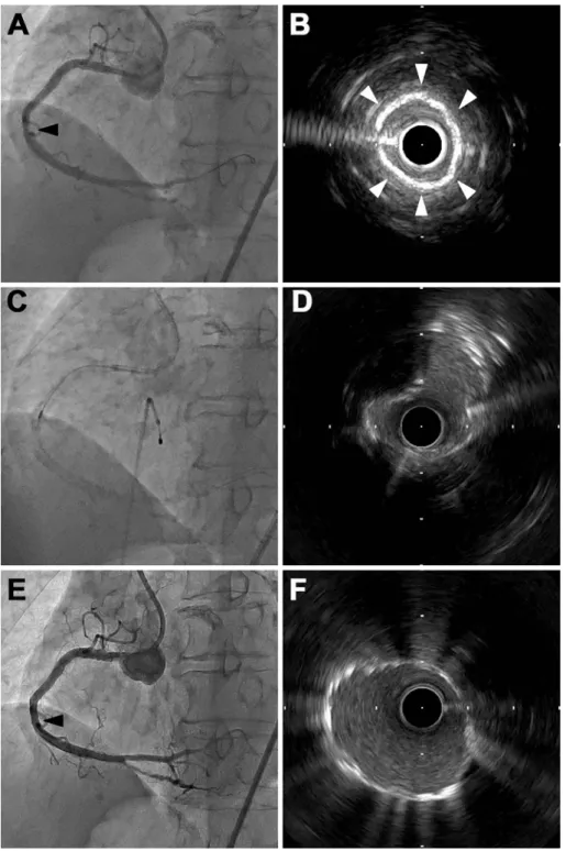
unsuccessfully inserted to the middle RCA because the proximal RCA stent generated high friction. Thus, the stent balloon was dilated in the Guideplus distal shaft to fix the balloon and Guideplus catheter (Figure 1C). We synchronously pulled out the stent balloon shaft and the Guideplus shaft, then, the Guideplus was retrieved leaving the Guideplus tip in the middle RCA (Figure 1D). In-travascular ultrasound (IVUS) showed encircling high echogenicity with acoustic shadowing in the middle RCA (Figure 2B). Even though a post-dilation was carried out with 2.0, 2.5, and 3.5 mm noncompliant balloons, the bal-loon indentation remained. RA was performed with a 1.25 mm burr to fracture the Guideplus tip (Figure 2C). The IVUS after RA procedure showed a crack formation in the Guideplus tip (Figure 2D). After further post-dilation with noncompliant balloons, a 4.0 × 24 mm Ultimaster DES was successfully deployed in the middle RCA. The final angiography and IVUS study showed sufficient stent expansion with a minimum luminal area (MLA) of 6.6 mm2 (Figure 2E, F). Four-week follow-up computed
to-mography coronary angiography showed no in-stent restenosis in the RCA.
Discussion
Guide support extension catheters can make complex
From the1
Department of Cardiovascular Medicine, Okamura Memorial Hospital, Shizuoka, Japan and2
Division of Cardiovascular Medicine, Stanford Cardiovascular Institute, Stanford University School of Medicine, Stanford, USA.
Address for correspondence: Shinji Imura, MD, Department of Cardiovascular Medicine, Okamura Memorial Hospital, 293-1 Kakita, Shimizu, Suntou, Shi-zuoka 411-0904, Japan. E-mail: imura.the@gmail.com
Received for publication November 20, 2020. Revised and accepted January 12, 2021. doi: 10.1536/ihj.20-748
All rights reserved by the International Heart Journal Association.
Int Heart J
May 2021 BAILING OUT GUIDE EXTENSION CATHETER ENTRAPMENT 683
Figure 1. Entrapment and subsequent fracture of the guide extension catheter. A: A diagnostic angiogra-phy showed the diffuse 75% lesion from the proximal to middle RCA segment and 90% lesion in the distal RCA segment with severe calcifications. B: After a Xience Xpedition DES and a Ultimaster DES were de-ployed in the RCA, a Xience Sierra DES was advanced into the middle RCA using a Guideplus guide exten-sion catheter. C: The stent delivery balloon was dilated in the Guideplus distal shaft to anchor the Guideplus catheter. D: The Guideplus catheter above was fractured at the Guideplus tip in this case.
PCI procedures easy to facilitate PCI equipment deliv-ery.5,6)The design of the Guideplus guide support
exten-sion catheter, which is similar to that of the GuideLinerV2 and GuideZilla, has a 250 mm distal catheter segment with a stainless steel hypo tube shaft (Figure 3A). The Guideplus is easily advanced to a deep seating position, although we sometimes overlook the hypotube shaft be-cause of reduced visibility.7) Our case describes how the
Guideplus extension catheter was entrapped by the de-ployed DES in the middle RCA.
There are some verified previous reports describing the successful percutaneous retrieval of a RA burr, IVUS catheter and balloon,8-11) but few cases of a guide
exten-sion catheter. A case report described how a Guideplus extension catheter was entrapped in the proximal RCA and was then retrieved when the stent balloon was with-drawn.7)
Since we concluded it was not certain whether this method would be effective in the middle RCA, we simulated the same situation in which the Guideplus ex-tension catheter was entrapped by the deployed DES in the middle RCA using a silicone-tube RCA model. First,
the Guideplus was not successfully retrieved in the middle RCA when the stent balloon was withdrawn, as shown in a previous case report.7) While withdrawing a stent
bal-loon should be an effective method, it appears to depend on the entrapped length and location. Subsequently, we pulled out the Guideplus shaft vigorously resulting in fracturing of the Guideplus catheter at the rapid exchange collar transition (Figure 3B). Based on this result, the Guideplus catheter shaft should not be pulled out alone because it seems to have a weak structural characteristic against the horizontal force at the point of rapid exchange collar transition. Secondly, we performed a “deep seating method”, in which the guide catheter is deeply inserted passing across the Guideplus tip. The stent balloon was dilated distal to the stent and the 7 Fr guide catheter was deeply inserted and covered the whole Guideplus tip. Al-though this method resulted in successful retrieval of the whole Guideplus catheter, it involved a high risk of stent deformation when passing across the guiding catheter (Figure 4). Finally, we implemented an “anchoring guide extension catheter method”, in which the stent balloon
Int Heart J May 2021
684 IMURA, ET AL
Figure 2. The procedures after leaving the tip of the guide extension catheter. A, B: Angiography showed a stenosis at the Guideplus tip in the middle RCA. Intravascular ultrasound (IVUS) showed encircling high echogenicity with acoustic shadowing. C, D: Rotational atherectomy (RA) was performed with a 1.25 mm burr. The IVUS showed a crack at 2 o’clock after RA. E, F: The final angiography and IVUS showed a suffi-cient stent expansion with a minimum luminal area of 6.6 mm2.
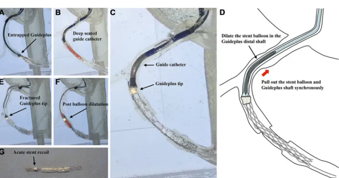
and the Guideplus shaft were pulled out synchronously while the stent balloon was dilated within the Guideplus distal shaft (Figure 5C, D). Our results suggested that this method should be performed after trying the “deep seating method” because even if the other structure could success-fully be retrieved out of the body, the tip of the Guideplus
was left in the coronary artery after the procedure (Figure 5A, B, E). The Guideplus was not often fractured in the distal shaft body made of a metal blade, but was often fractured in the rapid exchange collar transition or the Guideplus tip. The Guideplus tip was frequently fractured using an “anchoring guide extension catheter method”
Int Heart J
May 2021 BAILING OUT GUIDE EXTENSION CATHETER ENTRAPMENT 685
Figure 3. Structure of Guideplus catheter. A: Guideplus K Catheter (Nipro Japan granted permission for reproduction). B: The Guideplus catheter was fractured at the rapid exchange collar transition with an excessive horizontal force.
Figure 4. Deep seating method in vitro. A: A Guideplus was entrapped in the middle RCA. B: A 7 Fr Guide catheter was deeply inserted while the stent delivery balloon was dilated distal to the Guideplus. C: The 7 Fr Guide catheter was further inserted and covered the whole Guideplus tip.
D: The Guideplus was successfully retrieved, but with stent deformation.
(Figure 5E). Even though the stent balloon dilation was performed in the Guideplus tip, acute recoil was observed (Figure 5F, G). The balloon post-dilatation alone hardly prevents acute recoil since the Guideplus tip is made from elastic polyamide resin. RA and a cutting balloon may be effective if acute recoil occurs. In the present case, the “deep seating method” was hardly performed because the RCA proximal stent generated a high degree of friction. Thus, we chose the “anchoring guide extension catheter method” and successfully retrieved the Guideplus catheter leaving the Guideplus tip in the middle RCA. RA was performed since the balloon post-dilation alone hardly achieved adequate balloon expansion. In the end, the final angiography demonstrated no residual stenosis.
This in vitro experiment has limitations. The
silicon-tube model is different from a coronary artery in terms of vessel friction though previous reports described the suc-cessful percutaneous retrieval of a RA burr, IVUS cathe-ter, and balloon using similar methods in each case.8-11)
Also, we did not perform in vitro experiments to verify the bailout methods using other guide extension catheters. However, this result could apply to others because all guide extension catheters currently in use have a similar design. Further case reports will help to clarify the feasi-bility of these methods.
Conclusion
This case describes methods to deal with the possible complication in which the guide extension catheter was
Int Heart J May 2021
686 IMURA, ET AL
Figure 5. Anchoring guide extension catheter method in vitro. A: A Guideplus was entrapped in the middle RCA. B: Although a “deep seating method” was performed, the guide catheter was unable to cover the entire Guideplus tip. C, D: The stent balloon and the Guideplus shaft were pulled out synchronously while the stent balloon was dilated within the Guideplus distal shaft. E: The Guideplus was successfully retrieved while leaving the Guideplus tip. F, G: Even though the stent balloon dilation was performed in the Guideplus tip, acute recoil was observed.
entrapped in the middle RCA. According to our in vitro study, while the “deep seating method” and the “anchor-ing guide extension catheter method” were useful, with-drawing the guide extension catheter alone was risky. This knowledge would enhance the safe and reliable perform-ance of PCI in complex lesions.
Disclosure
Conflicts of interest: All authors have no conflicts of
in-terest relevant to the contents of this paper.
References
1. Chang Y, Fang H, Chen T, Wu C. Left main coronary artery bidirectional dissection caused by ejection of GuideLiner cathe-ter from the guiding cathecathe-ter. Cathecathe-ter Cardiovasc Incathe-terv 2013; 82: E215-20.
2. Seto A, Kern M. The GuideLiner: keeping your procedure on track or derailing it? Catheter Cardiovasc Interv 2012; 80: 451-2.
3. Thomas W, Harit D, Kintur S. A unique complication of the GuideZilla guide extension support catheter and the risk of stent stripping in interventional & endovascular interventions. Indian
Heart J 2015; 67: 381-4.
4. Jin WK, You HL, Jae HP, Jae HL. Difficult Stentablation with an Episode of Stuck and Entrapped Burr within the Underex-panded Stent. Int Heart J 2018; 59: 413-6.
5. Zehan H, Bin Z, Weilu C, et al. Usefulness and Safety of a Novel Modification of the Retrograde Approach for the Long Tortuous Chronic Total Occlusion of Coronary Arteries. Int Heart J 2017; 58: 351-6.
6. Papayannis AC, Michael TT, Brilakis ES. Challenges associated with use of the GuideLiner catheter in percutaneous coronary interventions. J Invasive Cardiol 2012; 24: 370-1.
7. Higuchi R, Yokoyama H, Hagiya K, Takamisawa I, Tobaru T. Bailout from Guideplus entrapment by coronary stent. Cardio-vasc Interv Ther 2019; 34: 375-6.
8. Sakakura K, Taniguchi Y, Tsukui T, Yamamoto K, Momomura SI, Fujita H. Successful Removal of an Entrapped Rotational Atherectomy Burr Using a Soft Guide Extension Catheter. JACC Cardiovasc Interv 2017; 10: e227-9.
9. Yamamoto K, Sakakura K, Taniguchi Y, et al. Trapping Balloon Technique for Removal of the Burr in Rotational Atherectomy. Int Heart J 2018; 59: 399-402.
10. Brett MS, James AB, Rakesh S, et al. Intravascular ultrasound catheter entrapment after coronary artery stenting. Catheter Car-diovasc Interv 2002; 57: 229-33.
11. Kawamura A, Asakura Y, Ishikawa S, et al. Extraction of previ-ously deployed stent by an entrapped cutting balloon due to the blade fracture. Catheter Cardiovasc Interv 2002; 57: 239-43.