INTRODUCTION
Proton pump inhibitors (PPIs) are more potent than histamine H2receptor antagonists (H2
block-ers) in terms of their ability to inhibit gastric acid secretion while also promoting the healing of
gas-ORIGINAL
Gastric mucosal levels of prostaglandins and leukotrienes
in patients with gastric ulcer after treatment with
ra-beprazole in comparison to treatment with ranitidine
Michiyo Okazaki
1, Ichiro Shimizu
2, Momoko Ishikawa
3, Soichiro Fujiwara
3,
Hirofumi Yamamoto
3, Tatsuhiko Shiraishi
3, Takahiro Horie
3, Arata Iuchi
3, and
Susumu Ito
2 1Department of Gastroenterology, Kochi Red Cross Hospital, Kochi, Japan,2
Department of Digestive and Cardiovascular Medicine, Institute of Health Biosciences, The University of Tokushima Graduate School, Tokushima, Japan ; and3
Department of Internal Medicine, Tokushima Prefectural Miyoshi Hospital, Tokushima, Japan
Abstract :
AIM : Prostaglandins (PGs) and leukotrienes (LTs) are major factors involved in the defense of the gastric mucosa against ulcer formation. However, little is still known about the gastromucosa-protecting action of proton pump inhibitors (PPIs) and histamine H2
receptor antagonists (H2blockers) in patients with gastric ulcer. We therefore examined
the effectiveness of a PPI in protecting the gastric mucosa.
METHODS : We compared the PGE2and LTB4levels and the expression levels of
cyclooxy-genase (COX)-1 and COX-2 mRNA in the gastric mucosa in gastric ulcer patients between the group treated for 8 weeks with a PPI, rabeprazole (PPI group ; n=5), and the group treated for 8 weeks with an H2blocker, ranitidine (H2blocker group ; n=6), as well as in
nonulcer subjects (control group ; n=5).
RESULTS : The mucosal levels of PGE2and COX-2 mRNA expression were significantly
lower in the ulcer patients than those in the nonulcer patients, whereas the LTB4 level
was significantly higher in the ulcer patients than that in the nonulcer patients, and it was also significantly lower in the ulcerated mucosa than that in the nonulcerated mu-cosa. The PPI group had a significantly increased PGE2 and decreased LTB4 levels in
comparison to the H2blocker group during the ulcer-healing stage. The COX-1 mRNA
ex-pression showed no difference among the PPI and H2blocker groups or between before
and after the treatment. However, the COX-2 mRNA expression increased in the PPI group more than that in the H2blocker group during the ulcer-healing stage.
CONCLUSION : These findings demonstrated the significant gastric-mucosa-protecting effect of PPI by increasing the PGE2production and reducing the LTB4production. J. Med.
Invest. 54 : 83-90, February, 2007
Keywords : rabeprazole, COX-2, PGE2, LTB4, H2blocker
Received for publication November 30, 2006 ; accepted December 20, 2006.
Address correspondence and reprint requests to Ichiro Shimizu, M.D., Department of Digestive and Cardiovascular Medicine, Institute of Health Biosciences The University of Tokushima Graduate School, Kuramoto-cho, Tokushima 770-8503, Japan and Fax : 81-88-633-9235
The Journal of Medical Investigation Vol. 54 2007
tric ulcers(1). PPIs block the final step in acid se-cretion from gastric parietal cells. Rabeprazole is a substituted benzimidazole PPI. In addition to its inhibitory effect on gastric acid secretion, rabeprazole has been reported to prevent gastric mucosal dam-age such as that resulting from ethanol or aspirin administration in animal models(2-4). Lansopra-zole, one of PPIs, has also been reported to induce gastric mucosal protection through up-regulation of cyclooxygenase (COX)-2 with endogenous prostaglandin (PG) synthesis in an animal model of gastric mucosal injury(5). COX is the enzyme that catalyzes the committed step in the metabo-lism of arachidonic acid to PGs(6). Two isoforms of COX exist : including constitutively expressed COX-1 and inducible COX-2 in stomach. PGs play an important role in the gastric mucosal defense (7). It should be noted that increased PG synthe-sis via the COX-2 expression and decreased leu-kotriene (LT) synthesis in the gastric mucosa may be involved in mucosal cytoprotection(8). LTs be-long to a group of proinflammatory mediators de-rived from arachidonic acid(9, 10). However, little is still known about the gastromucosa-protecting action of PPIs and H2blockers in patients with gastric ulcer.
We experienced a case who demonstrated a very large gastric ulcer which perforated the abdominal wall and had been caused by diclofenac, a nonster-oidal anti-inflammatory drug (NSAID)(11). In this case, since conservative treatment by the oral ad-ministration of the H2blocker ranitidine and a PG
preparation did not show any sign of closure of the perforation, ranitidine was replaced by rabeprazole. Thereafter, the mucosal regeneration progressed rapidly, the perforation closed, and the ulcer com-pletely healed. These findings suggested that not only the PG preparation but also rabeprazole thus appears to promote cytoprotection against the gas-tric mucosal injury. Therefore, in order to clarify the differences between the gastromucosa-protecting actions of rabeprazole and ranitidine in patients with gastric ulcer, we administered either rabeprazole or ranitidine to patients having their first episodes
of gastric ulcer for 8 weeks and then comparatively evaluated the expression of gastric mucosal COX and the secretory kinetics of PGs and LTs. To bal-ance the background of damage to the gastric mu-cosa, the subjects were limited to patients who were positive for Helicobacter pyroli (Hp).
SUBJECTS AND METHODS
SubjectsFrom September 2000 to December 2002, the study was conducted at Tokushima Prefecture Miyoshi Hospital. The subjects consisted of 11 pa-tients who demonstrated their first episode of gastric ulcer with no previous history of the disease in whom a single acute stage (A1 or A2) ulcer was located at the gastric angle without any bleeding, and the presence of Hp was confirmed by the13
C-urea breath test (Otsuka Pharmaceutical Co., Tokyo) at the first endoscopy. The patients with acute gas-tric ulcers were divided at random into age- and sex-matched 2 groups. Five patients (4 males and 1 fe-male ; mean age±SD, 54.4±12.4 years ; PPI group)
were administered rabeprazole (10 mg/day), while 6 (5 males and 1 female ; 53.3 ± 12.6 years ; H2
blocker group) were administered ranitidine (300 mg/day) for 8 weeks. The control group without any episode of gastric ulcer comprised 5 subjects (4 males and 1 female ; 55.2±11.9 years) who were
all positive for Hp. In the PPI group, one had dia-betes mellitus, and one had bronchial asthma as complications of gastric ulcer. In the H2 blocker
group, one had hypertension, one had diabetes mel-litus, and one had chronic bronchitis as complica-tions. In the control group without any episode of gastric ulcer, one had hypertension and one had dia-betes mellitus. The oral treatments for these com-plications were continued. The ulcer diameters and the blood hemoglobin levels at the first endoscopy were similar in all groups (Table 1). No patient had taken NSAIDs, PPIs, H2blockers, PG preparations,
or antibiotics prior to enrollment in this study.
Pa-Table 1. Clinical characteristics of gastric ulcer patients (PPI and H2blocker groups) and nonulcer subjects (control group) with
Hpinfection
PPI H2blocker Control Pvalues
Male/female 4/1 5/1 4/1 NS
Age (years) 54.4±12.4 53.3±12.6 55.2±11.9 NS
Diameter of gastric ulcer (cm) 1.2±0.4 1.1±0.4 Not detected NS Blood hemoglobin level (g/dl) 15.9±2.7 15.8±2.4 15.5±3.1 NS Values are the means±SD (n = 5 or 6).
M. Okazaki, et al. PPI on COX-2, PGE2and LTB4in gastric ulcer
tients with gastric or other cancers, liver disease, re-nal dysfunction, or duodere-nal ulcer were excluded. Informed consent was obtained from all of the sub-jects.
METHODS
At endoscopic examinations performed in the control group and in the antisecretory agents-groups before and at 8 and 12 weeks after beginning the ad-ministration of the antisecretory agents, tissue sam-ples were collected both from the edge of the ulcer on the gastric angle (ulcerated areas) and the lower gastric body on the greater curvature (nonulcer-ated areas), and were immediately stored at -80C until assayed. The concentrations of PGE2, 6-keto
PGF1a, which is a metabolic product of stable PGI2,
and LTB4, and the gene expressions of COX-1 and
COX-2 were examined in the gastric mucosa. Hp elimination treatment was performed at 12 weeks or more after beginning the administration of the antisecretory agents.
The biopsy specimens were homogenized in a Polytron PCU homogenizer (Kinematica, Luzern, Switzerland), the PGE2 and 6-keto PGF1a
concen-trations were determined using radioimmunoas-say kits (Amersham, Little Chalfont, UK), and the LTB4 concentration was determined using an
en-zyme immunoassay kit (Amersham). The protein concentration was determined by the Lowry pro-tein assay. For RNA extraction from gastric mucosal tissue specimens after pooling the samples from the ulcerated and nonulcerated areas, 10 mg of the fro-zen gastric mucosal tissue was homogenized with a glass homogenizer in 200μl of RNA zolTM B (Biotex Laboratories, Friendswood, TX, USA) at 4C. Twenty microliters of phenol/chloroform were then added to the mixture and the aqueous phase was collected after centrifugation at 12,000 X g at 4C for 15 min-utes. The RNA was precipitated with an equal vol-ume of isopropanol and then it was washed with 75% ethanol. RNA pellets were resuspended in 20 μl of diethylpyrocarbonate-treated distilled water and stored at -80C. For the analysis of 2 mRNA and COX-1 mRNA expression, RT-PCR was performed using a one-step RNA PCR kit (AMV). Briefly, 1 μg of total RNA for each sample was added to a PCR re-action. After reverse transcription (30 min at 50 C ; 2 min at 94 C), 30 cycles of amplifications (1 min at 94 C ; 1 min at 50 C ; 1 min at 72 C) were per-formed. The linear range for PCR was found to be
between 20 and 40 cycle. The PCR products were separated on 2% agarose gel containing 0.5 μg/ml of ethidium bromide. The oligonucleotides, TGCCCAGCTCCTGGCCCGCCGCTT-3’ and 5’-GTGCATCAACAGGCGCCTCTTC-3’ were used for amplification of the 303 bp fragment of human COX-1 mRNA(12). The oligonucleotides,
5’-TTCAAATGAGATTGTGGGAAAAT-3’ and
5’-AGATCATCTCTGCCTGAGTATCTT-3’ were used for amplification of the 305 bp fragment of human COX-2 mRNA(12). Yeast transfer RNA was used as negative controls. Beta-actin gene expression was monitored for the control of mRNA loading. The ex-pression intensities of COX-2 mRNA and COX-1 mRNA were evaluated by scanning densitometry (Gel Doc 1000UV, Richmond, CA, USA) in compari-son to the intensity of beta-actin gene expression.
Statistical analysis
The results are expressed as the means ± SD.
Differences between the groups were assessed by Student’s t test. P values of <0.05 were considered to be significant.
RESULTS
In the PPI and H2 blocker groups of gastric ulcer patients with Hp infection before treatment with the antisecretory agents, the concentrations of PGE2
(Fig. 1) and 6-keto PGF1a(Fig. 2) in the gastric
mu-cosa were significantly lower than those in the con-trol group of nonulcer subjects with Hp infection, although the 6-keto PGF1a concentration in the
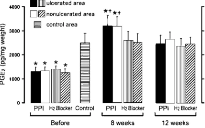
Fig. 1. Changes in the gastric mucosal levels of PGE2after treatment with rabeprazole (PPI group) or ranitidine (H2blocker group)
The gastric ulcer base disappeared at 8 − 12 weeks after treat-ment. The samples were obtained from the gastric mucosa in ulcer patients and nonulcer subjects (control group). Values are the means±SD (n = 5 or 6). *P <0.05 in comparison with the control group.+P <0.05 in comparison with the H2blocker group. The Journal of Medical Investigation Vol. 54 February 2007 85
nonulcerated gastric mucosa in ulcer patients did not show a significant decrease (Fig. 2). All of gas-tric ulcers had been already scarred (S2) 8 weeks after treatment with the antisecretory agents. Dur-ing gastric ulcer healDur-ing, the PGE2 concentration
in the PPI group after 8 weeks increased significantly more than that in the H2blocker and control groups,
and then returned after 12 weeks to a level similar to that in the H2blocker and control groups (Fig. 1).
The 6-keto PGF1a concentration in the ulcerated
gastric mucosa in ulcer patients returned after 8 weeks to a level similar to that in the nonulcerated mucosa in both the ulcer patients and nonulcer sub-jects (Fig. 2). Whereas the LTB4 (Fig. 3)
concen-tration in ulcer patients before the antisecretory agent treatment was significantly higher than in that in nonulcer subjects, and it was significantly lower in the ulcerated mucosa than that in the nonul-cerated mucosa. During gastric ulcer healing, the LTB4concentration of the ulcer-scarred and
nonul-cerated mucosa in the PPI group after 8 weeks de-creased significantly more than that in the H2blocker
and control groups without any significant differ-ence between in the ulcer-scarred and nonulcerated mucosae, and then it returned after 12 weeks to a level similar to that in the H2 blocker and control
groups (Fig. 3).
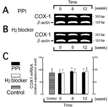
The COX-1 mRNA expression level in the gas-tric mucosa showed no difference among the PPI, H2 blocker and control groups or between before
and after the treatment (Fig. 4). However, the COX-2 mRNA expression level in the gastric mucosa in ulcer patients was significantly reduced before the treatment in comparison with that in nonulcer sub-jects. Moreover, the relative densities of COX-2 PCR bands at 8 weeks were 2.8 times and 2.1 times increased in the PPI group and in the H2 blocker
group, respectively, and the increase was
signifi-Fig. 2. Changes in the gastric mucosal levels of 6-keto PGF1α
after treatment with rabeprazole (PPI group) or ranitidine (H2 blocker group)
The gastric ulcer base disappeared at 8 − 12 weeks after treat-ment. The samples were obtained from the gastric mucosa in ul-cer patients and nonulul-cer subjects (control group). Values are the means±SD (n = 5 or 6). *P <0.05 in comparison with the control group.
Fig. 3. Changes in the gastric mucosal levels of LTB4after treatment with rabeprazole (PPI group) or ranitidine (H2 blocker group)
The gastric ulcer base disappeared at 8 - 12 weeks after treat-ment. The samples were obtained from the gastric mucosa in ulcer patients and nonulcer subjects (control group). Values are the means±SD (n = 5 of 6). *P <0.05 in comparison with the control group.+P <0.05 in comparison with the H2blocker group.#P <0.05 in comparison with the nonulcerated area in the same group of ulcer patients.
Fig. 4. Changes in the gastric mucosal expressions of COX-1 mRNA after treatment with rabeprazole (PPI group, A) or rani-tidine (H2blocker group, B)
The samples were obtained from the gastric mucosa in ulcer patients and nonulcer subjects (control group). Both tissue speci-mens obtained from the ulcerated and nonulcerated areas were mixed for RNA extraction. The levels of COX-1 gene expres-sion (303 bp) andβ-actin gene expression (125 bp) were ana-lyzed by RT-PCR. The results of a densitometric analysis are pre-sented as the mean percentages of the signal intensity ofβ-actin for COX-1 mRNA expression (C). Values are the means±SD (n = 5 or 6).
M. Okazaki, et al. PPI on COX-2, PGE2and LTB4in gastric ulcer
cantly greater in the PPI group. The COX-2 gene expression level in the PPI group returned after 12 weeks to a level similar to that in the H2 blocker
and control groups (Fig. 5).
DISCUSSION
PGs, which are mucosal protective factors, have an antisecretory effect as well as a mucosa-protecting effect(7, 13). PGs are produced from arachidonic acid released from phospholipids of biological mem-branes by the action of phospholipase. Phospholip-ids metabolize into PGs and LTs by the COX and lipoxygenase pathways, respectively. While COX-1 acts as a housekeeping enzyme in the gastric mu-cosa and platelets, COX-2, the expression of which is induced by agents including cytokines in such cells as macrophages and neutrophils, is known to play a role in inflammation and cell proliferation. NSAIDs reduce the PG production by inhibiting the expression of COX-1 and COX-2 in the gastric mucosa and by increasing LTs in relative terms
(14). A decrease in PGs depresses various factors in the gastric mucosal defense system, and an in-crease in LTs enhances the radical production while also exacerbating damage to the gastric mucosa (15). This study was initiated from based on the hypothesis that PPI promotes the healing mecha-nism of the gastric mucosa via the secretory kinet-ics of PGs and LTs by exerting some effect on the COX expression in not only NSAID-induced gastric ulcers, which we experienced(11), but also in com-mon gastric ulcers. The mucosal protecting effects of PGs against various agents that induce damage to the gastric mucosa are called cytoprotection. PGs produce effects such as directly protecting cells and promoting cytotaxis in the luminal cavity, epithe-lial, and subepithelial levels(14). Particularly, PGE2
increases the mucosal blood flow, promotes mu-cus secretion, and increases bicarbonate secretion (16), while PGI2suppresses gastric acid secretion
(17). In addition, Hp infection is associated with an increased production of PGE2in the gastric mucosa
(18). Hp infection elicits persistent neutrophil infil-tration in the gastric mucosa. The COX-2 expres-sion by the neutrophils results in PGE2 synthesis
(19). In this study, the PGE2 and 6-keto PGF1a (a
metabolic product of stable PGI2) concentrations in
ulcer patients with Hp infection decreased during the active-ulcer stage, and thereafter increased dur-ing the ulcer-healdur-ing stage after treatment with PPI and H2blocker to a level similar to those in
nonul-cer subjects with Hp infection. Moreover, the in-crease in the PGE2 concentration after 8 weeks,
when the gastric ulcers had been already scarred, was significantly greater in the PPI group than in the H2blocker group. In contrast, the 6-keto PGF1a
con-centration after 8 weeks showed no significant dif-ference between the PPI, H2 blocker and control
groups, when comparing the findings before and after the treatment.
Kobayashi, et al. reported the PG levels to in-crease at 4 and 7 days post-polypectomy in patients in whom gastric ulcers were produced by an elec-tric burning resection of gaselec-tric polyps, while the most remarkable increase took place in the mucosa along the ulcer margin rather than the mucosa far from the ulcer site(20). In the present study, how-ever, the PGE2 and 6-keto PGF1a levels were the
lowest during the active-ulcer stage with no signifi-cant difference between the ulcerated and nonul-cerated mucosae, although the 6-keto PGF1a level
in the ulcerated mucosa was significantly lower than that in the control group. In addition, Kobayashi, et
Fig. 5. Changes in the gastric mucosal expressions of COX-2 mRNA after treatment with rabeprazole (PPI group, A) or rani-tidine (H2blocker group, B)
The samples were obtained from the gastric mucosa from both ulcer patients and nonulcer subjects (control group). Both tis-sue specimens obtained from the ulcerated and nonulcerated areas were mixed for RNA extraction. The levels of COX-2 gene expression (305 bp) andβ-actin gene expression (125 bp) were analyzed by RT-PCR. The results of a densitometric analysis are presented as the mean percentages of the signal intensity of
β-actin for COX-2 mRNA expression (C). Values are the means
±SD (n = 5). *P <0.05 in comparison with the control group. +P <0.05 in comparison with the H2blocker group.
al. also reported that the PGE2 level in chronic
gastric ulcer patients was not significantly differ-ent from that in normal subjects(21). Since chronic Hp infection is accompanied by a persistent mu-cosal production of PGE2(18), the PG production
mechanisms might therefore be depleted when a gastric ulcer breaks out, although this study could not show any comparative information on the de-gree of gastric mucosal inflammation between the gastric ulcer groups.
LTs are synthesized in leukocytes as arachidonic acid is metabolized by lipoxygenase. An increase in LTs enhances the radical production and aggra-vates damage to the gastric mucosa(22). LTC4, LTD4,
and LTE4 enhance capillary permeability and
pro-mote airway mucosal secretion. LTB4promotes
leu-kocytotaxis(23). In our study, the LTB4
concentra-tion increased during the active-ulcer stage in ulcer patients, and it decreased during the ulcer-healing stage after the treatment. The decrease in the LTB4
concentration of the ulcer-scarred and nonulcerated mucosae was significantly greater in the PPI group than in the H2blocker group after 8 weeks, thus
sug-gesting that PPI could reduce the LTB4production
in the stomach.
COX-1 is routinely expressed in the normal gas-tric mucosa, but COX-2 is expressed in the lamina propria mucosae in the gastric mucosa infected by Hp(24, 25). In experimental erosion or ulcer cre-ated in animals, COX-2 mRNA and COX-2 protein have been reported to be expressed in ulcer mar-gins, while a COX-2 inhibitor was said to delay the cure of ulcers(26, 27). Although whether selective COX-2 inhibitors could induce directly gastrointes-tinal ulceration remains to be elucidated, these find-ings suggest that COX-2 could play an important role in the repair of gastric ulcers by regulating PG biosynthesis. In this study, gastric ulcer patients with Hp infection showed significant difference in the COX-2 mRNA expressions between the acute and scarring phases during gastric ulcer healing. The mucosal expression of COX-2 mRNA increased 2.8 times at 8 weeks after the beginning of the treat-ment in the PPI group, and this increase was sig-nificantly greater than that in the H2blocker group.
The COX-1 gene expression showed no significant difference between the PPI and H2blocker groups
or when comparing the findings before and after the treatment.
These results suggest that rabeprazole has a mucosa-protecting effect on the healing process of ulcers by increasing the PGE2 production and
re-ducing the LTB4 production. In addition,
rabepra-zole has an unique pharmacological ability to aug-ment the production of gastric mucus and mucin which thus generate the so-called mucus buffer layer covering the gastric mucosa(28). PPIs have been reported to inhibit radical production and inflammation by suppressing the neutrophil activ-ity(29-31). Akimoto, et al. reported that rabeprazole is able to promote vessel regeneration and matura-tion, thereby facilitating ulcer healing(32). In ulcer patients, the increase of basic fibroblast growth factor was reported to be greater with PPIs than H2
blockers(33). Although the mechanisms by which rabeprazole induces COX-2 and PGE2production and
reduces LTB4production remain to be elucidated in
gastric ulcer patients with Hp infection, it is con-sidered to promote the healing of ulcers due to these mucosa-protecting actions in addition to its antise-cretory action.
REFERENCES
1. Sachs G : Proton pump inhibitors and acid-related diseases. Pharmacotherapy 17 : 22-37, 1997 2. Mattsson H, Andersson K, Larsson H :
Ome-prazole provides protection against experi-mentally induced gastric mucosal lesions. Eur J Pharmacol 91 : 111-114, 1983
3. Konturek SJ, Brzozowski T, Radecki T : Pro-tective action of omeprazole, a benzimidazole derivative, on gastric mucosal damage by as-pirin and ethanol in rats. Digestion 27 : 159-164, 1983
4. Watanabe T, Higuchi K, Tominaga K, Fujiwara Y, Arakawa T : Cytoprotective effect of rabepra-zole against ehtanol-induced gatric mucosal damage : possible involvement of nitric oxide. Drugs Exp Clin Res 26 : 41-45, 2000
5. Tsuji S, Sun WH, Tsujii M, Kawai N, Kimura A, Kakiuchi Y, Yasumaru S, Komori M, Murata H, Sasaki Y, Kawano S, Hori M : Lan-soprazole induces mucosal protection through gastrin receptor-dependent up-regulation of cyclooxygenase-2 in rats. J Pharmacol Exp Ther 303 : 1301-1308, 2002
6. Smith WL, Garavito RM, DeWitt DL : Prosta-glandin endoperoxide H synthases (cyclooxy-genases)-1 and -2. J Biol Chem 271 : 33157-33160, 1996
7. Robert A, Nezamis JE, Lancaster C, Davis JP, Field SO, Hanchar AJ : Mild irritants prevent
M. Okazaki, et al. PPI on COX-2, PGE2and LTB4in gastric ulcer
gastric necrosis through ”adaptive cytoprotec-tion” mediated by prostaglandins. Am J Physiol 245 : G113-G121, 1983
8. Mizuno H, Sakamoto C, Matsuda K, Wada K, Uchida T, Noguchi H, Akamatsu T, Kasuga M : Induction of cyclooxygenase 2 in gastric mucosal lesions and its inhibition by the spe-cific antagonist delays healing in mice. Gas-troenterology 112 : 387-397, 1997
9. Samuelsson B, Dahlen SE, Lindgren JA, Rouzer CA, Serhan CN : Leukotrienes and lipoxins : structures, biosynthesis, and biological effects. Science 237 : 1171-1176, 1987
10. Serhan CN, Haeggstrom JZ, Leslie CC : Lipid mediator networks in cell signaling : update and impact of cytokines. FASEB J 10 : 1147-1158, 1996
11. Shimizu I, Horie T, Inoue H, Oshima T, Fujimoto M, Ozaki Y, Yamamoto K, Iuchi A, Ito S : Penetration by a giant gastric ulcer in-duced by a nonsteroidal anti-inflammatory drug. Endoscopy 32 : 539-541, 2000
12. Hla T, Neilson K : Human cyclooxygenase-2 cDNA. Proc Natl Acad Sci USA 89 : 7384-7388, 1992
13. Miller TA : Protective effects of prostagland-ins agaprostagland-inst gastric mucosal damage : current knowledge and proposed mechanisms. Am J Physiol 245 : G601-G623, 1983
14. Wallace JL, Tigley AW : Review article : new in-sights into prostaglandins and mucosal defence. Aliment Pharmacol Ther 9 : 227-235, 1995 15. Asako H, Kubes P, Wallace J, Gaginella T,
Wolf RE, Granger DN : Indomethacin-induced leukocyte adhesion in mesenteric venules : role of lipoxygenase products. Am J Physiol 262 : G903-G908, 1992
16. Gudis K, Sakamoto C : The role of cyclooxy-genase in gastric mucosal protection. Dig Dis Sci 50 (Suppl 1) : S16-S23, 2005
17. Kauffman GL, Zhang L, Xing LP, Seaton J, Colony P, Demers L : Central neurotensin pro-tects the mucosa by a prostaglandin-mediated mechanism and inhibits gastric acid secretion in the rat. Ann NY Acad Sci 597 : 175-190, 1990 18. Hudson N, Balsitis M, Filipowicz F, Hawkey
CJ : Effect of Helicobacter pylori colonisation on gastric mucosal eicosanoid synthesis in patients taking non-steroidal anti-inflammatory drugs. Gut 34 : 748-751, 1993
19. Kim JS, Kim JM, Jung HC, Song IS : Expres-sion of cyclooxygenase-2 in human
neutro-phils activated by Helicobacter pylori water-soluble proteins : possible involvement of NF-kappaB and MAP kinase signaling pathway. Dig Dis Sci 46 : 2277-2284, 2001
20. Kobayashi K, Arakawa T, Nakamura H, Chono S, Yamada H, Satoh H, Kamata T, Ono T : Pro-tective action of endogenous prostacyclin (PGI2)
and prostaglandin E2 (PGE2) in endoscopic
polypectomy-induced human ulcers. Gastro-enterol Jpn 17 : 430-433, 1982
21. Kobayashi K, Arakawa T, Nakamura H, Chono S, Yamada H, Kamata T, Ono T : Role of prostaglandin E2on humna gastric ulcers.
Gas-troenterol Jpn 17 : 21-24, 1982
22. Peskar BM : Leukotrienes in mucosal damage and protection. J Physiol Pharmacol 42 : 135-145, 1991
23. Pihan G, Rogers C, Szabo S : Vascular injury in acute gastric mucosal damage. Mediatory role of leukotrienes. Dig Dis Sci 33 : 625-632, 1988 24. Takahashi S, Shigeta J, Inoue H, Tanabe T,
Okabe S : Localization of cyclooxygenase-2 and regulation of its mRNA expression in gastric ulcers in rats. Am J Physiol 275 : G1137-G1145, 1998
25. Tatsuguchi A, Sakamoto C, Wada K, Akamatsu T, Tsukui T, Miyake K, Futagami S, Kishida T, Fukuda Y, Yamanaka N, Kobayashi M : Localisation of cyclooxygenase 1 and cyclooxy-genase 2 in Helicobacter pylori related gastri-tis and gastric ulcer gastri-tissues in humans. Gut 46 : 782-789, 2000
26. Shigeta J, Takahashi S, Okabe S : Role of cyclooxygenase-2 in the healing of gastric ul-cers in rats. J Pharmacol Exp Ther 286 : 1383-1390, 1998
27. Brzozowski T, Konturek PC, Konturek SJ, Schuppan D, Drozdowicz D, Kwiecien S, Majka J, Nakamura T, Hahn E : Effect of local appli-cation of growth factors on gastric ulcer heal-ing and mucosal expression of cyclooxygenase-1 and -2. Digestion 64 : cyclooxygenase-15-29, 200cyclooxygenase-1
28. Skoczylas T, Sarosiek I, Sostarich S, McElhinney C, Durham S, Sarosiek J : Significant enhance-ment of gastric mucin content after rabepra-zole administration : its potential clinical sig-nificance in acid-related disorders. Dig Dis Sci 48 : 322-328, 2003
29. Wandall JH : Effects of omeprazole on neutro-phil chemotaxis, super oxide production, de-granulation, and translocation of cytochrome b-245. Gut 33 : 617-621, 1992
30. Suzuki M, Mori M, Miura S, Suematsu M, Fukumura D, Kimura H, Ishii H : Omeprazole attenuates oxygen-derived free radical produc-tion from human neutrophils. Free Radic Biol Med 21 : 727-731, 1996
31. Lapenna D, de GS, Ciofani G, Festi D, Cuccurullo F : Antioxidant properties of omeprazole. FEBS Lett 382 : 189-192, 1996
32. Akimoto M, Hashimoto H, Shigemoto M, Maeda A, Yamashita K : Effects of antisecre-tory agents on angiogenesis during healing of gastric ulcers. J Gastroenterol 40 : 685-689, 2005 33. Tsuji S, Kawano S, Tsujii M, Michida T, Masuda E, Gunawan ES, Hori M : Mucosal microcir-culation and angiogenesis in gastrointestinal tract. Jpn J Clin Med 56 : 2247-2252, 1998
M. Okazaki, et al. PPI on COX-2, PGE2and LTB4in gastric ulcer