Emphysematous Pyelonephritis with M assive Renal
Parenchymal Gas and Fluid Collection :
A Case Report
Naoyuki Harada,
Nobuhiro Akuzawa,
Toshimitsu Hayama,
Noriko Hasegawa,
Hidenori Seki,
Yuko Oku,
Masayuki Totsuka,
Takashi Hatori,
Jun Murakami,
Kunihiko Imai,
Younosuke Kitahara,
Takeshi Miyao,
Souta Kurihara,
Kazunari Ohki,
Kouhei Okamoto,
Kouichi Suzuki,
Yutaka Kubota,
Yasushige Matsuo
and Masahiko Kurabayashi
A 76-year-old female patient with diabetes was referred to our hospital for evaluation of a 1-week history of left abdominal pain with loss of appetite. Laboratory data showed markedly elevated neutrophils. Abdominal X-ray showed massive gas and fluid collection in the left pararenal space, resembling an intestinal obstruction. Abdominal computed tomography revealed renal parenchymal gas and fluid and led to the correct diagnosis of emphysematous pyelonephritis. Based on these findings, this patient was immediately transferred to the intensive care unit. She underwent emergent ne-phrectomy and was discharged 28 days after surgery without complications.(Kitakanto Med J 2013; 63:141∼145)
Key words: computed tomography,diabetes mellitus,emphysematous pyelonephritis,nephrectomy
Introduction
Emphysematous pyelonephritis (EPN)is a severe, acute necrotizing infection of the renal parenchyma and perirenal tissue and is characterized by gas forma-tion. Emphysematous pyelonephritis is associated with high mortality in the absence of surgical interven-tion. This illness predominantly affects female patients with diabetes and can occur in patients with poorly controlled insulin-dependent and non-insulin-dependent diabetes in the absence of ureteric obstruc-tion. The clinical manifestations of EPN appear to be similar to those of common upper urinary tract infections and include abdominal pain, septic shock, vomiting, and fever. Confusion might be seen in
severe cases of EPN. In addition, EPN tends to preferentially affect the left kidney; bilateral EPN is uncommon.
Causative microorganisms detected from patients with EPN may be cultured from urine, blood, and aspirated pus. Escherichia coli is the most common microorganism and is isolated from 90% of patients with EPN. Klebsiella pneumoniae is also a com-mon pathogen. Other causative organisms include Proteus mirabilis, Pseudomonas aeruginosa, and Acinetobacter species, suggesting that involvement of anaerobic bacteria is uncommon in patients with EPN. Microbial infection and rapid catabolism causes the production of gas,including carbon dioxide, nitrogen, and hydrogen, because the ischemic kidney
1 Department of Internal Medicine, Social Insurance Gunma Chuo General Hospital,1-7-13 Koun-cho,Maebashi,Gunma 371-0025, Japan 2 Department of Urology, Maebashi Red Cross Hospital, 3-21-36 Asahi-cho, Maebashi, Gunma 371-0014, Japan
3 Department of Medicine and Biological Science,Gunma University Graduate School of Medicine,3-39-22 Showa-machi,Maebashi, Gunma 371-8511, Japan
Received : February 4, 2013
Address: NAOYUKI HARADA Social Insurance Gunma Chuo General Hospital, 1-7-13 Koun-cho, Maebashi, Gunma 371-0025, Japan
receives inadequate blood flow for removal of these gases. Three conditions are believed to be important in the development of gases: the presence of gas-forming bacteria,high levels of glucose in tissues,and impaired tissue perfusion. These gases are revealed by radiological examination. Abdominal X-ray is useful,but cannot be used to diagnose EPN at the first presentation. Abdominal gases detected on X-ray may be misdiagnosed as perforation of the gastrointes-tinal tract. Conversely, computed tomography (CT)has played a pivotal role in the definitive diagno-sis of EPN.
We experienced a case of EPN with a giant vol-ume of abdominal gas on abdominal X-ray,similar to acute gastric dilatation. Abdominal CT revealed a large quantity of gas inside the left subrenal capsule, which led to the correct diagnosis of EPN.
Case Presentation
A 76-year-old female patient with diabetes was referred to our hospital. She had a 1-week history of left-sided abdominal pain with appetite loss. Ini-tially,she felt vague nausea and abdominal distension.
Although these symptoms gradually progressed, she had no chills,vomiting,lumbago,or pollakiuria. She had no remarkable past medical history or family history. She had been prescribed glibenclamide(1.25 mg/day),alogliptin (25 mg/day),and pindrol (15 mg/ day) for type 2 diabetes mellitus and hypertension during the past 5 years,and had no diabetic complica-tions. Prior to medical examination in our hospital, she had undergone a gastroscopic exam in another clinic because of progressive appetite loss. She was told by the physician at the clinic that her stomach may have been compressed by an extramural tumor-like mass from the posterior side. Nevertheless,her gastric mucosal findings were almost normal.
On admission, she was alert. Her height and body weight were 148 cm and 61 kg,respectively. Her blood pressure was normal (118/70 mmHg), and her pulse rate was increased at 115 bpm. She had a slight fever (37.2℃). Physical examination showed mild distension and tenderness in her left middle abdomen. The results of an arterial blood gas analysis revealed marked acidosis and hypocapnea (pH, 7.280; PaCO , 21 Torr). The laboratory data on admission indicated an increased white blood cell count (51,800/μL; nor-mal, 3500-9000/μL) and elevated serum C-reactive protein level (32.6 mg/dL; normal, <0.30 mg/dL). Her serum urea nitrogen and serum creatinine levels were also elevated at 38.4 mg/dL (normal, 8.0-22.0 mg/dL) and 0.94 mg/dL (normal, 0.4-0.8 mg/dL), respectively (Table 1). Urinalysis was positive for protein, sugar, and bacteria. The urinary sediment indicated elevation of both red blood cells (50-99/
Table 1 Laboratory data on admission Hematology
White blood cells 51,800/mm Red blood cells 489×10 /mm
Hemoglobin 14.9 g/dL Hematocrit 42.7% Platelets 18.6×10 /mm Blood chemistry Total protein 6.2 g/dL Albumin 2.0 g/dL Total bilirubin 0.4 mg/dL Aspartate aminotransferase 13 IU/L Alanine aminotransferase 26 IU/L Lactate dehydrogenase 400 IU/L Blood urea nitrogen 38.4 mg/dL
Creatinine 0.94 mg/dL
Amylase 10 IU/L
Free blood sugar 535 mg/dL
Hemoglobin A1c 10.1%
Creatine phosphokinase 23 IU/L C-reactive protein 32.6 mg/dL Na 141 mEq/L K 3.0 mEq/L Cl 112 mEq/L Ca 7.9 mg/dL Urinalysis Urine protein 1+ Urine sugar 4+ Uric blood 3+
White blood cells 10-19/HPF Red blood cells 50-99/HPF Blood gas analysis
pH 7.280
PaCO 22.7 Torr
PaO 71.2 Torr
HCO 10.7 mmol/L
Base excess -13.8 mmol/L
These values are out of the normal range.
Fig. 1 Abdominal X-ray of the present case. A massive vol-ume of gas with niveau formation was present. The gas image was surrounded by a vague outline(white arrow) that probably represented other gas components.
HPF)and white blood cells(10-19/HPF). Although chest X-ray findings were normal, abdominal X-ray showed a massive volume of gas with niveau formation resembling acute gastric dilatation (Fig.1). Strange-ly, the gas image was surrounded by a vague outline that probably represented the other extramural gases. At this time,we considered the possibility that the gas comprised intramural stomach or intestinal gases. We performed an abdominal ultrasonographic examina-tion,but we could not acquire enough images because
of the patients obesity. Hence,we performed abdom-inal CT, which revealed some key findings for a definite diagnosis: (i) a massive gas image with slight leakage beyond the renal fascia and inner fluid reten-tion, strongly suggesting the possibility of EPN; (ii) markedly compressed left renal parenchyma by a massive volume of gas; (iii) fluid retention showing a density slightly higher than that of water; and (iv) preserved perirenal fatty tissue in spite of huge gas production, presumably by EPN (Fig.2).
Fig. 2 Abdominal computed tomography image of the present case. A massive volume of gas with fluid collection was observed within the left renal fascia.The left renal paren-chyma was markedly compressed (white arrow) by a massive volume of gas.
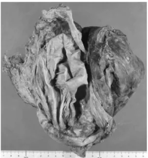
Fig. 3 Surgically resected kidney. A gross monocystic lesion was present in the left kidney parenchyma.
Fig. 4 Clinical course of the patient
te m p er a tu re (℃ )
Accordingly,the patient was immediately transfer-red to the intensive care unit to undergo left ne-phrectomy. The surgical treatment was successfully finished on that day by urologists. During the proce-dure,the upper aspect and lateral side of the left renal capsule were partially damaged, and pus exudation around the fascia was observed; these findings had not been detected by preoperative CT imaging. Patholog-ical findings showed that the renal pelvis was signifi-cantly expanded and that the renal capsule was separat-ed from the renal parenchyma, forming a huge cavity (Fig.3). Histopathological examination of the removed specimen revealed that inflammatory cells, mainly neutrophils, had infiltrated the renal pelvis to the parenchyma. Escherichia coli was detected in urine,blood,and pus cultures. Considering all of the above findings, we diagnosed the patient with EPN.
Following the operation, the patient was treated with intravenous antibiotics; initially meropenem(1.0 g/day for 5 days)and subsequently ceftazidime(2.0 g/ day for 7 days). Her white blood cell count and C-reactive protein level immediately normalized. Furthermore, her blood sugar level improved to approximately 150 mg/dL by additional dietary ther-apy(1400 kcal/day)with oral medication that she had taken before admission (Fig.4). She showed no fur-ther degeneration of renal function requiring hemodialysis or other postoperative complications. She was discharged 28 days after hospitalization.
Discussion
EPN predominantly affects female patients with diabetes. This may be related to the increased suscep-tibility of females to urinary tract infection. The left kidney is more frequently involved than the right, although the reason is unclear. The initial diagnosis of EPN may be difficult because the symptoms are vague, nonspecific, and similar to those of acute pyelonephritis. Fever and abdominal or back pain are reportedly the most common symptoms of EPN. Although some patients may show nausea, vomiting, confusion, or septic shock, these symptoms cannot provide clues for a definitive diagnosis of EPN. The present patient had nonspecific symptoms and a rela-tively low fever. Abnormal laboratory data showed the possibility of severe infectious disease; however, these data were not enough to diagnose EPN.
For the correct diagnosis of EPN in the early stage, radiological investigations are reportedly use-ful. The presence of abnormal gas on abdominal X-ray may suggest the diagnosis of EPN. In particular, huge volumes of gas may be observed in progressed cases, but such cases are rare. Notably, the gas images on abdominal X-ray are not clear in the
onset or early stage of EPN. Previous reviews have reported that early diagnosis of EPN using abdominal X-ray alone might be quite difficult. Conversely, abdominal CT is the most effective tool for the identifi-cation of EPN. It is useful for both the severity and diagnosis of EPN. EPN is also known to have a high mortality rate and may result in a critical condition if left untreated. Thus,proper treatment in the early stage is essential to avoid a detrimental outcome. As a staging method to predict the mortality and progno-sis of patients with EPN,the Classification System has been proposed by Huang et al. As shown in Table 2, this system is based on CT imaging findings. This system classifies the patients into four classes in pro-portion to the distribution of gases inside or outside the renal parenchyma. This system has also report-ed close relationships between high mortality rates and extrarenal gases,and nephrectomy rather than medica-tion or continuous drainage should be recommended in such cases. The present case was classified as class IIIa based on the findings of extrarenal gases in the perinephric space. According to a previous review, the recommended treatment for patients with class IIIa disease is nephrectomy. This review also reported a high mortality rate of patients with class IIIa disease (92%)when surgical treatment is not selected. Thus, rapid diagnosis using abdominal CT may be indispens-able at the initial examination of patients with EPN. However,this case also shows the limitation of abdom-inal CT: macroscopic findings during the surgical procedure showed pus retention beyond the renal fas-cia that was not detected during the initial CT exami-nation. This may suggest the possibility of underes-timating the severity of patients with EPN. Even information obtained from CT should be carefully interpreted.
In conclusion, we herein reported a case of EPN with massive gas and fluid collection in the renal parenchyma. Abdominal CT played a key role in the prompt diagnosis and decision-making for proper treatment. The possibility of EPN should be taken into consideration when we see patients with diabetes who show symptoms of general infectious diseases.
Table 2 Classification System of EPN Class I : Gas in collecting system only Class 11: Parenchymal gas only
Class IIIa:Extension of gas into perinephric space Class IIIb :Extension of gas into pararenal space Class IV: EPN in solitary kidney, or bilateral disease
Recommended treatment
Class I, II: Medication or continuous drainage Others: Nephrectomy
References
1. Schultz EH,Klorfein EH. Emphysematous pyelonephritis. J Urol 1962; 87: 762-766.
2. Michaeli J, Mogle P, Perlberg S, et al. Emphysematous pyelonephritis. J Urol 1984; 131: 203-208.
3. Pontin AR, Barnes RD. Current management of em-physematous pyelonephritis. Nat Rev Urol 2009 ; 6: 272-279.
4. Huang JJ, Tseng CC. Emphysematous pyelonephritis: clinicoradiological classification, management, prognosis and pathogenesis. Arch Intern Med 2000; 160: 797-805. 5. Shokeir AA, El-Azab M, Mohsen T, et al.
Em-physematous pyelonephritis: a 15-year experience with 20 cases. Urol 1997; 49 : 343-346.
6. Tang HJ,Li CM,Yen MY,et al. Clinical characteristic of emphysematous pyelonephritis. J Microbiol Immunol Infect 2001; 34: 125-130.
7. Yang WH,Shen NC. Gas-forming infection of the urinary tract: an investigation of fermentation as a mechanism. J Urol 1990; 143: 960-964.
8. Abdul-Halim H, Kehinde EO, Abdeen S, et al. Severe
emphysematous pyelonephritis in diabetic patients: diagno-sis and aspects of surgical management. Urol Int 2005; 75: 123-128.
9. Samuel RV, Ranjit S, James AF, et al. Emphysematous pyelonephritis in type II diabetes: a case report of an undiagnosed ureteric colic. Cases J 2008; 1: 192. 10. Wan YL,Lee TY,Bullard MJ,et al. Acute gas-producing
bacterial renal infection : correlation between imaging find-ings and clinical outcome. Radiology 1996; 198: 433-438.
11. Sathyanathan VP, Gomathy S, Potty RN, et al. Em-physematous pyelonephritis. J Assoc Physicians India 1998; 46: 562-563.
12. Pontin AR, Barnes RD, Joffe J, et al. Emphysematous pyelonephritis in diabetic patients. Br J Urol 1995; 75: 71-74.
13. Wan YL,Lo SK,Bullard MJ,et al. Predictors of outcome in emphysematous pyelonephritis. J Urol 1998; 159 : 369-373.
14. Tajima K, Kurabayashi M. Medical mystery-abnormal abdominal radiograph. N Engl J Med 2006; 355: 2467.