Evaluation of intrathoracic tracheal narrowing
in patients with obstructive ventilatory
impairment using dynamic chest radiography: A
preliminary study.
著者
WATASE Sayaka, SONODA Akinaga, MATSUTANI
Noritsugu, MURAOKA Shintarou, HANAOKA Jun,
NITTA Norihisa, WATANABE Yoshiyuki
journal or
publication title
European journal of radiology
volume
129
page range
109141
year
2020-06-19
URL
http://hdl.handle.net/10422/00012749
doi: 10.1016/j.ejrad.2020.109141(https://doi.org/10.1016/j.ejrad.2020.109141)
This is an open access article under the CC BY-NC-ND license
(http://creativecommons.org/licenses/BY-NC-ND/4.0/).
Contents lists available atScienceDirect
European Journal of Radiology
journal homepage:www.elsevier.com/locate/ejradResearch article
Evaluation of intrathoracic tracheal narrowing in patients with obstructive
ventilatory impairment using dynamic chest radiography: A preliminary
study
Sayaka Watase
a,*
, Akinaga Sonoda
a, Noritsugu Matsutani
b, Shintarou Muraoka
b, Jun Hanaoka
c,
Norihisa Nitta
a, Yoshiyuki Watanabe
aaDepartment of Radiology, Shiga University of Medical Science, Seta Tsukinowa-cho, Otsu, Shiga, 520-2192, Japan bHealthcare Business Headquarters, Konica Minolta, Inc, 2970 Ishikawa-machi, Hachioji-shi, Tokyo, 192-8505, Japan cDepartment of Thoracic Surgery, Shiga University of Medical Science, Seta Tsukinowa-cho, Otsu, Shiga, 520-2192, Japan
A R T I C L E I N F O
Keywords:
Dynamic digital radiography Flat panel detector Trachea
Expiratory central airway collapse Excessive dynamic airway collapse Tracheobronchomalacia
A B S T R A C T
Purpose: Dynamic chest radiography (DCR) can observe the dynamic structure of the chest using continuous pulse fluoroscopy irradiation. However, its usefulness remains largely undetermined. The purpose of this study was to examine the relationship between changes in tracheal diameter during deep breathing and obstructive ventilation disorders using DCR.
Method: Twelve participants with obstructive ventilatory impairment and 28 with normal pulmonary function underwent DCR during one cycle of deep inspiration and expiration. Three evaluators blinded to pulmonary function test results independently measured lateral diameters of the trachea in DCR images to determine whether there was a difference in the amount of change in tracheal diameter depending on the presence or absence of pulmonary dysfunction. Tracheal narrowing was defined as a decrease in the lateral tracheal diameter of more than 30 %. Participants were divided into a narrowing group and a non-narrowing group, and it was examined whether each group correlated with values of pulmonary function tests.
Results: Tracheal diameter was significantly narrowed in subjects with obstructive ventilatory impairment compared to normal subjects (P < 0.01). When subjects were divided into narrowing (tracheal narrowing rate [TNr] = 41.5 ± 7.7 %, n = 9) and non-narrowing groups (TNr = 9.1 ± 7.0 %, n = 31, p < 0.01), FEV1%-G, and %V25 were significantly smaller in the narrowing group than in the non-narrowing group (p < 0.01). Conclusions: Changes in tracheal diameter during deep breathing were easily evaluated using DCR. DCR may, therefore, be useful for evaluating obstructive ventilation disorders.
1. Introduction
In recent years, Yamashiro et al. and Kipritidis et al. have reported that dynamic 4-dimensional computed tomography (4D-CT) and dual-energy CT allow estimation of lung ventilation based on Hounsfield unit (HU) values [1,2]. Advances in CT and magnetic resonance imaging (MRI) technology have made it possible to obtain functional images of the lung.
Meanwhile, Tanaka et al. have recently reported that dynamic chest
radiography (DCR) could obtain ventilation-related information during an entire respiratory cycle [3] (Fig. 1). While this is a novel system, it can be examined like a conventional chest X-ray. The chest structure can be dynamically observed by irradiating weak continuous pulse X-rays. To perform DCR, in addition to commercially available computed radiography (CR) that can irradiate continuous pulse X-rays, digital radiography (DR) and a dedicated analyzer are required. The DCR system is reasonable, as the equipment is smaller, and the operation is easier compared to that of CT or MRI. Furthermore, unlike CT and MRI,
https://doi.org/10.1016/j.ejrad.2020.109141
Received 1 April 2020; Received in revised form 3 June 2020; Accepted 13 June 2020
Abbreviations: 4D-CT, 4-dimensional computed tomography; HU, Hounsfield unit; MRI, magnetic resonance imaging; DCR, dynamic chest radiography; CR, computed radiography; DR, digital radiography; PFTs, pulmonary function tests; ECAC, expiratory central airway collapse; TBM, tracheobronchomalacia; EDAC, excessive dynamic airway collapse; COPD, chronic obstructive pulmonary disease; FPD, flat panel detector; TNr, tracheal narrowing rate
⁎Corresponding author.
E-mail addresses:[email protected](S. Watase),[email protected](A. Sonoda),
[email protected](N. Matsutani),[email protected](S. Muraoka),[email protected](J. Hanaoka),
[email protected](N. Nitta),[email protected](Y. Watanabe).
European Journal of Radiology 129 (2020) 109141
0720-048X/ © 2020 The Author(s). Published by Elsevier B.V. This is an open access article under the CC BY-NC-ND license (http://creativecommons.org/licenses/BY-NC-ND/4.0/).
it is possible to obtain a rapid overview of the entire lung field from a single breath with DCR. It has been suggested that this system could evaluate pulmonary blood flow and ventilation volume using changes in lung field concentration values during respiration [4]. However, it is still unclear how useful this device will be compared to conventional chest X-ray, CT, MRI, nuclear medicine tests, and pulmonary function tests (PFTs).
In general, expiratory central airway collapse (ECAC) is defined as excessive airway collapse during expiration. ECAC describes two dif-ferent pathophysiologic entities, tracheobronchomalacia (TBM) and excessive dynamic airway collapse (EDAC) [5]. Acquired ECAC is idiopathic or secondary to other obstructive ventilation disorders such as chronic obstructive pulmonary disease (COPD) and asthma [5].
COPD patients show various degrees of tracheal narrowing [6]. However, it is reported that the symptoms, severity, and PFTs did not correlate well with the degree of tracheal narrowing at end-expiration [7]. However, using DCR, the degree of tracheal narrowing could be evaluated not only at end-expiration, but dynamically throughout the whole expiration phase. Therefore, we thought that the DCR might lend new insights between the degree of tracheal narrowing and symptoms, PFTs, etc. for subjects with obstructive ventilation disorders.
2. Materials and methods
2.1. Study population
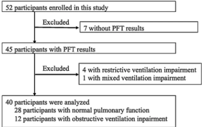
This study was approved by the ethics review board of Shiga University of Medical Science, and each participant provided written informed consent. Among the patients scheduled for thoracic surgery at our hospital between May and October 2017, 52 participants, who were 20 years or older with normal cognitive function, were selected. Among these 52 participants, 12 participants had obstructive ventilatory im-pairment (FEV1/FVC < 70 %), four participants had restrictive venti-latory impairment (TLC < 80 %), one had mixed ventiventi-latory impair-ment (FEV1/FVC < 70 % and TLC < 80 %), and 28 participants had normal (FEV1/FVC ≥ 70 % and TLC ≥ 80 %) parameter values. Seven of the 52 participants were excluded due to a lack of PFT results. PFTs were performed using a pulmonary function measurement device with computer processing (FUDAC-77; FUKUDA DENSHI Co. Tokyo, Japan). Of the 40 remaining participants, 30 were men and 10 women. The average age was 64.9 ± 12.2 years. Eleven participants were current smokers, 18 participants were ex-smokers, and 11 participants had never smoked. The average body mass index was 21.5 ± 3.7 kg/m2. Thirty-six participants had resectable lung tumors, and four participants
had mediastinum tumors. No participants had large tumors, and no tumor infiltrated the trachea. The flowchart of the study is shown in Fig. 2.
2.2. DCR imaging protocol
DCR was performed using a prototype X-ray system (Konica Minolta, Inc., Tokyo, Japan), composed of a flat panel detector (FPD) (PaxScan, 4343CB, Varex Imaging Corporation, Salt Lake City, UT, USA), X-ray tube (RAD-94/B-130H, Varian Medical Systems, Inc., Palo Alto, CA, USA), and X-ray generator (Epsilon, EPS45RF, EMD technol-ogies, Quebec, Canada). The X-ray exposure conditions were as follows: tube voltage, 100 kV; tube current, 20 mA; pulse duration of pulsed X-ray, 5.0 ms; source-to-image distance, 1.5 m; additional filter, 0.5 mm Al + 0.1 mm Cu for filtering out soft X-rays. The exposure time was approximately 15 s. The pixel size was 388 × 388 μm, and the matrix size was 1024 × 1024. Dynamic image data was captured at 15 frames/ s. The entrance surface dose was approximately 1.3 mGy for 15 s.
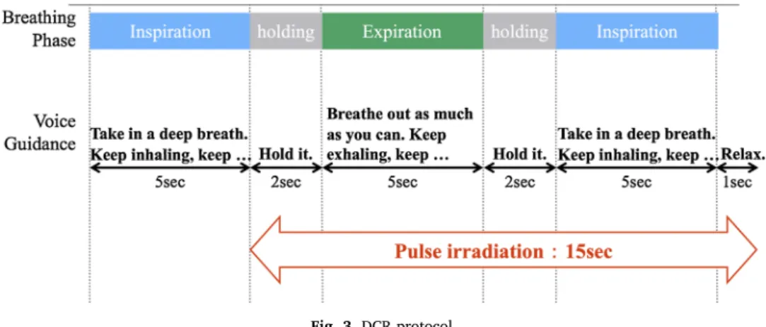
DCR was performed with participants in the supine position. A radiologist was present at all instances to instruct and monitor parti-cipant breathing. Partiparti-cipants performed deep breathing, including maximal expiration and maximal inspiration, following the audio guide, as shown inFig. 3. The imaging range was similar to that of a normal chest X-ray.
2.3. Image analysis
Fig. 4 shows an example of the difference between the tracheal diameter at maximum inspiration and maximum expiation on a DCR image. A prototype software (Konica Minolta, Inc., Tokyo, Japan) in-stalled in an independent workstation (Operating system: Windows 7 Pro SP1, Microsoft, Redmond WA; CPU: Intel® Core™ i5−4590, 3.30 GHz; memory 16 GB) was used for all tracheal diameter mea-surements.
The first item to be examined was whether there was a difference in the degree of tracheal narrowing between participants with normal pulmonary function and those with obstructive ventilatory impairment. Images were independently evaluated by three evaluators (two thoracic radiologists and one general radiologist) blinded to the PFT results. We manually measured the tracheal lateral diameter at the start of ex-piration and at maximum exex-piration and calculated the change in tra-cheal diameter according to the following equation:
Tracheal diameter change = Maximum tracheal diameter at the start of expiration – Minimum tracheal diameter at maximum expiration
Measurements were obtained at the inner diameter of the trachea. The tracheal measurement level was at the level of the intrathoracic Fig. 1. Dynamic Chest Radiography System.
The system is composed of an indirect dynamic flat panel detector (FPD) and a pulsed X-ray generator/tube. Sequential chest radiography at 15 frames per second can be conducted using much lower radiation doses than required for CT.
Fig. 2. Study flowchart shows participants selection.
Fifty-two people participated in this study. Those without PFT results and those with restrictive or mixed ventilation were excluded. A total of 40 participants were analyzed.
S. Watase, et al. European Journal of Radiology 129 (2020) 109141
trachea, and the narrowest part was measured at the time of maximum expiration. At the start of expiration, the most dilated part was mea-sured at the same level.
The second item to be examined was whether there was a correla-tion between the presence of tracheal narrowing and PFT result para-meters (%FEV1, FEV1%-G, %VC, %V25, %V50, and %V75). Measurements of tracheal diameter were performed by a thoracic radiologist blinded to the PFT results (normal, obstructive). The mea-surement method was the same as in the first analysis. The timing of measurement of tracheal diameter was seven frames before and after the start of expiration and maximum expiration (15 frames in total, approximately 1 s each), and average values were used (Fig. 5). A tra-cheal narrowing rate of 30 % or more was defined as significant nar-rowing, and a narrowing rate of less than 30 % was defined as no-narrowing. This cutoff was defined based on a previous CT report that narrowing of more than 50 % of the tracheal area was regarded as ECAC [8] (Fig. 6). The tracheal narrowing rate was calculated using the following equation:
Tracheal narrowing rate (TNr)=100 × (Maximum tracheal diameter at
the start of expiration – Minimum tracheal diameter at maximum ex-piration)/Maximum tracheal diameter at the start of expiration
2.4. Statistical analysis
Statistical analysis was performed using IBM SPSS statistics 25 (IBM, Tokyo, Japan) for Macintosh. The Student's t-test was used to determine whether there was a difference in the degree of tracheal narrowing between normal functioning subjects and those with obstructive ven-tilatory impairment. The correlation between tracheal narrowing and PFT results was evaluated using the Mann-Whitney U test All p values were two-sided, and p < 0.05 was considered to be significant.
3. Results
3.1. Tracheal diameter change
In the first item to be examined, all evaluators had significantly large changes in tracheal diameter in subjects with obstructive Fig. 3. DCR protocol.
Fig. 4. Representative DCR images during maximum inspiration and maximum expiration.
The concentration was adjusted easily to evaluate the trachea. A1 and A2 are images of a 69-year-old man with tracheal narrowing. B1 and B2 are images of a 42-year-old man without tracheal narrowing. The respiratory phase can be seen from the position of the diaphragm. The red arrows indicate the measured tracheal lateral diameter.
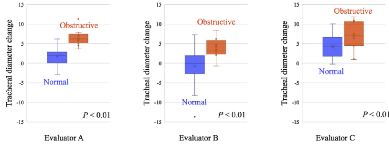
impairment than in normal pulmonary function. In the tracheal dia-meter change, the results of each of the evaluators A, B, and C were as follows. Normal vs. Obstructive = 1.73 ± 4.52 mm vs. 6.53 ± 3.84 mm (p < 0.01), -0.75 ± 17.63 mm vs. 3.74 ± 5.74 mm (p < 0.01), and 4.29 ± 8.10 mm vs. 6.94 ± 13.56 mm (p < 0.01) (Fig. 7).
3.2. Relationship between tracheal narrowing and pulmonary function tests
In the second item to be examined, the participants were divided into two groups based on the method described above; no-narrowing group (TNr = 9.1 ± 7.0 %, n = 31) and narrowing group (TNr = 41.5 ± 7.7 %, n = 9, p < 0.01). %FEV1, FEV1%-G, %V50, and %V25 were significantly lower in the narrowing group than those in the no-narrowing group; %FEV1: 101.1 ± 18.6 % vs. 78.5 ± 24.4 % (p < 0.05), FEV1%-G: 75.6 ± 10.6 % vs. 62.5 ± 16.4 % (p < 0.01), %V50: 90.4 ± 38.3 % vs. 52.9 ± 33.7 % (p < 0.05), %V25: 77.3 ± 46 % vs. 31.7 ± 16.5 % (p < 0.01). There was no significant difference in %VC and %V75 among the two groups. Tracheal narrowing occurred in 33.3 % of participants whose FEV1%-G is less than 70 % (n = 4/12) (Fig. 8).
4. Discussion
In our evaluation using DCR, changes in tracheal diameter were
significantly greater in the obstructive ventilation disorder group than in the normal pulmonary function group.
Factors associated with changes in tracheal diameter include tra-cheal strength, endotratra-cheal pressure, extratratra-cheal pressure (mainly intrathoracic pressure), and resting tracheal diameter. Of these, the intrathoracic pressure is considered to have the greatest effect on tra-cheal narrowing. Intrathoracic pressure, rises slowly during forced ex-piration in healthy subjects. On the other hand, intrathoracic pressure rises sharply in patients with obstructive ventilation, and changes in tracheal and bronchial diameters are linked to changes in intrathoracic pressure [9]. In obstructive ventilatory impairment, the peripheral bronchial diameter decreases sharply with increasing intrathoracic pressure, and air trapping occurs. This also causes the trachea to be compressed from the outside. As another factor, pursed-breathing seen in COPD can increase endotracheal pressure and affect tracheal dia-meter. Furthermore, chronic inflammation of the trachea occurs in obstructive ventilation disorders such as COPD and asthma. As a result, ECAC in which the elastic membrane or tracheal cartilage is damaged also reduces the strength of the tracheal wall and affects the tracheal diameter [5]. In addition, saber sheath trachea is common in COPD patients. This morphological change also affects airway resistance and can also affect changes in tracheal diameter. Therefore, multiple fac-tors, such as the fragility of the trachea itself, rapid change in Fig. 5. Timing of the tracheal diameter
mea-surement in the second analysis.
The graph shows a simulated change in tra-cheal diameter when tratra-cheal narrowing is indicated. The timing of measurement of tra-cheal diameter was seven frames before and after the start of expiration and maximum ex-piration (15 frames in total, about 1 s each). The measurement timing is as shown in the figure (↔︎).
Fig. 6. Criteria for narrowing tracheal diameter.
A narrowing rate of 30 % or more was defined as significant tracheal narrowing. This was based on the CT report showing that the narrowing of 50 % or more of the tracheal area was regarded as ECAC.
S. Watase, et al. European Journal of Radiology 129 (2020) 109141
intrathoracic pressure, and pulmonary hyperinflation accompanying peripheral airway obstruction, may have influenced tracheal narrowing in patients with obstructive ventilation disorders.
In this study, the value of FEV1%-G was significantly lower in the tracheal narrowing group than in the no-narrowing group. FEV1%-G has been reported to be strongly associated with the narrowing of the diameter of the peripheral airway but not the central airway [10]. This suggests that finding tracheal narrowing using DCR can predict per-ipheral airway diameter, and DCR may help screen patients who are prone to obstructive ventilation. However, it is known that even a healthy person can undergo a tracheal collapse of 50 % or more during expiration, and in extreme cases, about 70–80 % of healthy people have narrowing [8]. Furthermore, it has been reported that tracheal nar-rowing was observed in COPD in which airway lesions were superior to those in the emphysema-predominant type [11]. Previous studies using DCR have also shown that patients with COPD have characteristic diaphragmatic movements [12]. The combination of DCR findings may enable early screening of patients who are truly prone to obstructive ventilatory impairment.
Previous studies have used bronchoscopy, chest X-ray, CT, and MRI to evaluate tracheal narrowing [8,13,14]. DCR is superior to all these methods in that dynamic evaluation can be performed more easily. Previous studies have pointed out that the tracheal diameter measured at the time of breath holding during maximum inspiration was not the maximum value [15]. Similarly, in our study, tracheal diameter re-ex-panded during breath-hold with expiration in some cases. In other words, there is a possibility that the tracheal diameter of forced ex-piration, at rest may differ from the tracheal diameter at the end of expiration (immediately before breath-holding) adopted in this study.
Another advantage is that, unlike ordinary CT or MRI, DCR can be imaged in various body positions. Although our examinations were performed in a supine position in order to resolve room installation space and exclude the effect of body movement, activities in daily life are often performed in a standing or sitting position, and evaluating the movement of the chest in these states seems to be of great clinical significance.
Our study had several limitations. First, this was a preliminary study of a small number of cases before thoracic surgery. Participants with malignant tumors were included, although no tumor affected the tra-cheal or rib cage movement. It is necessary to increase the number of cases and analyze whether changes in tracheal diameter vary de-pending on each participants' background, such as normal participants, COPD by severity, and asthma. In this study, only participants with normal and obstructive disorders were studied, but studies on re-strictive disorders and mixed disorders are also necessary. Second, the tracheal measurement method is controversial. It was difficult to de-termine the measurement site uniformly because the trachea moves in the craniocaudal direction due to breathing, and the tracheal narrowing site differs depending on the subject. In addition, the evaluation can be oversized or undersized due to slight differences in the measurement position and timing. Possible solutions include averaging the tracheal diameters within a certain period and evaluating the intrathoracic tracheal area and its rate of change. It is still unclear what kind of measurement method is suitable, and further research is expected. Third, under the assumption that the trachea was reduced to a circular shape, 30 % or more was defined as a significant tracheal lateral nar-rowing rate. It has been reported that when the cross-sectional area of the trachea was reduced by about 55 %, the narrowing rate of the Fig. 7. Tracheal diameter change.
The results of each of the evaluators A, B, and C were as follows. Normal vs. Obstructive = 1.73 ± 4.52 mm vs. 6.53 ± 3.84 mm (p < 0.01), -0.75 ± 17.63 mm vs. 3.74 ± 5.74 mm (p < 0.01) and 4.29 ± 8.10 mm vs. 6.94 ± 13.56 mm (p < 0.01).
Fig. 8. Relationship between tracheal narrowing and pulmonary function tests.
lateral diameter was 29 % [8]. Therefore, 30 % was judged to be within the acceptable range. However, the trachea often narrows in a crescent or horseshoe shape [8]. Only the frontal view was examined in this study, and cases of anterior-posterior narrowing might not have been noticed. In the future, it will be necessary to take a lateral view and evaluate it. Fourth, measurement of the tracheal diameter was per-formed manually, and objectivity was poor. Although the trachea was judged by the presence of air, it was sometimes difficult to recognize the area of the trachea due to the obesity or overlap of blood vessel shadows and air of the esophagus. Therefore, it is necessary to develop tracheal measurement software that automatically extracts the tracheal data and measures the diameter.
In conclusion, DCR could be used to assess tracheal diameter. In our evaluation using DCR, tracheal narrowing tended to be stronger in obstructive impairment than in the normal group. The group showing tracheal narrowing showed a correlation with parameters such as re-duced FEV1%-G, V50, and V25 compared to the no-narrowing group. This indicated that DCR might help to assess obstructive ventilation disorders.
Funding
This research did not receive any specific grant from funding agencies in the public, commercial, or not-for-profit sectors.
CRediT authorship contribution statement
Sayaka Watase: Writing - original draft. Akinaga Sonoda:
Methodology, Validation, Investigation, Writing - review & editing.
Noritsugu Matsutani: Software, Resources, Formal analysis, Data
curation. Shintarou Muraoka: Software, Resources. Jun Hanaoka: Resources. Norihisa Nitta: Conceptualization, Investigation.
Yoshiyuki Watanabe: Supervision. Declaration of Competing Interest
This research was supported by Konica Minolta, Inc.
DCR and Image analysis system were provided free of charge from Konica Minolta, Inc.
Acknowledgement
None.
References
[1] T. Yamashiro, H. Moriya, S. Matsuoka, Y. Nagatani, M. Tsubakimoto, N. Tsuchiya,
S. Murayama, Asynchrony in respiratory movements between the pulmonary lobes in patients with COPD: continuous measurement of lung density by 4-dimensional dynamic-ventilation CT, Int. J. Chron. Obstruct. Pulmon. Dis. 12 (2017) 2101–2109,https://doi.org/10.2147/COPD.S140247.
[2] J. Kipritidis, M.S. Hofman, S. Siva, J. Callahan, P.Y. Le Roux, H.C. Woodruff, W.B. Counter, P.J. Keall, Estimating lung ventilation directly from 4D CT Hounsfield unit values, Med. Phys. 43 (2016) 33–43,https://doi.org/10.1118/1. 4937599.
[3] R. Tanaka, T. Tani, N. Nitta, T. Tabata, N. Matsutani, S. Muraoka, T. Yoneyama, S. Sanada, Pulmonary function diagnosis based on respiratory changes in lung density with dynamic flat-panel detector imaging: an animal-based study, Invest. Radiol. 53 (2018) 417–423,https://doi.org/10.1097/RLI.0000000000000457. [4] R. Tanaka, Dynamic chest radiography: flat-panel detector (FPD) based functional
X-ray imaging, Radiol. Phys. Technol. 9 (2016) 139–153,https://doi.org/10.1007/ s12194-016-0361-6.
[5] S. Murgu, H. Colt, Tracheobronchomalacia and excessive dynamic airway collapse, Clin. Chest Med. 34 (2013) 527–555,https://doi.org/10.1016/j.ccm.2013.05.003. [6] N. Sverzellati, A. Rastelli, A. Chetta, V. Schembri, L. Fasano, A.M. Pacilli,
V.D. Scioscio, T. Bartalena, M.D. Filippo, M. Zompatori, Airway malacia in chronic obstructive pulmonary disease: prevalence, morphology and relationship with emphysema, bronchiectasis and bronchial wall thickening, Eur. Radiol. 19 (2009) 1669–1678,https://doi.org/10.1007/s00330-009-1306-9.
[7] T. Yamashiro, R.S.J. Estépar, S. Matsuoka, B.J. Bartholmai, J.C. Ross, A. Diaz, S. Murayama, E.K. Silverman, H. Hatabu, G.R. Washko, Intrathoracic tracheal vo-lume and collapsibility on inspiratory and end-expiratory CT scans correlations with lung volume and pulmonary function in 85 smokers, Acad. Radiol. 18 (2011) 299–305,https://doi.org/10.1016/j.acra.2010.11.005.
[8] P.M. Boiselle, C.R. O’Donnell, A.A. Bankier, A. Ernst, M.E. Millet, A. Potemkin, S.H. Loring, Tracheal collapsibility in healthy volunteers during forced expiration: assessment with multidetector CT, Radiology 252 (2009) 255–262,https://doi.org/ 10.1148/radiol.2521081958.
[9] Y. Orita, Airway dynamics during forced vital capacity maneuver with special re-ference to the study on dynamic bronchography and transmural pressure, Bull. Chest Dis. Res. Inst. Kyoto Univ. 10 (1977) 74–89 (in Japanese).
[10] P.T. Macklem, Airway obstruction and collateral ventilation, Physiol. Rev. 51 (1971) 368–436,https://doi.org/10.1152/physrev.1971.51.2.368. [11] G. Camiciottoli, S. Diciotti, F. Bigazzi, S. Lombardo, M. Bartolucci, M. Poletti,
M. Mascalchi, M. Pstolesi, Is intrathoracic tracheal collapsibility correlated to clinical phenotypes and sex in patients with COPD? Int. J. Chron. Obstruct. Pulmon. Dis. 10 (2015) 843–852,https://doi.org/10.2147/COPD.S80558.
[12] Y. Yamada, M. Ueyama, T. Abe, T. Araki, T. Abe, M. Nishino, M. Jinzaki, H. Hatabu, S. Kudoh, Difference in diaphragmatic motion during tidal breathing in a standing position between COPD patients and normal subjects: time-resolved quantitative evaluation using dynamic chest radiography with flat panel detector system ("dy-namic X-ray phrenicography"), Eur. J. Radiol. 87 (2017) 76–82,https://doi.org/10. 1016/j.ejrad.2016.12.014.
[13] P. Ciet, P.M. Boiselle, B. Heidinger, E.R. Andrinopoulou, C. O’Donnel, D.C. Alsop, D.E. Litmanovich, Cine MRI of tracheal dynamics in healthy volunteers and patients with tracheobronchomalacia, AJR Am J Roentgenol. 209 (2017) 757–761,https:// doi.org/10.2214/AJR.17.17802.
[14] M.O. Wielpütz, R. Eberhardt, M. Puderbach, O. Weinheimer, H.U. Kauczor, C.P. Heussel, Simultaneous assessment of airway instability and respiratory dy-namics with low-dose 4D-CT in chronic obstructive pulmonary disease: a technical note, Respiration 87 (2014) 294–300,https://doi.org/10.1159/000357448. [15] R.H. Baroni, D. Feller-Kopman, M. Nishino, H. Hatabu, S.H. Loring, A. Ernst,
P.M. Boiselle, Tracheobronchomalacia: comparison between end-expiratory and dynamic expiratory CT for evaluation of central airway collapse, Radiology 235 (2005) 635–641,https://doi.org/10.1148/radiol.2352040309.
S. Watase, et al. European Journal of Radiology 129 (2020) 109141