T
he insertion of a self-expandable metal stent (SEMS) is the first-line option for an unresectable malignant distal biliary stricture. Compared to tradi-tional plastic stents, SEMSs allow for good drainage and high patency rates [1-3]. A SEMS costs more than a plastic stent, but its use results in fewer re-interven-tions and hospital stays. A SEMS is also a cost-effective palliative treatment for individuals with unresectable diseases [4,5]. However, SEMSs are associated with some serious complications, e.g., cholecystitis, cholan-gitis, and pancreatitis. Many mechanisms of post-endoscopic retrograde cholangiopancreatography creatitis (PEP) have been suggested; one is main pan-creatic duct (MPD) obstruction due to papillary swell-ing durswell-ing the procedure [6-10]. In order to solve this problem and prevent PEP, endoscopic pancreatic stent-ing (EPS) for individuals at high risk of developstent-ing PEP is recommended by international guidelines [11-14].
We have performed SEMS insertions for malignant distal biliary strictures with biliary sphincterotomy, and we sometimes observe PEP in patients without MPD obstruction. Shimizu et al. and Kawakubo et al. revealed that nonpancreatic cancer is a risk factor predicting PEP
CopyrightⒸ 2020 by Okayama University Medical School.
http ://escholarship.lib.okayama-u.ac.jp/amo/
Original Article
Prevention of Post-Endoscopic Retrograde Cholangiopancreatography
Pancreatitis by Endoscopic Pancreatic Stenting after Insertion of
Self-Expandable Metal Stent for Malignant Distal Biliary Stricture
Ryo Harada
*, Ryosuke Sato, Tomoaki Tsutsui, Nao Hattori,
Masafumi Inoue, and Haruhiko Kobashi
Department of Gastroenterology and Hepatology, Japanese Red Cross Okayama Hospital, Okayama 700-8607, Japan
The insertion of a self-expandable metal stent (SEMS) for nonpancreatic cancer is a factor predicting the risk of post-endoscopic retrograde cholangiopancreatography pancreatitis (PEP). We evaluated the efficacy of endo-scopic pancreatic stenting (EPS) to prevent PEP after SEMS insertion in patients with malignant distal biliary stricture and without main pancreatic duct (MPD) obstruction. We performed a single-center, retrospective, historically controlled investigation to assess the outcomes of 33 consecutive patients who underwent SEMS insertion. From March 2013 to June 2015, 13 patients did not undergo EPS (Non-EPS group). The other 20 patients underwent EPS (EPS group) between July 2015 and August 2018. The background data demonstrated no significant differences. Except for one patient in the Non-EPS group, all patients underwent biliary sphinc-terotomy. The EPS group’s PEP incidence was significantly lower (n=1, 5%) than that of the Non-EPS group (n=4, 31%) (p=0.04). The median serum amylase and lipase levels after the procedure were significantly lower in the EPS group than in the Non-EPS group (amylase: 104 vs. 262 U/L; p<0.01, lipase: 102 vs. 666 U/L;
p=0.01). The use of EPS decreased the incidence of PEP after SEMS insertion in individuals with malignant
distal biliary stricture and without MPD obstruction.
Key words: post-endoscopic retrograde cholangiopancreatography pancreatitis, endoscopic pancreatic stent, main
pancreatic duct obstruction, self-expandable metal stent, distal biliary stricture
Received May 8, 2020 ; accepted July 14, 2020.
*Corresponding author. Phone : +81-86-222-8811; Fax : +81-86-222-8841
the SEMS from compressing the MPD and of decreas-ing PEP risk. We performed the present investigation to determine the efficacy of EPS for the prevention of PEP after SEMS insertion in patients without MPD obstruc-tion.
Patients and Methods
Patients. All of the patients in this study were identified from our endoscopic retrograde cholan-giopancreatography (ERCP) database. Between March 2013 and August 2018, 83 patients underwent a SEMS insertion for a distal biliary stricture. All of these patients underwent an examination including a com-puted tomography (CT) scan and/or magnetic reso-nance imaging before undergoing the ERCP. By using the findings of those examinations, we excluded patients with MPD obstruction and/or dilatation of the MPD >4 mm (n=38). Patients with a history of SEMS insertion (n=9) and patients with a benign biliary stric-ture (n=3) were excluded as well. We thus retrospec-tively investigated the cases of 33 patients.
Of the 33 patients, 13 underwent a SEMS insertion between March 2013 and June 2015 and did not undergo the insertion of a pancreatic stent (Non-EPS group). The other 20 patients who underwent SEMS insertion between July 2015 and August 2018 under-went a pancreatic stent insertion (EPS group) (Fig.1). These two periods were mutually exclusive. The Ethics Committee of our institute approved the study design (Japanese Red Cross Okayama Hospital, 2018-84), and the study adhered to the Declaration of Helsinki. Before each ERCP, the patient provided written informed consent to undergo the procedure and for the data to be used.
SEMS procedures. The deployed stent was a 5-French straight pancreatic stent with various lengths (3-7 cm) (Geenen: Cook Endoscopy, Winston-Salem,
NC, USA). Four SEMS were used: WallFlex (Boston Scientific; Natick, MA, USA); X-Suit NIR (Olympus; Tokyo); BONA Stent (Medicos-Hirata; Osaka, Japan); and Niti-S (Taewoong Medical; Seoul, Korea). Each ERCP was conducted with a JF-260V or TJF-260V video endoscope (Olympus Medical; Tokyo) with the patient under conscious sedation (midazolam, pethi-dine hydrochloride). Selective MPD or common bile duct cannulation was first attempted using an ERCP Catheter (MTW Endoscopie; Wesel, Germany) and a 0.025-inch guidewire (VisiGlide or VisiGlide2: Olympus Medical Systems, Tokyo). A guidewire-assisted cannu-lation technique was attempted as the first option; if this proved difficult, a more conventional contrast- assisted cannulation method was used. After biliary cannulation, the guidewire was passed through the bil-iary stricture and then placed in the intrahepatic bile duct. A sphincterotomy with a moderate incision was achieved with a blended current. A moderate incision was defined as one that exceeded the transverse fold and did not reach the superior margin of the papillary bulge, according to the Japan Gastroenterological Endoscopy Society guidelines for endoscopic sphincterotomy [17]. The SEMS was then inserted over the guidewire by using a fluoroscopic control.
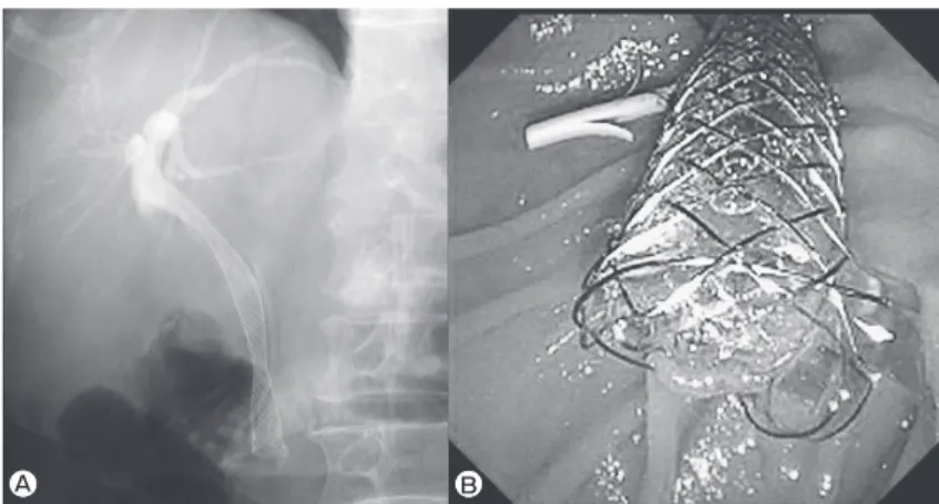
In the EPS group, generally, the patients were nulated by the double-guidewire method in biliary can-nulation. If pancreatic cannulation proved difficult, we cannulated to a pancreatic duct after a biliary sphincter-otomy or a precut papillsphincter-otomy. The pancreatic stent was inserted (under fluoroscopic control) over the guide-wire before the SEMS insertion (Fig.2). In the Non-EPS group, we tried not to cannulate MPD and completed the procedure without EPS. If the ERCP training fellow
could not obtain an endoscopic retrograde pancreatog-raphy (ERP) or endoscopic retrograde cholangiogpancreatog-raphy (ERC) image within 10 min, two senior endoscopists with a total career length of more than 10 years and experience with 2,000 ERCPs helped obtain the ERP or ERC image. When it took longer than 10 min to achieve a successful selective cannulation, the procedure was categorized as difficult.
The stent type was chosen by the operator to achieve the safest and most successful procedure. The pancre-atic stent was not removed unless and until it clogged and caused pancreatitis. We infused 150,000 U of uli-nastatin (Mochida Pharmaceutical, Tokyo, Japan) dis-solved in 100 mL of 0.9% saline solution immediately after the ERCP throughout the study period. In both patient groups, diclofenac was administered at the dis-cretion of each endoscopist. We defined ʻadverse
eventsʼ in accord with the lexicon of endoscopic adverse
events issued by the American Society Gastrointestinal Endoscopy [18]; ERCP-related adverse events were defined in accord with the European Society of Gastrointestinal Endoscopy Guideline [11].
Definitions of post-ERCP pancreatitis. We defined ʻPEPʼ based on the standard criteria [11]: new
abdominal pain or worsening abdominal pain persisting for ≥24 h after the ERCP, in addition to a serum amy-lase level or a serum lipase level >3 times the upper limit of normal. We also graded pancreatitis according to the standard criteria [11]: ʻmildʼ, no organ failure
and no local or systemic complications; ʻmoderateʼ,
transient (<48 h) organ failure and/or local or systemic complications without persistent organ failure; and
ʻsevereʼ, persistent (48 h) organ failure.
Statistical analysis. The study’s primary end-point was the development of PEP. We compared cate-gorical variables and continuous variables between the EPS and Non-EPS groups by using the χ2 test and the
Mann-Whitney U-test. P-values <0.05 were accepted as significant. All statistical analyses were conducted using the JMP ver. 12.2.0 statistical software package (SAS Institute, Cary, NC, USA).
Results
Patient characteristics and procedures. The background data demonstrated no significant between-group differences (Table 1). In both between-groups, the pri-mary diseases included pancreatic cancer, biliary can-cer, gastric cancan-cer, and colon cancer. One ovarian cancer was included in the EPS group. Four patients (20%) in the EPS group and 4 (31%) in the Non-EPS group were given analgesic drugs for abdominal pain before ERCP. Fifteen patients (75%) had naïve papilla in the EPS group, as did 6 (46%) in the Non-EPS group (p=0.14). In the EPS group, pancreatography was per-formed in all patients for the deployment of the pancre-atic stents, whereas only 6 (46%) patients in the Non-EPS group underwent pancreatography (p<0.01). In the Non-EPS group, only one patient did not undergo bili-ary sphincterotomy and 2 patients underwent insertion of uncovered SEMS. Regarding the marker for proce-dure difficulty, more than 10 min elapsed for successful selective cannulation in 4 patients (20%) in the EPS group and in 3 patients (23%) in the Non-EPS group (Table 2). EPS was successful in all 20 patients in the EPS group.
A B
Table 3 Incidence of pancreatitis and hyperamylasemia and serum levels of amylase and lipase
EPS (n=20) Non-EPS (n=13) p-value
Pancreatitis 1 (5%) 4 (31%) 0.04
Mild 0 4
Moderate 1 0
Severe 0 0
Hyperamylasemia 3 (15%) 7 (54%) 0.11
Serum amylase, U/L; median (IQR)
Before 72 (40-84) 65 (55-94) 0.52
18 h after procedure 104 (54-169) 262 (147-1,392) <0.01
Serum lipase, U/L; median (IQR)
Before 42 (20-75) 62 (28-101) 0.63
18 h after procedure 102 (33-216) 666 (134-1,850) 0.01
IQR, interquartile range.
Primary disease (Pancreatic cancer/Biliary cancer/
Gastric cancer/Colon cancer/Ovarian cancer) 4/10/1/4/1 3/6/3/1/0 0.46 Administration of analgesic drugs for abdominal pain 4 (20%) 4 (31%) 0.68
Previous post ERCP pancreatitis 0 1 0.39
Mean main pancreatic duct dia., mm (range) 2.4 (1-4) 2.0 (1-4) 0.23
Administered diclofenac 3 (15%) 2 (15%) 0.98
Observation period, days; median (IQR) 159 (85-373) 231 (35-283) 0.47 ERCP, endoscopic retrograde cholangiopancreatography; IQR, interquartile range.
Table 2 ERCP findings and endoscopic procedure
EPS (n=20) Non-EPS (n=13) p-value
Naïve papilla 15 (75%) 6 (46%) 0.14
Duodenal diverticulum 1 (5%) 1 (8%) 0.75
Previous biliary plastic stent 5 (25%) 7 (54%) 0.14
Pancreatography 20 (100%) 6 (46%) <0.01
Precut* 2 (10%) 1 (8%) 0.82
Biliary sphincterotomy 20 (100%) 12 (92%) 0.40
More than 10 minutes elapsed for the successful
selective cannulation 4 (20%) 3 (23%) 0.99
Mean procedure time, min (range) 28 (15-36) 30 (16-73) 0.46
Trainee participation 8 (40%) 3 (23%) 0.31
Stent type (covered/uncovered) 20/0 11/2 0.15
*Precut include precut papillotomy and transpancreatic sphincterotomy.
The median serum amylase level and the median serum lipase level at 18 h after the procedure were both signifi-cantly lower in the EPS group than in the Non-EPS group (amylase: 104 vs. 262 U/L; p<0.01, lipase: 102 vs. 666 U/L; p=0.01) (Table 3, Fig.3).
Other complications. No other major early com-plications were observed. SEMS dislodgement was observed after approx. 18 months in one EPS group patient. Mild pancreatitis was observed in one EPS group patient after 1 month, but this resolved with con-servative therapy and without the need for pancreatic stent extraction.
Discussion
A biliary SEMS is the first option for an unresectable malignant distal biliary stricture. Previous studies showed a comparatively high frequency of acute adverse events caused by mechanical stent compression, such as cholecystitis and pancreatitis [15,16,19]. Some cases of pancreatitis have required the immediate removal of a stent after its insertion. Itoi et al. reported the case of a patient with lethal PEP after SEMS insertion in a distal biliary stricture due to unresectable cholangiocarci-noma [20]; the SEMS was removed just 2 days after the procedure. However, the PEP worsened and the patient died. Itoi et al. suggested 2 major mechanisms of PEP after SEMS insertion. One is MPD opacification during the ERCP procedure: namely, common PEP. The other mechanism is impaired drainage from the MPD due to SEMS compression toward the papilla. Based on their patient’s clinical course, Itoi et al. concluded that the expansive force of the SEMS caused the patient’s PEP.
The present report is the first to reveal that using EPS decreased the rate of PEP in individuals without MPD obstruction, even though all 20 of the patients in the EPS group underwent pancreatography. This may sup-port the theory that MPD obstruction caused by the expansive force of the SEMS is the most important fac-tor for PEP after the insertion of a SEMS in a distal bili-ary stricture.
Shimizu et al. and Kawakubo et al. revealed that nonpancreatic cancer was one of the predictive risk fac-tors for PEP after SEMS insertion in a distal biliary stricture [15,16]. Isayama et al. and Nakai et al. showed that the rate of PEP in individuals with MPD obstruc-tion by their tumors was low [21,22]. Usually, most patients with pancreatic head cancer also show MPD involvement by the tumor, and the pancreatic paren-chyma of the distal part is atrophic. They thus have impaired exocrine function. However, in patients with nonpancreatic cancer, the MPD is not completely obstructed and exocrine function of the pancreas is pre-served. In accord with this theory, we conducted the present study with patients without MPD obstruction.
Insertion to a pancreatic duct is sometimes difficult, and a failure in pancreatic duct cannulation presents a risk of PEP. In our patient series, EPS was successfully inserted in all EPS group patients. Fifteen patients were cannulated to the pancreatic duct first; one patient required a precut papillotomy and 4 required a biliary sphincterotomy for cannulation into the pancreatic duct. It is widely known that cannulation after a biliary sphincterotomy is a comparatively easy way to cannu-late a pancreatic duct. After a biliary sphincterotomy, it is easy to identify the pancreatic duct.
Serum amylase level (U/L)
5,000 4,000 3,000 2,000 1,000 0 -1,000 p<0.01 EPS Non-EPS
Serum lipase level (U/L)
10,000 8,000 6,000 4,000 2,000 0 -2,000 p=0.01 EPS Non-EPS
Fig. 3 Comparison of serum amylase and lipase levels between EPS and Non-EPS groups after endoscopic retrograde cholangiopan-creatography. Both serum amylase and lipase levels were significantly lower in the EPS group than in the Non-EPS group (p<0.01, p=0.01, respectively).
patients is necessary. However, it may be easy to exchange a pancreatic stent because we can easily iden-tify the pancreatic duct orifice after pancreatic stent extraction. When there is no pancreatic stent, it might be difficult to find the pancreatic duct orifice after SEMS placement.
Our study has some limitations. The number of patients was relatively low, and the study design was retrospective. Due to the small number of patients, it may not be possible to conclude whether EPS can prevent PEP in patients without MPD obstruction. Second, the imaging confirmation of PEP was not done by CT scan due to our investigation’s retrospective design. The abdominal pain associated with PEP can be difficult to distinguish from the abdominal pain related to SEMS expansion. We used standard criteria: abdominal pain lasting ≥24 h and a high level of serum amylase or serum lipase. Abdominal pain may include the pain associated with stent expansion. We therefore also compared the serum amylase and lipase levels in both groups. Third, the PEP rate in the Non-EPS patients was high (31%), but this value is similar to the 28% in another study of nonpancreatic cancer patients [15].
In summary, our analyses demonstrated that the PEP incidence was reduced and the serum amylase and serum lipase levels 18 h post-ERCP were lower in EPS patients than in non-EPS patients. The use of EPS decreased the rate of PEP in patients without MPD obstruction after the insertion of a SEMS. Although additional studies with larger numbers of patients and prospective randomized controlled studies are needed, we recommend the use of endoscopic pancreatic stent-ing before SEMS insertion in patients without MPD obstruction for the prevention of PEP.
Wallstent vs. double-layer stent. Dig Endosc (2011) 23: 310-315. 4. Yeoh KG, Zimmerman MJ, Cunningham JT and Cotton PB:
Comparative costs of metal versus plastic biliary stent strategies for malignant obstructive jaundice by decision analysis. Gastrointest Endosc (1999) 49: 466-471.
5. Arguedas MR, Heudebert GH, Stinnett AA and Wilcox CM: Biliary stents in malignant obstructive jaundice due to pancreatic carci-noma: A cost-effectiveness analysis. Am J Gastroenterol (2002) 97: 898-904.
6. Freeman ML, Overby C and Qi D: Pancreatic stent insertion: Consequences of failure and results of a modified technique to maximize success. Gastrointest Endosc (2004) 59: 8-14.
7. Takenaka M, Fujita T, Sugiyama D, Masuda A, Shiomi H, Sugimoto M, Sanuki T, Hayakumo T, Azuma T and Kutsumi H: What is the most adapted indication of prophylactic pancreatic duct stent within the high-risk group of post-endoscopic retrograde cholangiopancreatography pancreatitis? Using the propensity score analysis. J Hepatobiliary Pancreat Sci (2014) 21: 275-280. 8. Singh P, Das A, Isenberg G, Wong RC, Sivak MV Jr, Agrawal D
and Chak A: Does prophylactic pancreatic stent placement reduce the risk of post-ERCP acute pancreatitis? A meta-analysis of con-trolled trials. Gastrointest Endosc (2004) 60: 544-550.
9. Sofuni A, Maguchi H, Itoi T, Katanuma A, Hisai H, Niido T, Toyota M, Fujii T, Harada Y and Takada T: Prophylaxis of post-endoscopic retrograde cholangiopancreatography pancreatitis by an endoscopic pancreatic spontaneous dislodgement stent. Clin Gastroenterol Hepatol (2007) 5: 1339-1346.
10. Smithline A, Silverman W, Rogers D, Nisi R, Wiersema M, Jamidar P, Hawes R and Lehman G: Effect of prophylactic main pancreatic duct stenting on the incidence of biliary endoscopic sphincterotomy-induced pancreatitis in high-risk patients. Gastrointest Endosc (1993) 39: 652-657.
11. Dumonceau JM, Kapral C, Aabakken L, Papanikolaou IS, Tringali A, Vanbiervliet G, Beyna T, Dinis-Ribeiro M, Hritz I, Mariani A, Paspatis G, Radaelli F, Lakhtakia S, Veitch AM and van Hooft JE: ERCP-related adverse events: European Society of Gastro-intestinal Endoscopy (ESGE) Guideline. Endoscopy (2020) 52: 127-149.
12. Dumonceau JM, Andriulli A, Deviere J, Mariani A, Rigaux J, Baron TH and Testoni PA; European Society of Gastrointestinal Endoscopy: European Society of Gastrointestinal Endoscopy (ESGE) Guideline: prophylaxis of post-ERCP pancreatitis. Endoscopy (2010) 42: 503-515.
13. Dumonceau JM, Andriulli A, Elmunzer BJ, Mariani A, Meister T, Deviere J, Marek T, Baron TH, Hassan C, Testoni PA and Kapral C; European Society of Gastrointestinal Endoscopy: Prophylaxis of post-ERCP pancreatitis: European Society of Gastrointestinal Endoscopy (ESGE) Guideline − updated June 2014. Endoscopy (2014) 46: 799-815.
14. Mine T, Morizane T, Kawaguchi Y, Akashi R, Hanada K, Ito T, Kanno A, Kida M, Miyagawa H, Yamaguchi T, Mayumi T, Takeyama Y and Shimosegawa T: Clinical practice guideline for post-ERCP pancreatitis. J Gastroenterol (2017) 52: 1013-1022. 15. Shimizu S, Naitoh I, Nakazawa T, Hayashi K, Miyabe K, Kondo H,
Yoshida M, Yamashita H, Umemura S, Hori Y, Ohara H and Joh T: Predictive factors for pancreatitis and cholecystitis in endoscopic covered metal stenting for distal malignant biliary obstruction. J Gastroenterol Hepatol (2013) 28: 68-72.
16. Kawakubo K, Isayama H, Nakai Y, Togawa O, Sasahira N, Kogure H, Sasaki T, Matsubara S, Yamamoto N, Hirano K, Tsujino T, Toda N, Tada M, Omata M and Koike K: Risk factors for pancreatitis following transpapillary self-expandable metal stent placement. Surg Endosc (2012) 26: 771-776.
17. Ryozawa S, Itoi T, Katanuma A, Okabe Y, Kato H, Horaguchi J, Fujita N, Yasuda K, Tsuyuguchi T and Fujimoto K: Japan Gastroenterological Endoscopy Society guidelines for endoscopic sphincterotomy. Dig Endosc (2018) 30: 149-173.
18. Cotton PB, Eisen GM, Aabakken L, Baron TH, Hutter MM, Jacobson BC, Mergener K, Nemcek A Jr, Petersen BT, Petrini JL, Pike IM, Rabeneck L, Romagnuolo J and Vargo JJ: A lexicon
for endoscopic adverse events: Report of an ASGE workshop. Gastrointest Endosc (2010) 71: 446-454.
19. Nakai Y, Isayama H, Kawakubo K, Kogure H, Hamada T, Togawa O, Ito Y, Matsubara S, Arizumi T, Yagioka H, Takahara N, Uchino R, Mizuno S, Miyabayashi K, Yamamoto K, Sasaki T, Yamamoto N, Hirano K, Tada M and Koike K: Metallic stent with high axial force as a risk factor for cholecystitis in distal malignant biliary obstruction. J Gastroenterol Hepatol (2014) 29: 1557-1562. 20. Itoi T, Tsuchiya T, Tanaka R, Ikeuchi N and Sofuni A: Lethal
post-endoscopic retrograde cholangiopancreatography pancreatitis following fully covered metal stent placement in distal biliary obstruction due to unresectable cholangiocarcinoma. Dig Endosc (2013) 25 Suppl 2: 117-121.
21. Isayama H, Kawabe T, Nakai Y, Komatsu Y and Omata M: Covered metallic stents for management of distal malignant biliary obstruc-tion. Dig Endosc (2004) 16: S104-106.
22. Nakai Y, Isayama H, Komatsu Y, Tsujino T, Toda N, Sasahira N, Yamamoto N, Hirano K, Tada M, Yoshida H, Kawabe T and Omata M: Efficacy and safety of the covered Wallstent in patients with distal malignant biliary obstruction. Gastrointest Endosc (2005) 62: 742-748.