Effect of Clarithromycin on the Expression of UL16-Binding Protein 2 in
Human Cells
Kensaku Okada,* Hiroki Chikumi,*† Miyako Takata,* Kosuke Yamaguchi,* Haruhiko Makino,* Tsuyoshi Kitaura,* Masaki Nakamoto,*† Akira Yamasaki,* Tadashi Igishi,† Naoto Burioka‡ and Eiji Shimizu*
*Division of Medical Oncology and Molecular Respirology, Department of Multidisciplinary Internal Medicine, School of Medicine, Tottori University Faculty of Medicine, Yonago 683-8504, Japan, †Center for Infectious diseases, Tottori University Hospital, Yonago 683-8504, Japan and ‡Department of Pathological Science and Technology, School of Health Science, Tottori University Faculty of Medicine, Yonago 683-8503, Japan
ABSTRACT
Background Clarithromycin is a macrolide antibiotic that possesses anti-inflammatory and immunomodu-latory properties. Although recent data suggests that macrolide antibiotics enhance Pseudomonas aeruginosa clearance from the lung, involving natural killer (NK) T cells in this process by activating the NKG2D-NKG2D ligand system, the precise underlying mechanism is still unclear. In this study, we examined the effect of clar-ithromycin on a potent NKG2D ligand, UL16-binding protein 2 (ULBP2), in the lung and its shedding mecha-nism.
Methods The gene expressions of ULBP2 and the shredder proteinases of ULBP2, a disintegrin and metal-loproteinase domain 10 (ADAM10) and ADAM17, were measured using real-time PCR. The cell surface ULBP2 expression was measured by flow cytometry. The amount of solubilized ULBP2 (sULBP2) was measured using an ELISA. The activity of ADAM17 was exam-ined by measurement of fluorescence intensity from the fluorescence resonance energy transfer peptide substrate cleaved by ADAM17.
Results Clarithromycin significantly induced tran-scription of ULBP2 and ADAM17 in both A549 and LCSC #2 cells, which endogenously express minimal and abundant levels of ULBP2, respectively. How-ever, there was no significant change on transcription of ADAM10. The same tendency was observed when LCSC #2 cells were treated with tumor necrosis factor-alpha processing inhibitor-2 to inhibit ADAM17 activity. The amount of sULBP2 was significantly decreased in Corresponding author: Hiroki Chikumi, MD
chikumi@grape.med.tottori-u.ac.jp. Received 2014 December 8 Accepted 2015 January 5
Abbreviations: ADAM, a disintegrin and metalloproteinase do-main; FRET, fluorescence resonance energy transfer; GAPDH, glyceraldehyde-3-phosphate dehydrogenase; HB-EGF, heparin-binding epidermal growth factor-like growth factor; IL, inter-leukin; MFI, mean fluorescence intensity; NK, natural killer; sULBP2, solubilized ULBP2; TAPI-2, TNF-alpha processing in-hibitor-2; TGF, transforming growth factor; TNF, tumor necrosis factor; ULBP2, UL16-binding protein 2
both A549 and LCSC #2 cells by treatment with clar-ithromycin. Finally, clarithromycin significantly inhib-ited the activity of ADAM17 in LCSC #2 cells.
Conclusion These findings suggest that clarithromy-cin induces ULBP2 expression and reduces the amount of sULBP2, by possibly inhibiting the activity of the po-tent ULBP2-shedding enzyme ADAM17. Because these changes in ULBP2 and sULBP2 levels could activate NKT cells, this finding might indicate a novel mecha-nism by which clarithromycin improves the clearance of P. aeruginosa in chronic respiratory diseases.
Key words a disintegrin and metalloprotease domain 17 protein; clarithromycin; Pseudomonas aeruginosa; UL16 binding protein 2
Clarithromycin is a member of the macrolide antibiotic class, potent and well-established antimicrobials with 14- and 15-member lactone rings that also possess anti-inflammatory and immunomodulatory properties.1–3 The
effect of clarithromycin is mainly attributed to inhibition of inflammatory cytokine and chemokine production. The most proverbial demonstrations of the anti-inflam-matory and immunomodulatory effects of macrolide antibiotics were reported in the successful treatment of diffuse panbronchiolitis4 and cystic fibrosis.5 These
suc-cesses expanded the evaluation of macrolide antibiotics for the treatment of other chronic inflammatory diseases in the lung.6
Chronic infection of the lung by pathogens such as Pseudomonas aeruginosa is a life-threatening problem for patients with diffuse panbronchiolitis,7 cystic
fibro-sis8 and other chronic inflammatory lung diseases.
Re-cent data suggests that macrolide antibiotics enhanced the clearance of P. aeruginosa from lung, although they intrinsically do not have antimicrobial activity for this bacterium.9 This result is thought to be another
benefi-cial effect of macrolide antibiotics for the treatment of lung diseases; however, the precise mechanism of clar-ithromycin-induced clearance of P. aeruginosa in these chronic diseases remains unclear.
Table 1. Primers used in this study
Amplicon size
Gene (bp) Sequences of primers
ULBP2 108 Forward: ccgctaccaagatccttctg Reverse: ggatgacggtgatgtcatagc ADAM10 60 Forward: atattacggaacacgagaagctg
Reverse: tcaatcgctttaacatgactgg ADAM17 69 Forward: cctttctgcgagagggaac
Reverse: caccttgcaggagttgtcagt GAPDH 66 Forward: agccacatcgctgagaca
Reverse: gcccaatacgaccaaatcc ADAM, a disintegrin and metalloproteinase domain; GAPDH, glyceraldehyde-3-phosphate dehydrogenase; ULBP2, UL16-binding protein 2.
Recent data also suggests that natural killer (NK) T cells play a central role in clearing P. aeruginosa from the lungs.10 NKT cells are a specialized type of T cell
that share properties of both T cells and NK cells and are emerging as critical regulators of the immune re-sponse to infectious agents.11, 12 Additionally, NKT cells
are thought to play a role in managing human infections such as cystic fibrosis.13, 14 Previous studies showed that
the activities of NKT cells, CD8+T cells and NK cells are tightly controlled by the activation receptor NKG2D that is expressed on the cell surface of these immune effector cells.15 The ligands for NKG2D are generally
not expressed in normal cells, but their expression is in-duced in infected16 or transformed17 cells that should be
eliminated by the host immune system. NKG2D on the surface of immune effector cells recognizes its ligands expressed on the surfaces of target cells and subsequent-ly augments the cytosubsequent-lytic activity of the immune effector cells to promote destruction and clearance of pathogen-infected cells. In line with this, recent data suggests that expression of NKG2D contributes to the pulmonary clearance of P. aeruginosa.16, 18 Therefore, this
NKG2D-NKG2D ligand system is thought to play an essential role in host immunity during chronic lung infections.
UL16-binding protein 2 (ULBP2) is one of the li-gands for NKG2D that is expressed on infected human lung epithelial cells.19 Recently, we demonstrated that
cell surface ULBP2 is released in soluble form from the surface and the resultant soluble ULBP2 (sULBP2) reduces the cytotoxic activity of human peripheral blood mononuclear cells.19 Therefore, ULBP2 on the
cell surface and sULBP2 may have crucial but opposite effects on the infection immunity of NKT, CD8+T and NK cells in the lung. However, the effect of clarithro-mycin on these molecules has not been explored. In the present study, we evaluated the effect of clarithromycin on ULBP2, sULBP2 and their candidate shedder pro-teinase, a disintegrin and metalloproteinase domain 10 (ADAM10) and ADAM17,20 in human cells originated
from the lung.
MATERIALS AND METHODS Cells
A549 lung cancer cell lines were obtained from the RIKEN cell bank (Tsukuba, Japan). LCSC #2 lung can-cer cell lines were provided by the Cell Resource center for Biomedical Research (Institute of Development, Aging and Cancer, Tohoku University, Sendai, Japan). A549 cells were maintained in Dulbecco’s Modified Eagles Medium (Wako, Osaka, Japan) with 10% heat-inactivated fetal bovine serum (FBS), 50 units/mL penicillin and 50 units/mL streptomycin. LCSC #2 cells
were maintained in RPMI 1640 (Wako) with 10% heat-inactivated FBS, 50 units/mL penicillin and 50 units/mL streptomycin. Both cell lines were cultured in humidi-fied air with 5% CO2 at 37 ˚C.
Reagents
Clarithromycin was obtained from Tokyo Chemical Industry (Tokyo, Japan). Tumor necrosis factor (TNF)-alpha processing inhibitor-2 (TAPI-2) was obtained from Enzo Life Science (Farmingdale, NY). Anti-ULBP2 (clone 165903) and recombinant Anti-ULBP2-Fc were obtained from R&D Systems (Minneapolis, MN). Anti-ULBP2 (BUMO1) was purchased from Bamomab (Munich, Germany) and goat anti-mouse IgG2a was pur-chased from SouthernBiotech Associates (Birmingham, AL). Isotype control IgG2a was obtained from Sigma-Aldrich (St. Louis, MO).
RNA extraction and cDNA synthesis
Total RNA was extracted from cells using a QIAGEN RNeasyPlus Mini Kit (Qiagen, Venlo, Netherlands) ac-cording to the manufacturer’s instructions. RNA was dissolved in 50 mL of Rnase-free water and stored at –80 ˚C until use in a cDNA preparation reaction. An aliquot was removed to determine the concentration of RNA in a spectrophotometer at 260 nm. For cDNA synthesis, each 20 mL reaction contained 1 mg of total RNA, as described in the QIAGEN QuantiTect Reverse Transcription Kit (Qiagen). Synthesis of cDNA was performed in a TaKaRa PCR Thermal Cycler (TaKaRa, Kyoto, Japan) according to the following procedure: the RNA samples were incubated in gDNA Wipeout Buffer (Qiagen) at 42 ˚C for 2 min; Quantiscript Reverse Tran-scriptase, Quantiscript RT Buffer and RT Primer Mix (Qiagen) were added and incubated at 42 ˚C for 15 min; and the reverse transcriptase was inactivated for 3 min at 95 ˚C. The cDNA was stored at –30 ˚C until use in real-time PCR.
Quantitative real-time PCR
A LightCycler 480 (Roche Diagnostics, Mannheim, Germany) was used for all quantitative PCRs. Table 1 shows the gene-specific primers used. All PCR am-plification reactions were performed with 0.5 mM forward primers, 0.5 mM reverse primers (Sigma-Genosys, Ishikari, Japan), 0.4 mM hydrolysis probes for each target gene from a universal probe library (Roche Diagnostics), 1× LightCycler 480 Probes Master (Roche
Diagnostics) and 4 mL diluted cDNA (1:10). The cy-cling parameters used were as follows: one denaturation cycle for 600 s at 95 ˚C and 45 amplification cycles (temperature transition rate of 4.4 ˚C s–1) of 10 s at 95
˚C, annealing for 25 s at 55 ˚C and extension for 1 s at 72 ˚C. Fluorescence readings were taken after each cycle following the extension step. The LightCycler 480 soft-ware generated a standard curve from the standards and determined the gene copy number in each test sample.
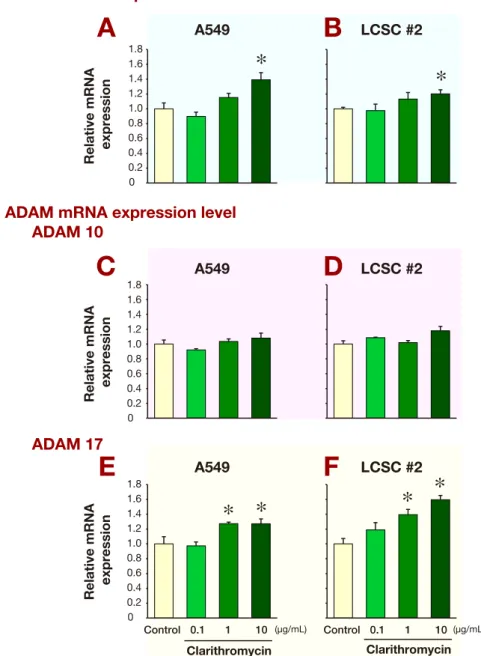
Fig. 1. The effect of clarithromycin on the mRNA expression of ULBP2, ADAM10 and ADAM17 in A549 and LCSC #2 cell lines.
A549 and LCSC #2 cells were treated with 0.1, 1 or 10 μg/mL clarithromycin for 24 h after serum starvation and harvested for total RNA extraction and quantitative real-time PCR. The ratios of gene expression between the target genes (ULBP2, ADAM10 and ADAM17) and internal standard (GAPDH) were expressed relative to those of A549 or LCSC #2 cells without clarithromycin treatment, which are set at 1.00. Data are expressed as mean values ± SD (n = 3 for each group). *P < 0.05 versus control. A: mRNA expression of ULBP2 in A549 cells. B: mRNA expression of ULBP2 in LCSC #2 cells. C: mRNA expression of ADAM10 in A549 cells. D: mRNA expression of ADAM10 in LCSC #2 cells. E: mRNA expression of ADAM17 in A549 cells. F: mRNA expression of ADAM17 in LCSC #2 cells. ADAM, a disintegrin and metalloproteinase domain; GAPDH, glyceraldehyde-3-phosphate dehydrogenase; ULBP2, UL16-binding pro-tein 2.
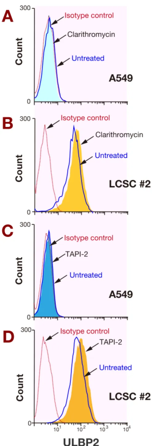
Fig. 2. The effect of clarithromycin on the cell surface expression
of ULBP2 in A549 and LCSC #2 cell lines. A549 and LCSC #2 cells were treated with or without 10 μg/mL clarithromycin or 50 μM TAPI-2 for 24 h after serum starvation and harvested for flow cytometry. A: A549 cells treated with or without 10 μg/mL clarithromycin. B: LCSC #2 cells treated with or without 10 μg/ mL clarithromycin. C: A549 cells treated with or without 50 μM 2. D: LCSC #2 cells treated with or without 50 μM TAPI-2. TAPI-2, TNF-alpha processing inhibitor-2; ULBP2, UL16-binding protein 2.
The ratios of gene expression between the target genes (ULBP2, ADAM10, and ADAM17) and the internal standard (GAPDH) were expressed relative to those of A549 or LCSC #2 without clarithromycin, which is set at 1.00.
Flow cytometry
Cells were incubated with monoclonal anti-ULBP2 antibodies or isotype controls. After washing, goat anti-mouse IgG labeled with FITC was added, and the fluorescence intensities of samples were analyzed on a FACSCalibur (BD Biosciences, La Jolla, CA). Data from 10,000 cells were collected, and the geometric mean fluorescence intensity (MFI) was calculated using CELLQuest software (BD Biosciences). MFI ratios were calculated by dividing the MFI obtained with a specific antibody by that for the isotype control.
ELISA
Two monoclonal anti-ULBP2 antibodies were used to detect sULBP2. Plates were coated with the anti-ULBP2 mAb BUMO1 at 2 mg/mL in coating buffer for 24 h at 4 ˚C, then blocked with blocking buffer for 18 h at 4 ˚C. The blocking buffer was prepared with a commercially available blocking agent (Applied Bioscience, Mumbai, India). Next, ULBP2-Fc (standard) and the samples were added, and the plates were incubated for 1 h at room temperature. For analysis of serum samples, ULBP2-Fc was diluted in blocking buffer with 25% horse serum, and sera were diluted 1:3 in blocking buffer before ad-dition to the plates. After incubation, the detector mAb (anti-ULBP2, clone 165903) was added at 0.5 mg/mL in blocking buffer for 1 h at room temperature, followed by incubation with anti-mouse IgG2a-HRP (1:10,000 in blocking buffer) for 1 h at 37 ˚C. Color was devel-oped with a Sumilon peroxidase ELISA kit (Sumitomo Chemical, Tokyo), and the absorbance was read at 490 nm on a Model 680 microplate reader (Bio-Rad Labora-tories, Hercules, CA).
TACE activity assay
The activity of ADAM17, which is also known as TACE, was determined using the SensoLyte 520 TACE Activity Assay Kit (AnaSpec, San Jose, CA) with 10 µg of cell lysate proteins from each sample according to the manufacturer’s protocol. This assay uses a 5-car-boxyfluorescein (5-FAM)-labeled fluorescence reso-nance energy transfer (FRET) peptide substrate. Upon cleavage of the FRET peptide by the active enzyme, the fluorescence of 5-FAM is recovered and can be moni-tored at excitation/emission wavelengths of 490/520 nm. Fluorescence of the cleavage product was measured in a fluorescence microplate reader (TECAN, Seestrasse, Switzerland). Results are expressed as the mean change in fluorescence intensity per min.
Statistical analysis
Statistical analyses were performed using the SPSS 21.0 software (International Business Machines Corporation,
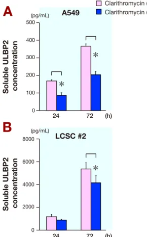
Fig. 3. The effect of clarithromycin on the level of sULBP2 in
A549 and LCSC #2 cell lines. Measurement of sULBP2 in culture supernatants using ELISA. A549 and LCSC #2 cells were treated with or without 10 μg/mL clarithromycin for 24 or 72 h after serum starvation. After treatment, the culture supernatants were harvested for ELISA. Data are expressed as mean values ± SD (n = 3 for each group). *P < 0.05. A: A549 cells treated with or with-out 10 μg/mL clarithromycin. B: LCSC #2 cells treated with or without 10 μg/mL clarithromycin. sULBP2, solubilized ULBP2. Armonk, NY) and P < 0.05 was regarded as statistically significant.
RESULTS
Effect of clarithromycin on ULBP2 mRNA expres-sion
To evaluate the influence of clarithromycin on ULBP2 and its shedding mechanism, we first assessed the ef-fect of clarithromycin on mRNA expression of ULBP2, ADAM10 and ADAM17. We used two cell lines, A549 and LCSC #2, which express ULBP2 at low and high levels, respectively. Transcript levels of ULBP2 were significantly up-regulated in A549 (Fig. 1A) and LCSC #2 (Fig. 1B) cells treated with 10 mg/mL clarithromycin for 24 h. Additionally, although there was no significant effect of clarithromycin on the mRNA expression of ADAM10 in A549 (Fig. 1C) or LCSC #2 (Fig. 1D) cells, ADAM17 mRNA expression was up-regulated in A549 (Fig. 1E) and LCSC #2 (Fig. 1F) cells treated with 1 or
10 mg/mL clarithromycin for 24 h. These data suggest that clarithromycin induces transcription of ULBP2 and ADAM17.
Effect of clarithromycin on cell surface and soluble ULBP2
Next, to identify the effect of clarithromycin on pro-tein expression of ULBP2, we evaluated the changes of cell surface ULBP2 expression in A549 and LCSC #2 cells after 24 h treatment with or without 10 mg/mL clarithromycin (Figs. 2A and B). Although there was no significant difference of cell surface ULBP2 expression in A549 cells with or without 10 mg/mL clarithromycin treatment (Fig. 2A, MFI ratio: 1.13 versus 1.11, respec-tively), the expression level of ULBP2 in LCSC #2 cells with 10 mg/mL clarithromycin was significantly higher than that without clarithromycin (Fig. 2B, MFI ratio: 22.6 versus 11.5, respectively). To test the contribution of ULBP2 shedding to this change, we next examined the effect of TAPI-2, an ADAM17 inhibitor, on the expres-sion of ULBP2. The same tendency was observed when LCSC #2 cells were treated with 50 mM TAPI-2 for 24 h (Figs. 2C and D). Therefore, clarithromycin may mim-ic the effect of TAPI-2, especially for cells with abun-dant expression of ULBP2 such as LCSC #2 cells. These data suggest that clarithromycin induces cell surface ULBP2 expression. In addition, the mechanism of this effect is presumably induction of ULBP2 gene transcrip-tion and inhibitranscrip-tion of ULBP2 shedding by ADAM17. Quantification of sULBP2
Previously, we showed that ULBP2 is shed into the cul-ture medium in proportion to the level of its cell surface expression by non-small cell lung cancer cell lines.19
Therefore, to reveal the effect of clarithromycin on sULBP2, we used ELISA to analyze culture superna-tants from A549 (Fig. 3A) and LCSC #2 cells (Fig. 3B) at 24 and 72 h after treatment with or without clarithro-mycin. The number of the cells did not differ significant-ly under each experimental condition (data not shown); however, the amount of sULBP2 increased over time in these cell lines. The amount of sULBP2 with clarithro-mycin was significantly decreased relative to that with-out clarithromycin in both A549 and LCSC #2 cells (Figs. 3A and B). These findings suggest an inhibitory effect of clarithromycin on the ULBP2 shedding mechanism. Evaluation of ADAM17 activity
To explore the effect of clarithromycin on the ULBP2 shedding mechanism, we next examined the effect of clarithromycin on the activity of ADAM17 because re-duction of sULBP2 was observed despite increased tran-scription of the shedder enzyme ADAM17. As shown in
Fig. 4, 10 mg/mL clarithromycin significantly inhibited the activity of ADAM17 in LCSC #2 cells with high cell surface ULBP2 expression. However, the effect of clarithromycin on A549 cells, which expressed lower levels of ULBP2, was not significant. This finding dem-onstrates that clarithromycin inhibits ADAM17 activity and, consequently, the ULBP2 shedding mechanism. DISCUSSION
In this study, we examined the effect of clarithromycin on ULBP2 expression. Our results showed that clarithro-mycin induced the transcription and expression of cell surface ULBP2 while reducing the amount of sULBP2 in the culture medium. Additionally, clarithromycin in-hibited the activity of ADAM17, a shedder enzyme for cell surface NKG2D ligands.21 Because immune effector
cells such as NKT cells, CD8+T cells and NK cells are activated by enhanced expression of ULBP2 on the sur-faces of target cells and are affected by only low levels of sULBP2, these pleiotropic effects of clarithromycin on ULBP2 expression might contribute to the enhanced function of immune effector cells, thereby augmenting P. aeruginosa clearance in the lung.
In this study, we first reported the up-regulation of ULBP2 and ADAM17 gene transcription in human cells derived from the lung. Previous studies reported that clarithromycin influences the gene expression of various proteins in human cells, including both reduction and in-duction of genes involved in inflammation. For example, studies have shown that clarithromycin inhibited gene
transcription of mucin,22 IL-8 23 and various cytokines
such as TNF-alpha, interferon gamma, interleukin (IL)-2, IL-4, IL-5 and IL-6.24 On the other hand, transcription
of IL-1025 and alpha1-acid glycoprotein was induced by
clarithromycin in hepatocytes.26 Toward
understand-ing the mechanism of gene transcription modulation by clarithromycin, a previous study reported that clarithro-mycin inhibits activator protein-1 transcription factor23
or potentiates glucocorticoid response element.26 The
precise mechanism leading to up-regulation of ULBP2 and ADAM17 is still unclear, but it may occur through the modulation of certain transcription factors. Because most previous reports focused on the effect of clarithro-mycin on the inflammatory cytokines, its effect on other proteins involved in the immune process and its mecha-nism of action must be explored in future studies.
In addition to its role in transcriptional regulation, we revealed that clarithromycin inhibits the enzymatic activity of ADAM17. The inhibitory effect of clar-ithromycin on enzyme activity has been reported in other enzymes such as cytochrome P450 3A,27 human
serum paraoxonase28 and glycosidase.29 This effect of
clarithromycin on ADAM17 activity represents a new mechanism that may explain the recent reports of broad pleiotropic activities of macrolide antibiotics for immu-nomodulatory,30 anti-inflammatory31 and antitumor32
ef-fects. Previous studies revealed that ADAM17, originally identified as an enzyme that proteolytically cleaves pro-TNF-alpha to generate soluble pro-TNF-alpha,33 cleaves and
solubilizes a wide range of membrane-bound proteins including transforming growth factor (TGF)-alpha34 and
heparin-binding epidermal growth factor-like growth factor (HB-EGF).35 The substrate proteins of ADAM17
make obvious contributions to various pathogeneses, for example, TNF-alpha is involved in inflammation, and both TGF-alpha and HB-EGF contribute to tumorigen-esis. Therefore, inhibition of the activity of ADAM17 by clarithromycin might represent a novel mechanism for its effect on these pathological conditions.
Recent reports suggested that P. aeruginosa infec-tion induced the surface expression of human NKG2D ligands such as ULBP2 in airway epithelial cells and signified the indispensability of induced NKG2D-mediated immune activation in the clearance of this bacterium.16, 18 In addition, we previously revealed that P.
aeruginosa is actively involved in this process by secret-ing a quorum senssecret-ing molecule, N-3-oxo-dodecanoyl homoserine lactone, that decreases cell surface ULBP2 and increases sULBP2.36 This intervention by P.
aerugi-nosa leads to attenuation of the host NKG2D-mediated immune response and is beneficial for its survival in the human lung. Our novel finding in this study that Fig. 4. The effect of clarithromycin on ADAM17 activity in A549
and LCSC #2 cell lines. ADAM17 activity was determined using the SensoLyte 520 TACE Activity Assay Kit with 10 µg of cell lysate proteins for each sample according to the manufacturer’s protocol. A549 and LCSC #2 cells were treated with or without 10 μg/mL clarithromycin for 24 h after serum starvation and harvested for the assay. Data are expressed as the mean change in fluorescence intensity per min ± SD (n = 3 for each group). *P < 0.05. ADAM, a disintegrin and metalloproteinase domain.
clarithromycin increases the cell surface ULBP2 expres-sion and decreases sULBP2 suggests a mechanism by which it counteracts the immunosuppressive activity of P. aeruginosa and might explain the effectiveness of clar-ithromycin in treating chronic P. aeruginosa infection in chronic lung diseases.37
In conclusion, we revealed that clarithromycin in-duces expression of ULBP2 and, through ADAM17, re-duces sULBP2. These findings suggest the possibilities that clarithromycin can activate NKT cells and decrease the colonization of bacteria in patients with chronic respiratory diseases such as diffuse panbronchiolitis. Therefore, further study toward augmenting the effect of clarithromycin on NKT cell activity through ULBP2 might be useful for developing new macrolide-based treatments for chronic respiratory diseases.
The authors declare no conflict of interest. REFERENCES
1 Culic O, Erakovic V, Parnham MJ. Anti-inflammatory effects of macrolide antibiotics. Eur J Pharmacol. 2001;429:209-29. PMID: 11698042.
2 Lopez-Boado YS, Rubin BK. Macrolides as immunomodula-tory medications for the therapy of chronic lung diseases. Curr Opin Pharmacol. 2008;8:286-91. PMID: 18339582.
3 Shinkai M, Henke MO, Rubin BK. Macrolide antibiotics as immunomodulatory medications: proposed mechanisms of action. Pharmacol Ther. 2008;117:393-405. PMID: 18289694. 4 Kudoh S, Azuma A, Yamamoto M, Izumi T, Ando M.
Im-provement of survival in patients with diffuse panbronchiolitis treated with low-dose erythromycin. Am J Respir Crit Care Med. 1998;157(6 Pt 1):1829-32. PMID: 9620913.
5 Clement A, Tamalet A, Leroux E, Ravilly S, Fauroux B, Jais JP. Long term effects of azithromycin in patients with cystic fibrosis: A double blind, placebo controlled trial. Thorax. 2006;61:895-902. PMID: 16809416.
6 Basyigit I, Yildiz F, Ozkara SK, Yildirim E, Boyaci H, Ilgazli A. The effect of clarithromycin on inflammatory markers in chronic obstructive pulmonary disease: preliminary data. Ann Pharmacother. 2004;38:1400-5. PMID: 15252191.
7 Homma H, Yamanaka A, Tanimoto S, Tamura M, Chijimatsu Y, Kira S, et al. Diffuse panbronchiolitis. A disease of the transitional zone of the lung. Chest. 1983;83:63-9. PMID: 6848335.
8 Emerson J, Rosenfeld M, McNamara S, Ramsey B, Gibson RL. Pseudomonas aeruginosa and other predictors of mortal-ity and morbidmortal-ity in young children with cystic fibrosis. Pedi-atr Pulmonol. 2002;34:91-100. PMID: 12112774.
9 Yanagihara K, Tomono K, Imamura Y, Kaneko Y, Kuroki M, Sawai T, et al. Effect of clarithromycin on chronic respiratory infection caused by Pseudomonas aeruginosa with biofilm formation in an experimental murine model. J Antimicrob Chemother. 2002;49:867-70. PMID: 12003986.
10 Nieuwenhuis EE, Matsumoto T, Exley M, Schleipman RA, Glickman J, Bailey DT, et al. CD1d-dependent macrophage-mediated clearance of Pseudomonas aeruginosa from lung. Nat Med. 2002;8:588-93. PMID: 12042809.
11 Tupin E, Kinjo Y, Kronenberg M. The unique role of natural killer T cells in the response to microorganisms. Nat Rev
Mi-crobiol. 2007;5:405-17. PMID: 17487145.
12 Matsuda JL, Mallevaey T, Scott-Browne J, Gapin L. CD1d-restricted iNKT cells, the ‘Swiss-Army knife’ of the im-mune system. Curr Opin Immunol. 2008;20:358-68. PMID: 18501573.
13 Silk JD, Salio M, Brown J, Jones EY, Cerundolo V. Structural and functional aspects of lipid binding by CD1 molecules. Annu Rev Cell Dev Biol. 2008;24:369-95. PMID: 18593354. 14 Rzemieniak SE, Hirschfeld AF, Victor RE, Chilvers MA,
Zheng D, van den Elzen P, et al. Acidification-dependent acti-vation of CD1d-restricted natural killer T cells is intact in cys-tic fibrosis. Immunology. 2010;130:288-95. PMID: 20102408. 15 Gonzalez S, Lopez-Soto A, Suarez-Alvarez B, Lopez-Vazquez
A, Lopez-Larrea C. NKG2D ligands: key targets of the im-mune response. Trends Immunol. 2008;29:397-403. PMID: 18602338.
16 Borchers MT, Harris NL, Wesselkamper SC, Zhang S, Chen Y, Young L, et al. The NKG2D-activating receptor mediates pulmonary clearance of Pseudomonas aeruginosa. Infect Im-mun. 2006;74:2578-86. PMID: 16622193.
17 Bauer S, Groh V, Wu J, Steinle A, Phillips JH, Lanier LL, et al. Activation of NK cells and T cells by NKG2D, a receptor for stress-inducible MICA. Science. 1999;285:727-9. PMID: 10426993.
18 Wesselkamper SC, Eppert BL, Motz GT, Lau GW, Hassett DJ, Borchers MT. NKG2D is critical for NK cell activation in host defense against Pseudomonas aeruginosa respiratory infection. J Immunol. 2008;181:5481-9. PMID: 18832705. 19 Yamaguchi K, Chikumi H, Shimizu A, Takata M, Kinoshita
N, Hashimoto K, et al. Diagnostic and prognostic impact of serum-soluble UL16-binding protein 2 in lung cancer patients. Cancer Sci. 2012;103:1405-13. PMID: 22587355.
20 Wolpert F, Tritschler I, Steinle A, Weller M, Eisele G. A dis-integrin and metalloproteinases 10 and 17 modulate the im-munogenicity of glioblastoma-initiating cells. Neuro Oncol. 2014;16:382-91. PMID: 24327582.
21 Waldhauer I, Goehlsdorf D, Gieseke F, Weinschenk T, Wittenbrink M, Ludwig A, et al. Tumor-associated MICA is shed by ADAM proteases. Cancer Res. 2008;68:6368-76. PMID: 18676862.
22 Kaneko Y, Yanagihara K, Seki M, Kuroki M, Miyazaki Y, Hirakata Y, et al. Clarithromycin inhibits overproduction of muc5ac core protein in murine model of diffuse panbronchi-olitis. Am J Physiol Lung Cell Mol Physiol. 2003;285:L847-53. PMID: 12818892.
23. Abe S, Nakamura H, Inoue S, Takeda H, Saito H, Kato S, et al. Interleukin-8 gene repression by clarithromycin is medi-ated by the activator protein-1 binding site in human bronchial epithelial cells. Am J Respir Cell Mol Biol. 2000;22:51-60. PMID: 10615065.
24 Morikawa K, Zhang J, Nonaka M, Morikawa S. Modula-tory effect of macrolide antibiotics on the Th1- and Th2-type cytokine production. Int J Antimicrob Agents. 2002;19:53-9. PMID: 11814768.
25 Umezawa M, Tanaka N, Takeda K, Ihara T, Sugamata M. Clarithromycin and telithromycin increases interleu-kin-10 expression in the rat endometriosis model. Cytokine. 2011;55:339-42. PMID: 21665488.
26 Komori T, Kai H, Shimoishi K, Kabu K, Nonaka A, Maruyama T, et al. Up-regulation by clarithromycin of alpha(1)-acid glycoprotein expression in liver and primary cultured hepatocytes. Biochem Pharmacol. 2001;62:1391-7. PMID: 11709199.
27 Quinney SK, Malireddy SR, Vuppalanchi R, Hamman MA, Chalasani N, Gorski JC, et al. Rate of onset of inhibition of gut-wall and hepatic CYP3A by clarithromycin. Eur J Clin Pharmacol. 2013;69:439-48. PMID: 22777148.
28 Sinan S, Kockar F, Gencer N, Yildirim H, Arslan O. Am-phenicol and macrolide derived antibiotics inhibit paraox-onase enzyme activity in human serum and human hepatoma cells (HepG2) in vitro. Biochemistry (Mosc). 2006;71:46-50. PMID: 16457617.
29 Sadeghi-Khomami A, Lumsden MD, Jakeman DL. Glycosi-dase inhibition by macrolide antibiotics elucidated by STD-NMR spectroscopy. Chem Biol. 2008;15:739-49. PMID: 18635010.
30 Giamarellos-Bourboulis EJ. Macrolides beyond the conven-tional antimicrobials: a class of potent immunomodulators. Int J Antimicrob Agents. 2008;31:12-20. PMID: 17935949. 31 Zalewska-Kaszubska J, Gorska D. Anti-inflammatory
capa-bilities of macrolides. Pharmacol Res. 2001;44:451-4. PMID: 11735349.
32 Wang L, Kitaichi K, Hui CS, Takagi K, Sakai M, Yokogawa K, et al. Reversal of anticancer drug resistance by macrolide antibiotics in vitro and in vivo. Clin Exp Pharmacol Physiol. 2000;27:587-93. PMID: 10901387.
33 McGeehan GM, Becherer JD, Bast RC Jr, Boyer CM, Champion B, Connolly KM, et al. Regulation of tumour ne-crosis factor-alpha processing by a metalloproteinase inhibitor. Nature. 1994;370:558-61. PMID: 8052311.
34 Peschon JJ, Slack JL, Reddy P, Stocking KL, Sunnarborg SW, Lee DC, et al. An essential role for ectodomain shedding in mammalian development. Science. 1998;282:1281-4. PMID: 9812885.
35 Jackson LF, Qiu TH, Sunnarborg SW, Chang A, Zhang C, Patterson C, et al. Defective valvulogenesis in HB-EGF and TACE-null mice is associated with aberrant BMP signaling. EMBO J. 2003;22:2704-16. PMID: 12773386.
36 Matsunaga S, Chikumi H. Pseudomonas aeruginosa quorum-sensing molecule N-3-oxo-dodecanoyl homoserine lactone down-regulate cell surface ULBP2 on lung cancer cell lines. Yonago Igaku Zasshi. 2011;62:77-90. Japanese.
37 Tateda K, Ishii Y, Kimura S, Horikawa M, Miyairi S, Yamaguchi K. Suppression of Pseudomonas aeruginosa quorum-sensing systems by macrolides: a promising strategy or an oriental mystery? J Infect Chemother. 2007;13:357-67. PMID: 18095083.