Mallory Bodies in Hepatocytes of Alcoholic Liver Disease
and Primary Biliary Cirrhosis Contain
-(Carboxymethyl)lysine-Modified Cytokeratin,
but not those in Hepatic Carcinoma Cells
Masako Kato, Shinsuke Kato*, Seikoh Horiuchi†, Ryoji Nagai†, Yasushi Horie and Kazuhiko Hayashi‡
Pathology Division, Tottori University Hospital, Yonago 683-8504, *Department of Neuropathol-ogy, Institute of Neurological Sciences, Tottori University Faculty of Medicine, Yonago 683-8504, † Department of Medical Biochemistry, Graduate School of Medical and Pharmaceutical Sci-ences, Kumamoto University, Department of Biochemistry, Kumamoto University School of Med-icine, Kumamoto 860-0811 and ‡Division of Molecular Pathology, Department of Microbiology and Pathology, School of Medicine, Tottori University Faculty of Medicine, Yonago 683-8503 Japan
Mallory bodies (MBs) are intracytoplasmic bodies seen in hepatocytes of alcoholic liver disease, primary biliary cirrhosis and hepatocellular carcinoma. However, the mecha-nism of MB formation has not been fully understood. Proteins could be modified to ad-vanced glycation end products (AGEs) after long-term incubation with reducing sugar. AGEs are known to accumulate in several tissues in aging and age-enhanced disorders. To study the possible glycation process in the formation of MBs, hepatocytes of 80 human liver tissues with MBs were subjected to immunohistochemical analyses with five AGEs, two markers for oxidative stress proteins (OSPs) and four stress-response proteins (SRPs). MBs in hepatocytes of primary biliary cirrhosis and alcoholic liver disease were strongly positive for -(carboxymethyl)lysine (CML) and weakly positive for pyrraline. MBs in hepatocellular carcinomas were negative for both CML and pyrraline. No significant immunoreactivity was detected in MBs for other AGEs, such as -(carboxyethyl)lysine, pentosidine, and 3DG-imidazolone, or for OSPs and SRPs. Stainings for cytokeratin, a major protein component of MBs, and CML were co-localized. Furthermore, immu-noblot analysis suggested that cytokeratin of MBs was modified to AGE, since a single protein band detected by a monoclonal anti-CML had a molecular weight identical to cy-tokeratin. The absence of the CML signal in MBs of hepatocellular carcinoma cells could be explained by scarce content of cytokeratin in carcinoma MBs.
Key words: advanced glycation end product; cytokeratin; immunohistochemistry; Mallory
body; -(carboxymethyl)lysine
Abbreviations: ABC, avidin-biotin-immunoperoxidase complex; AEC, 3-amino-9-ethylcarbazole; AGE, advanced gly-cation end product; BSA, bovine serum albumin; CEL, -(carboxyethyl)lysine; CML, -(carboxymethyl)lysine; DAB, 3,3'-diaminobenzidine tetrahydrochloride; HCC, hepatocellular carcinoma; H&E staining, hematoxylin and eosin stain-ing-; HNE, 4-hydroxy-2-nonenal; MB, Mallory body; PAS, periodic acid Schiff; PBC, primary biliary cirrhosis; SDS, sodium dodecyl sulfate; SRP, stress-response protein
Mallory first described cytoplasmic hyaline de-generation in hepatocytes of alcoholic liver cir-rhosis (Mallory, 1911). This cytoplasmic hyaline inclusion has been called Mallory body (MB). MB had been thought as specific to alcoholic liver disease (Edmondson, 1986). However, they are also associated with a number of non-alcoholic hepatobiliary diseases, such as Indian childhood cirrhosis, primary biliary cirrhosis (PBC), Wil-son’s disease, hepatocellular carcinoma (HCC) and adenomatous hyperplasia (Jensen and Gluud, 1994a; Terada et al., 1989). Although several different theories have been proposed for the formation of MBs (Jensen and Gluud, 1994b), the mechanism of MB formation as well as their developmental and pathological significance has remained unknown.
Ultrastructurally MBs consist of aggregates of filaments (Yokoo et al., 1972), and aberrant in-termediate filaments of cytokeratin polypeptides (Katsuma et al., 1987). Recently, hyperphos-phorylation of cytokeratin 8 and 18 (Stumptner et al., 2000) or ubiquitination of cytokeratin pro-teins was revealed in MB formation (Yuan et al., 1996). MBs were also known to be positive for B-crystallin immunohistochemically (Lowe et al., 1992). Namely, MBs contain protein components such as cytokeratin, ubiquitin or B-crystallin. Furthermore, the other chemical analyses dem-onstrated that MBs possess carbohydrates (Lyon and Christoffersen, 1971) and reducing sugar (Luisada-Opper et al., 1977) in addition to cyto-keratin as the major protein component.
Long-term incubation of proteins with glucose leads, through the formation of early products such as Schiff base and Amadori rearrangement products, to the formation of advanced glyca-tion end products (AGEs). Since monoclonal or polyclonal antibodies against these AGEs have been newly produced, immunological studies us-ing anti-AGE antibodies have demonstrated ac-cumulation of AGE-modified proteins in several human tissues in association with aging (Araki et al., 1992; Kimura et al., 1996) and several dis-orders such as diabetic complications (Makino et al., 1995), atherosclerosis (Kume et al., 1995) and
Alzheimer’s disease (Smith et al., 1994). AGE-modification is known to occur in many proteins, especially long-lived proteins, which makes them insoluble, thus enhancing the deposition of modi-fied proteins inside the cells or in the extracellular space (Makino et al., 1995; Giardino et al., 1996). Finally, AGE-modified proteins that are accumu-lated in the tissues exhibit direct toxic effects on cells biologically (Vlassara et al., 1994).
AGE-structures reported so far include fluo-rescent and cross-linking structures such as pen-tosidine (Sell and Monnier, 1990) and crossline (Nakamura et al., 1992), and nonfluorescent and non-cross-linked structures such as imidazolone (Niwa et al., 1997), -(carboxymethyl)lysine (CML) (Ahmed et al., 1986) and pyrraline (Hayase et al., 1989). The purpose of the present study was to investigate whether AGE-modification could play a role in MB formation. To do this end, human liver tissues with MBs were examined immunohistochemically using antibodies against CML, -(carboxyethyl)lysine (CEL), pyrraline, pentosidine and 3DG-imidazolone. Our results revealed that chemical modification of cytokera-tin by CML, a major antigenic AGE-structure, is involved in MB formation in hepatocytes in alco-holic liver disease and primary biliary cirrhosis in contrast to no involvement of CML in MBs in HCC.
Materials and Methods
Patients
Eight hundred liver tissues from autopsy, biopsy and surgical files kept in our Department and Di-vision were surveyed from 1973 to 2000, and MBs were found histologically in 80 cases (8 autopsy cases, 11 biopsy cases and 61 surgical cases). The 80 cases with MBs included three PBC cases (fe-males, aged 37 to 68 years), 17 cases of alcoholic liver disease (15 males and 2 females, aged 33 to 66 years) and 60 cases of HCC (52 males and 8 females, aged 29 to 80 years). Surgical and au-topsy specimens of normal liver tissues and other
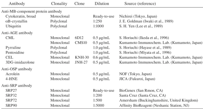
Table 1. Sources of primary antibodies and dilutions
Antibody Clonality Clone Dilution Source (reference) Anti-MB-component protein antibody
Cytokeratin, broad Monoclonal Ready-to-use Nichirei (Tokyo, Japan)
B-crystallin Polyclonal 1:250 J. E. Goldman (Iwaki et al., 1989) Ubiquitin Polyclonal 1:1000 S. H. Yen (Lee et al., 1989) Anti-AGE antibody
CML Monoclonal 6D12 0.5 μg/mL S. Horiuchi (Ikeda et al., 1996)
Monoclonal CMS10 0.5 μg/mL Kumamoto Immunochem. Lab. (Kumamoto, Japan) Pyrraline Polyclonal 1.0 μg/mL S. Horiuchi (Hayase et al., 1989)
Pentosidine Polyclonal 1.0 μg/mL S. Horiuchi (Miyata et al., 1996)
CEL Monoclonal KNH-30 0.6 μg/mL Kumamoto Immunochem. Lab. (Kumamoto, Japan) 3DG-imidazolone Monoclonal JNH-27 0.5 μg/mL Kumamoto Immunochem. Lab. (Kumamoto, Japan) Anti-OSP antibody
Acrolein Monoclonal 0.5 μg/mL NOF (Tokyo, Japan) 4-HNE Monoclonal 0.5 μg/mL JICA (Fukuroi, Japan) Anti-SRP antibody
SRP27 Monoclonal Ready-to-use BioGenex (San Rmon, CA) SRP32 Monoclonal 1:200 Santa Cruz (Santa Cruz, CA)
SRP72 Monoclonal 1:500 Amersham (Buckinghamshire, United Kingdom) SRP90 Monoclonal 1:5000 Affinity BioReagent (Neshanic Station, NJ)
AGE, advance glycation end product; CEL, -(carboxyethyl)lysine; CML, -(carboxymethyl)lysine; 4-HNE, 4-hydroxy-2-nonenal; MB, Mallory body; OSP, oxidative stress protein; SRP, stress response protein.
organ tissues from 10 individuals (7 males and 3 females; aged 19 to 68 years) were also examined as controls. The protocols were approved by the Ethics Committee in Tottori University Faculty of Medicine (No. 761).
Histology and immunohistochemistry
After fixation in 10% buffered formalin, the spec-imens were embedded in paraffin, cut into 4-μm-thick sections and examined by light microscopy. Liver sections were stained by the following routine methods: hematoxylin and eosin (H&E), periodic acid-Schiff (PAS), digestive PAS, silver and azan stainings. The distribution patterns of MBs were classified according to the method of Nakanuma and Ohta (1986). HCCs were graded into grades I, II and III according to the histologi-cal grading system of Edmondson and Steiner (1954). Serial sections were used for immuno-histochemical analysis. The sources of primary antibodies and their dilutions used are listed in Table 1. Sections were deparaffinized and
endog-enous peroxidase activity was quenched for 30 min with 0.3% H2O2. Sections were then washed
in phosphate-buffered saline (PBS), pH 7.4. Nor-mal sera isologous with each secondary antibody were used as blocking reagents. Sections were incubated with one of the primary antibodies or PBS for 18 h at 4°C. As positive controls for CML-immunostaining, atherosclerotic lesions in the wall of aortae from control individuals were used, biopsy specimens of diabetic kidney for pyrraline or pentosidine staining and atheroscle-rotic tissues also served as positive controls for acrolein and HNE stainings. Some sections were incubated with anti-CML antibody that had been preabsorbed with 1 mg/mL of CML-modified bo-vine serum albumin (BSA). With respect to the preabsorption test, some sections were incubated with anti-CML, anti-pyrraline, or anti-pentosidine antibody that had been preabsorbed with excess CML-, pyrraline- or pentosidine-modified BSA, respectively. Bound antibodies were visualized by the avidin-biotin-immunoperoxidase complex (ABC) method using the appropriate Vectastain
Table 2. Clinicopathological findings of 20 cas-es of PBC and ALD with MBs
Case Age Sex Clinical Histopathology
number diagnosis
1 37 F PBC LC/biliary
2 61 F PBC LC/biliary
3 68 F PBC LC/biliary
4 56 M ALD Fatty liver
5 44 M ALD AH 6 54 M ALD AH 7 47 M ALD AH 8 61 M ALD AH 9 37 M ALD Fibrosis 10 47 M ALD Fibrosis 11 49 F ALD Fibrosis 12 57 M ALD Fibrosis 13 65 M ALD Fibrosis 14 66 M ALD Fibrosis 15 37 F ALD LC/septal 16 56 M ALD LC/septal 17 59 M ALD LC/septal 18 70 M ALD LC/septal 19 33 M ALD LC/septal 20 61 M ALD LC/septal
AH, alcoholic hepatitis; ALD, alcoholic liver disease; F, fe-male; LC/biliary, liver cirrhosis of biliary type (by Havana classification) (Sherlock, 1956); LC/septal, liver cirrhosis of septal type; M, male; MB, Mallory body; PBC, primary bili-ary cirrhosis.
ABC kit (Vector Laboratories, Burlingame, CA) and 3,3'-diaminobenzidine tetrahydrochloride (DAB) (Dako, Glostrup, Denmark) as the final chromogen. For labeling multiple antigens in the same tissue section, normal and abnormal structures in H&E-stained sections were identi-fied, mapped and photomicrographed. The H&E sections were then decolorized in 70% ethanol containing 1% HCl, after which the sections were rehydrated, quenched for 30 min with 0.3% H2O2,
rinsed in PBS and incubated with the first prima-ry antibody for 18 h at 4°C. Bound antibody was visualized by the ABC method using 3-amino-9-ethylcarbazole (AEC; Vector Laboratories) as the chromogen, yielding a red product. After pho-tographing the immunoreactive structures, AEC sections were decolorized using 100% ethanol, rinsed twice in PBS and incubated for 60 min at room temperature with glycine-HCl buffer (pH 2.2) to elute the immunoreactive products. The completeness of the elution process was veri-fied by obtaining a negative reaction after reap-plication of the appropriate ABC kit including a secondary antibody and AEC on the eluted sec-tions. The sections were subsequently incubated with the second primary antibody for 18 h at 4° C, and immunoreactivity was visualized by the ABC method using DAB as the chromogen. The proportion of positively-immunostained MBs was classified into five different categories; category – means negative staining, category +/– a few MBs were stained weakly, category + less than 10%, category ++ 10–50% and category +++ more than 50%.
Immunoblot analysis
This analysis was carried out on one fresh liver sample of a patient with PBC (Patient 2, a 61-year-old female) and on a liver tissue of age-matched normal individual (a 68-year-old female). In brief, specimens were homogenized in Laemmli sample buffer (Bio-Rad, Hercules, CA) containing 2% sodium dodecyl sulfate (SDS), 25% glycerol, 10% 2-mercaptoethanol, 0.01% bromophenol blue and 62.5 mM Tris-HCl, pH 6.8. The sample was
heated at 100°C for 5 min. Soluble protein ex-tracts from the samples were separated on a SDS-polyacrylamide gel (10%–20% gradient, Bio-Rad) and transferred by electroblotting onto Immobilon PVDF (Millipore, Bedford, MA). After blocking with 5% skimmed milk for 30 min at room tem-perature, the blots were incubated with anti-CML antibody (6D12) overnight at 4°C and visualized with the Vectastain ABC kit and DAB. Appro-priate molecular weight markers (Bio-Rad) were included in each run.
Results
Histology and immunohistochemistry
Three cases clinically diagnosed as PBC showed histopathologically liver cirrhosis, biliary (Table 2). The histopathological findings of 17 cases
clini-Fig. 1. Hematoxylin and eosin staining of liver tissue of Patient 2 with primary biliary cirrhosis (PBC) (A) and the patient with hepatocellular carcinoma (B), showing Mallory bodies (MBs) (arrows). Bar = 100 μm.
cally diagnosed as alcoholic liver disease, showed fatty liver (1 case), alcoholic hepatitis (4 cases), liver fibrosis (6 cases) and liver cirrhosis (sep-tal type, 6 cases) by routine stainings (Table 2). Sixty cases of HCCs were classified as the grade I (15 cases), the grade II (41 cases) and the grade III (4 cases). MBs were found predominantly in peripheral areas of regenerative nodules of liver cirrhosis in PBC (Fig. 1A) and alcoholic liver disease, and also in periportal areas of fibrotic al-coholic liver disease. MBs were scattered in fatty liver and in alcoholic hepatitis. MBs in HCCs were found in the cancer tissues (Fig. 1B). The ratio of MB-bearing hepatocytes to total hepato-cytes varied from a few to ~10 percent in liver fi-brosis, liver cirrhosis and HCCs. The distribution patterns of MBs of PBC were classified as diffuse type (2 cases) and sparse type (1 case). The pat-terns of MBs of alcoholic liver disease were clas-sified as diffuse type (2 cases) and as sparse type (15 cases). The patterns of MBs of HCCs were as clustering type (21 cases), diffuse type (26 cases) and sparse type (13 cases) (Table 3).
The shape of MBs in PBC and alcoholic liver disease showed granular (Fig. 2A), staghorn, or circlar pattern (Fig. 2D). In contrast, the shape of MBs in HCCs was irregular (Fig. 1B). MBs of alcoholic liver disease, PBC and HCCs were
Table 3. Histopathological and immunohistochemical findings of 80 cases of PBC, ALD and HCC with MBs
Distribution Number CML CML Pyrraline Pentosidine CEL
Cyto-pattern of cases (6D12) (CMS10) keratin
PBC Clustering 0
Diffuse 2 +~+++ +~+++ +/–~– – – ++
Sparse 1 + + – – – ++
Alcoholic liver disease Clustering 0
Diffuse 2 +~++ +~++ +/– – – ++
Sparse 15 + + +/–~– – – ++
Hepatocellular carcinoma Clustering 21
Diffuse 26 – – – – – +/–
Sparse 13 – – – – – +/–
CEL, -(carboxyethyl)lysine; CML, -(carboxymethyl)lysine; MB, Mallory body; PBC, primary biliary cirrhosis. Distribution patterns of MBs were divided according to Nakanuma and Ohta (1986).
Proportion of MBs stained positively: +++, > 50%; ++, 10–50%; +, < 10%; +/–, only minor portion.
1A
weakly positive for PAS histochemically (data not shown). The PAS-positivity was confirmed by the digestive PAS staining. MBs in PBC and alcoholic liver disease were confirmed immuno-histochemically by positive staining to ubiquitin and B-crystallin addition to H&E staining. The reaction products within MBs in PBC by the anti-CML antibody exhibited a granular pattern (Fig. 2B) or a circular pattern (Fig. 2E). MBs and MB-bearing hepatic cytoplasm were also stained with the anti-cytokeratin antibody (Figs. 2C and F). The positive reaction products for both CML and
Fig. 2. Light microscopic characteristics of the Mallory bodies (MBs) of Patient 2 with primary biliary cirrhosis.
A: An MB in hematoxylin and eosin (H&E) staining (arrows). Bar = 10 μm (A–F: same magnification).
B: The same section as A immunostained for -(carboxymethyl)lysine (CML) using 3-amino-9-ethylcarbazole (AEC) as chromogen (arrowheads). C: The section used in B was decolorized and restained for cytokeratin using
3,3'-diaminobenzidine tetrahydrochloride (DAB) as chromogen (arrows). D: An MB in H&E staining (arrows).
E: The section used in D was decolorized and immunostained for CML using AEC as chromogen. A doughnut-shaped MB is stained in red (arrowheads). F: The section used in E was decolorized and restained for cytokeratin using
DAB as chromogen showing the MB (arrows).
Fig. 3. Liver sections of the pa-tient with hepatocellular carcino-ma. Immunostaining for ubiquitin (A) and CML (with monoclonal antibody 6D12) (B). Bar = 100 μm (A and B: same magnification).
cytokeratin were co-localized on the same MBs (Figs. 2B and C; and Figs. 2E and F, respectively). MBs derived from three cases of PBC and 17 cases of alcoholic liver disease were posi-tively stained to CML (both clone of 6D12 and CMS-10) (Table 3). MBs in one case of PBC and nine cases of alcoholic liver disease were weakly stained with anti-pyrraline antibody. MBs of all cases of both PBC and alcoholic liver disease showed positive for cytokeratin. MBs were not stained by anti-pentosidine antibody, anti-CEL antibody or anti-3DG-imidazolone antibody.
A
2
B
C
D
E
F
2
2
2
2
2
A
B
3
3
MBs were not stained with antibodies against oxidative stress proteins (OSPs) such as acrolein and HNE nor against stress response proteins (SRPs) (27 kDa, 32 kDa, 72 kDa or 90 kDa) (data not shown). MBs in HCCs were also identified by positive staining to anti-ubiquitin antibody (Fig. 3A). MBs in all cases of HCCs were weakly stained by anti-cytokeratin antibody. By contrast, MBs in HCCs were not stained to CML (Fig. 3B), pyrraline, pentosidine, CEL, 3DG-imidazolone, two types of OSPs (acrolein and HNE) nor four types of SRPs (srp27, 32, 72 and 90). There were no significant differences in AGE-expressions among the histologically different specimens in HCCs.
The proportion of positively-immunostained MBs varied from one sample to another, rang-ing from less than 10% to more than 50% of total MBs (Table 3). Immunohistochemical stain-ings of CML of PBC and alcoholic liver disease showed that 1 out of 20 cases was of category +++; 4 out of 20 cases were of category ++, and 15 cases out of 20 cases were of category +. Im-munohistochemical stainings of pyrraline of PBC and alcoholic liver disease showed that one case of PBC was of category +/– and nine of 17 cases of alcoholic liver disease were of category +/–. MBs with diffuse distribution pattern were posi-tive for CML or pyrraline more than MBs with sparse type. All cases of PBC and alcoholic liver disease were of category ++ about cytokeratin. All cases of HCC were of category +/– about cy-tokeratin.
Hepatocytes from 10 control individuals were not stained by five anti-AGE antibodies against either of CML, pyrraline, pentosidine, CEL or 3DG-imidazolone. Similarly, normal he-patocytes showed no immunoreactivities to OSPs, SRPs, B-crystallin nor ubiquitin. No staining was detected when sections were incubated with PBS. The specificity and high affinity of these antibodies were confirmed by control tissues. As expected (Makino et al., 1995; Kume et al., 1995; Horie et al., 1997), CML-immunoreactivities were observed in smooth muscle cells of atherosclerotic
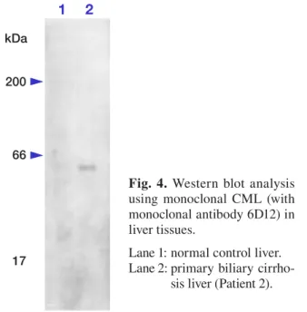
1 2
kDa 200
66
17
Fig. 4. Western blot analysis using monoclonal CML (with monoclonal antibody 6D12) in liver tissues.
Lane 1: normal control liver. Lane 2: primary biliary
cirrho-sis liver (Patient 2).
lesions, and immunoreactivities for pyrraline, pentosidine, CEL and 3DG-imidazolone were also noted in the thickening intimae of arteries. As re-ported earlier (Uchida et al., 1995), macrophage-derived foam cells in the atheromatous lesions were positive for HNE. Anti-CML antibody pre-treated with an excess amount of CML-modified BSA did not stain smooth muscle cells in the ath-erosclerotic lesions. Similarly, anti-pyrraline, or anti-pentosidine antibody pretreated with an ex-cess amount of pyrraline- or pentosidine-modified BSA did not stain the thickening intimae of arter-ies.
Immunoblot analysis
The results of immunoblot analyses are shown in Fig. 4. When the liver-tissue homogenate of PBC (Patient 2, a 61-year-old female), whose hepato-cytes were demonstrated to contain CML-positive MBs immunochemically, was subjected to immu-noblotting with anti-CML antibody, a single band with a molecular weight indistinguishable from that of cytokeratin was detected (Fig. 4). Immu-noblotting of the fresh autopsy liver specimen of a normal individual (a 68-year-old female) did not show any specific band (Fig. 4).
Discussion
Although the frequency and the distribution of MBs that were detected by H&E staining were various among 80 cases examined, all of the MBs were positive for ubiquitin, B-crystallin and cy-tokeratin immunohistochemically. MBs in HCCs were weakly positive for cytokeratin, but MBs in PBC and alcoholic liver disease were strongly positive for cytokeratin. The present immuno-histochemical results coincide with the previous reports which demonstrated that MBs consisted of aggregates of cytokeratin filaments (Yokoo et al., 1972) or that cytokeratin protein of MBs was ubiquitinated (Yuan et al., 1996). In the pres-ent study, the facts that MBs contain cytokeratin, ubiquitin and B-crystallin as protein components were confirmed immunohistochemically.
The novel finding of the present study is that MBs in PBC and alcoholic liver disease were positive for CML and pyrraline in contrast to those of HCCs. In cases of PBC and alcoholic liver disease, immunohistochemical results of the same paraffin sections using anti-CML and anti-cytokeratin antibodies, revealed that both CML and cytokeratin were co-localized on the same MBs. These results suggest that MBs in PBC and alcoholic liver disease have epitopes of CML addition to cytokeratin. No CML-positive reactivities except MBs were found in the liver sections of PBC, alcoholic liver disease, HCCs and normal controls. Furthermore, immunoblot analysis with anti-CML antibody supported the immunohistochemical findings. Single band with a molecular weight about 55 kDa was detected in the liver-tissue homogenate of PBC. This re-sult demonstrated that liver tissue-homogenate of PBC contain CML-combined protein with molecular weight about 55 kDa. Considering the fact that the molecular weight of ubiquitin, one of protein components of MBs is about 8 kDa (Lee et al., 1989; Lowe and Mayer, 1990) and that of B-crystallin is also 22 kDa (Iwaki et al., 1989), it
was suggested that CML-combined protein might be cytokeratin.
Glycation is one of biochemical reactions and it occurs when proteins were incubated with re-ducing sugars. Finally, CML, CEL or pyrraline-combined proteins through several steps by glyca-tion form AGEs. Although oxidaglyca-tion is necessary for the formation of AGEs in vitro (Nagai et al., 1997), the intention level of oxidation, the nature of oxidative processes, and the period of oxidative stresses are of different in vivo. At the cellular level, living cells can induce a diverse group of SRPs in response to different types of biological stresses, including oxidative damage (Morimoto et al., 1990). Since OSPs and SRPs in MBs were not detected in the present immunohistochemical studies, the amounts of these compounds in MBs might be, if any, negligible, suggesting that the oxidative stress that generates these marker com-pounds does not contribute to the process of MB formation in vivo.
Modification by glycation occurs in many proteins in relation to the pathogenesis of dis-eases such as atherosclerosis (Kume et al., 1995), diabetic complications (Makino et al., 1995), Alzheimers’ disease (Smith et al., 1994). Fur-thermore, AGEs are common to be long-lived, insoluble molecules, readily deposited in cells that have a direct cytotoxic effect (Vlassara et al., 1994). Although several hypotheses of MBs have been discussed (Jensen and Gluud, 1994a, 1994b) the results of the present study clarified that AGE-modification of cytokeratin, a major protein com-ponent of MBs, plays an important role in the for-mation of MBs in hepatocytes in the PBC and the alcoholic liver disease. Taken together with ab-normal cytokeratin aggregation toxicity, it is con-ceivable that the AGE modification of cytokeratin in MBs could amplify the aggregation of cyto-keratin and that the formation of the AGEs could result in greater toxicity in hepatocytes-bearing MBs in patients with the PBC and alcoholic liver disease. Considering the facts that MBs of HCCs contain less amount of cytokeratin protein, HCC cells form MBs for a short disease duration of
tu-morigenesis or have abnormal biological metabo-lism, AGE-modification do not contribute to the formation of the MBs in HCC cells. To elucidate the differences between AGE-expressions of alco-holic liver disease or PBC and AGE-expressions of HCCs, a further complete understanding of the molecular mechanisms of MB formation in hepa-tocytes will be necessary. Our results suggest that the formation of AGEs might be cytotoxic to MB-containing hepatocytes in PBC and alcoholic liver disease in contrast to HCCs.
Acknowledgments: The authors express their apprecia-tion to Dr. James E. Goldman (Department of Pathol-ogy, Columbia University), for providing the antibody to
B-crystallin and to Dr. S.-H. Yen (Department of Neuro-science, Mayo Clinic), for donating the antibody to ubiqui-tin.
This study was supported in part by a Grant-in-Aid for Scientific Research (c) from the Ministry of Education, Culture, Sports, Science and Technology of Japan (SK: 17500229), a Grant From Research on Psychiatric and Neu-rological Disease and Mental Health (SK) and a Research Grant on Measures for Intractable Diseases from the Minis-try of Health, Labour and Welfare of Japan (SK).
References
1 Ahmed MU, Thorpe SR, Baynes JW. Identification of -carboxymethyllysine as a degradation product of fructoselysine in glycated protein. J Biol Chem 1986;261:4889–4894.
2 Araki N, Ueno N, Chakrabarti B, Morino Y, Horiuchi S. Immunochemical evidence for the presence of ad-vanced glycation end products in human lens proteins and its positive correlation with aging. J Biol Chem 1992;267:10211–10214.
3 Edmondson HA. Alcoholic liver disease. In: Peters RL, Craig JR, eds. Liver pathology. New York: Churchill Livingstone; 1986. p. 255–283.
4 Edmondson HA, Steiner PE. Primary carcinoma of the liver. A study of 100 cases among 48900 nec-ropsies. Cancer 1954;7:462–503.
5 Giardino I, Edelstein D, Brownlee M. BCL-2 ex-pression or antioxidants prevent hyperglycemia-induced formation of intracellular advanced glyca-tion endproducts in bovine endothelial cells. J Clin Invest 1996;97:1422–1428.
6 Hayase F, Nagaraj RH, Miyata S, Njoroge FG, Monnier VM. Aging of proteins: immunologi-cal detection of a glucose-derived pyrrole formed
during Maillard reaction in vivo. J Biol Chem 1989;263:3758–3764.
7 Horie K, Miyata T, Maeda K, Miyata S, Sugiyama S, Sakai H, et al. Immunohistochemical colocaliza-tion of glycoxidacolocaliza-tion products and lipid peroxidacolocaliza-tion products in diabetic renal glomerular lesions. Impli-cation for glycoxidative stress in the pathogenesis of diabetic nephropathy. J Clin Invest 1997;100:2995– 3004.
8 Ikeda K, Higashi T, Sano H, Jinnouchi Y, Yoshida M, Araki N, et al. -(Carboxymethyl)lysine protein adduct is a major immunological epitope in proteins modified with advanced glycation end products of the Maillard reaction. Biochemistry 1996;35:8075– 8083.
9 Iwaki T, Kume-Iwaki A, Liem RKH, Goldman JE. alphaB-crystallin is expressed in non-lenticular tis-sues and accumulates in Alexander’s disease brain. Cell 1989;57:71–78.
10 Jensen K, Gluud C. The Mallory body: morpho-logical, clinical and experimental studies (part 1 of a literature survey). Hepatology 1994a;20:1061–1077. 11 Jensen K, Gluud C. The Mallory body: theories on
developmental and pathological significance (part 2 of a literature survey). Hepatology 1994b;20:1330– 1342.
12 Katsuma Y, Swierenga SHH, Khettry U, Marceau N, French SW. Changes in the cytokeratin intermediate filament cytoskeleton associated with Mallory body formation in mouse and human liver. Hepatology 1987;7:1215–1223.
13 Kimura T, Takamatsu J, Ikeda K, Kondo A, Miyakawa T, Horiuchi S. Accumulation of advanced glycation end products of the Maillard reaction with age in human hippocampal neurons. Neurosci Lett 1996;208:53–56. 14 Kume S, Takeya M, Mori T, Araki N, Suzuki H,
Horiuchi S, et al. Immunohistochemical and ultra-structural detection of advanced glycation end prod-ucts in atherosclerotic lesions of human aorta with a novel specific monoclonal antibody. Am J Pathol 1995;147:654–667.
15 Lee S, Park YD, Yen SH, Ksiezak-Reding H, Goldman JE, Dickson DW. A study of infantile motor neuron disease with neurofilament and ubiquitin immunocyto-chemistry. Neuropediatrics 1989;20:107–111.
16 Lowe J, Errington DR, Lennox G, Pike I, Spendlove I, Landon M, Mayer RJ. Balooned neurons in several neurodegenerative diseases and contain alphaB crys-tallin. Neuropathol Appl Neurobiol 1992;18:341–350. 17 Lowe J, Mayer RJ. Ubiquitin, cell stress and diseas-es of the nervous system. Neuropathol Appl Neuro-biol 1990;16:281–291.
18 Luisada-Opper AV, Kanagasundaram N, Leevy CM. Chemical nature of alcoholic hyalin. Gastroenterol-ogy 1977;73:1374–1376.
Mallory bodies. Acta Path Microbiol Scand 1971;79 (Section A):649–657.
20 Mallory FB. Cirrhosis of the liver. Five different types of lesions from which it may arise. Bull Johns Hopkins Hosp 1911;22:69–75.
21 Makino H, Shikata K, Hironaka K, Kushiro M, Yamasaki Y, Sugimoto H, et al. Ultrastructure of non-enzymatically glycated mesangial matrix in dia-betic nephropathy. Kidney Int 1995;48:517–526. 22 Miyata T, Taneda S, Kawai R, Ueda Y, Horiuchi S,
Hara M, et al. Identification of pentosidine as a na-tive structure for advanced glycation end products in 2-microglubulin-containing amyloid fibrils in
patients with dialysis-related amyloidosis. Proc Natl Acad Sci USA 1996;93:2353–2358.
23 Morimoto RI, Tissie’res A, Georgopoulos C. The stress response, function of the proteins, and perspec-tives. In: Morimoto RI, Tissie’res A, Georgopoulos C, eds. Stress Proteins in Biology and Medicine. Cold Spring Harbor: Cold Spring Harbor Laboratory Press; 1990. p. 1–36.
24 Nagai R, Ikeda K, Higashi T, Sano H, Jinnouchi Y, Araki N, et al. Hydroxyl radical mediates -(carboxymethyl)lysine formation from Ama-dori product. Biochem Biophys Res Commun 1997;234:167–172.
25 Nakamura K, Hasegawa T, Fukunaga Y, Ienaga K. Cross-lines A and B as candidates for the fluo-rophores in age- and diabetes-related cross-linked proteins, and their diacetates produced by Maillard reaction of -N-acetyl-L-lysine with D-glucose. J
Chem Soc Chem Commun 1992;14:992–994. 26 Nakanuma Y, Ohta G. Expression of Mallory bodies
in hepatocellular carcinoma in man and its signifi-cance. Cancer 1986;57:81–86.
27 Niwa T, Katsuzaki T, Ishizaki Y, Hayase F, Miyazaki T, Uematsu T, et al. Imidazolone, a novel advanced glycation end product, is present at high levels in kidneys of rats with streptozotocin-induced diabetes. FEBS Lett 1997;407:297–302.
28 Sell DR, Monnier VM. End-stage renal disease and
diabetes catalyze the formation of a pentose-derived crosslink from aging human collagen. J Clin Invest 1990;85:380–384.
29 Sherlock S. Report of the board for classifica-tion and nomenclature of cirrhosis of the liver. 5th Pan-American Congress of Gastroenterology. La Havana, Cuba. Gastroenterology 1956;31:213–216. 30 Smith MA, Taneda S, Richey PL, Miyata S, Yan
S-D, Stern D, et al. Advanced Maillard reaction end products are associated with Alzheimer disease pathology. Proc Natl Acad Sci USA 1994;91:5710– 5714.
31 Stumptner C, Omary MB, Fickert P, Denk H, Zatloukal K. Hepatocyte cytokeratins are hyper-phosphorylated at multiple sites in human alcoholic hepatitis and in a mallory body mouse model. Am J Pathol 2000;156:77–90.
32 Terada T, Hoso M, Nakanuma Y. Mallory body clustering in adenomatous hyperplasia in human cirrhotic livers: report of four cases. Hum Pathol 1989;20:886–890.
33 Uchida K, Itakura K, Kawakishi S, Hiai H, Toyokuni S, Stadtman ER. Characterization of epitopes rec-ognized by 4-hydroxy-2-nonenal specific antibodies. Arch Biochem Biophys 1995;324:241–248.
34 Vlassara H, Bucala R, Striker L. Biology of dis-ease. Pathogenic effects of advanced glycosylation: biochemical, biologic, and clinical implications for diabetes and aging. Lab Invest 1994;70:138–151. 35 Yokoo H, Minick OT, Batti F, Geoffrey K.
Mor-phologic variants of alcoholic hyalin. Am J Pathol 1972;69:25–40.
36 Yuan QX, Marceau N, French BA, Fu P, French SW. Mallory body induction in drug-primed mouse liver. Hepatology 1996;24:603–612.
Received September 4, 2006; accepted 10 November, 2006 Corresponding author: Masako Kato, MD