Abbreviation: CT, computed tomography
Retroperitoneal Laparoscopic Radical Nephrectomy for Renal Cell
Carcinoma: A Report on 2 Initial Cases
Tadahiro Isoyama, Takehiro Sejima, Hiroyuki Kadowaki, Shinji Hirakawa and Ikuo Miyagawa
Department of Urology, Tottori University Faculty of Medicine, Yonago 683-8504 Japan
We report our experience with retroperitoneal laparoscopic radical nephrectomy in 2 patients with renal cell carcinoma. In this procedure, a working space in the retroperi-toneum is created using the blunt balloon dissection technique. Carbon dioxide insuffla-tion is performed, and 4 trocars are inserted into the retroperitoneal cavity through the lateral abdominal wall. The kidney is removed together with the perirenal fat and Gerota’s fascia in a muscle-splitting fashion. Using this procedure, a right nephrectomy was performed in a 65-year-old man with a 2.4-cm tumor and in a 54-year-old woman with a 3.5-cm tumor. Operative time was 220 min and 195 min, respectively, and estimated blood loss was 10 mL and 115 mL, respectively. There were no major perioperative complications. Although a long-term follow-up is necessary to evaluate the efficacy of this procedure, it will probably become a standard treatment modality for localized renal cell carcinoma.
Key words: laparoscopy; nephrectomy; renal cell carcinoma
Recently, an increasing number of renal tumors have been detected incidentally in lower stages of growth due to the widespread use of ultrasonography and computed tomography (CT). Radical nephrectomy had previously been the standard treatment modality for localized renal cell carcinoma. In recent years though, laparoscopic nephrectomy has gained popularity in treating both benign and malig-nant renal diseases, and is usually performed via the transperitoneal or retroperitoneal approach.
We report on our experience using retro-peritoneal laparoscopic radical nephrectomy in 2 patients with renal cell carcinoma.
Patient Reports Case 1
A 65-year-old man with a left ureteral stone arrived at our hospital on May 11, 2001. He
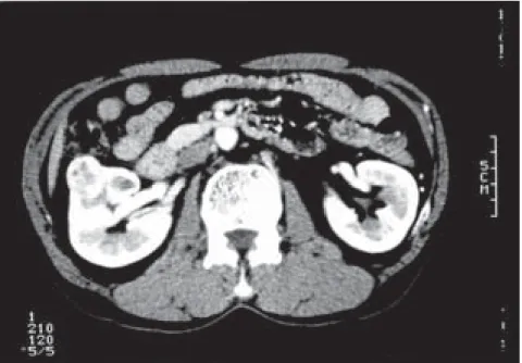
underwent shock wave lithotripsy on May 14 and June 4. During shock wave lithotripsy, a mass 2.4 cm in diameter was found incidentally in the right kidney by ultrasonography. CT revealed a tumor measuring 2.4 cm by 2.1 cm in the midportion of the right kidney, with no evi-dence of renal hilar lymphadenopathy or hepat-ic metastasis (Fig. 1). Chest CT and bone scin-tigraphy disproved the probability of metastatic disease. The patient was administered a laxa-tive the day before surgery, and an enema the morning of the surgery. Compression stockings were applied to both legs and upper thighs.
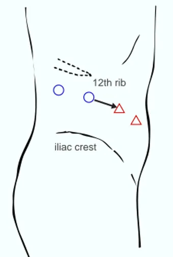
On August 24, under satisfactory general anesthesia combined with epidural anesthesia, a nasogastric tube and an urethral catheter were inserted. The patient was placed in the flank position, with the kidney bridge elevated, and the operating table was bent to widen the space between the 12th rib and the iliac crest. A 1.5-cm incision was made just below the tip of the 12th rib for the 1st trocar port. The flank muscle
Fig. 1. Early-phase enhanced computed tomogram demonstrating a renal tumor in the
right kidney (case 1).
fibers were bluntly separated. The thoracolum-bar fascia were gently pierced with a fingertip. A space for subsequent balloon dilator place-ment was created by gentle finger dissection of the retroperitoneum, anterior to the psoas mus-cle and posterior to Gerota’s fascia. A working space in the retroperitoneum was created by a trocar-mounted balloon device (PDB; United States Surgical Inc., Norwalk, CT). The balloon was inflated with approximately 800 mL of air. The dilation process was monitored laparo-scopically through the transparent balloon. Fol-lowing the balloon deflation and removal, a sec-ond 12-mm trocar was placed 7 cm dorsal to the 1st trocar port under bimanual guidance. A 12-mm blunt tip trocar was placed at the 1st trocar port. Carbon dioxide insufflation was perform-ed at a pressure of 8 mmHg to maintain the working space in the retroperitoneal cavity, and a 10-mm, 0-degree laparoscope was inserted via the 1st trocar. The peritoneum was dissect-ed off of the anterior abdominal wall to enable 2 additional trocars to be placed. The 3rd and fourth 5-mm trocars were placed under laparo-scopic vision (Fig. 2). Dissection and coagula-tion during the laparoscopic procedure were per-formed using the ultrasonically activated
scal-pel (Harmonic Scalscal-pel; Ethicon Inc., Cincinnati, OH) via the 3rd trocar. At first, the lateroconal fascia was incised along the quadratus lumbo-rum muscle, with exposure of the posterior lam-ina of Gerota’s fascia. The kidney was retract-ed anterolaterally by the surgical assistant, who used a grasping forceps via the 4th trocar. The leading edge of the psoas muscle was traced cephalad and renal hilar pulsation could be seen. The renal artery was identified and dis-sected, secured with five 10.5-mm laparoscopic clips (Ligaclip ERCA; Ethicon Inc.), and tran-sected with scissors, while 3 clips were left on the aorta side. The inferior vena cava and the renal vein were then identified and dissected. Its adrenal and gonadal branches were trans-ected with the Harmonic Scalpel to prevent them from being damaged when the renal vein was dissected. The renal vein was mobilized, stapl-ed and dividstapl-ed with a 35-mm laparoscopic sta-pler (Endocutter; Ethicon Inc.). The posterior aspect of the kidney was dissected upwards, and the anterior aspect of the kidney surrounded by Gerota’s fascia was dissected free from the peri-toneum. An incision was made in Gerota’s fas-cia at the upper pole of the kidney, and the ipsi-lateral adrenal gland was spared. The lower
Fig. 3. Macroscopic appearance of the specimen (case 1).
pole of the kidney was freed, and the ureter isolated during the procedure was secured with four 8.4-mm clips and transected. The speci-men was entirely freed upon the completion of the circumferential mobilization. Insufflation was lowered to confirm proper hemostasis in the surgical bed. A 5-mm drainage tube was then inserted through the 4th trocar, and left in the surgical bed. The trocars were removed under laparoscopic vision.
The specimen was removed in a muscle-splitting fashion through an additional skin incision, which was made between the 1st and 3rd tro-car ports. All wounds were closed using a synthetic ab-sorbable suture. Operative time was 220 min, and esti-mated blood loss was 10 mL. The specimen weighed 275 g (Fig. 3).
Pathological examination of the kidney re-vealed clear cell-type, grade-2 and stage-pT1a renal cell carcinoma. Postoperatively, the pa-tient began oral intake on postoperative day 1. He required analgesia for 3 days. Although a subcutaneous emphysema occurred at the right flank abdominal wall, it disappeared on post-operative day 2. Since the patient wanted to receive adjuvant therapy, he was treated with a daily intramuscular administration of 3 × 10⁶
-IU/mL interferon-alpha for 2 weeks. He was discharged from the hospital on postoperative day 31.
Case 2
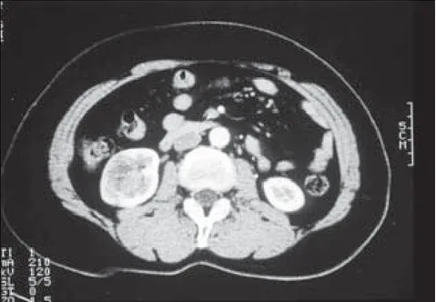
A 54-year-old woman visited our hospital with an asymptomatic gross hematuria on June 26, 2001. Urological examination including ultrasono-graphy and abdominal CT, revealed a tumor measuring 3.5 cm by 3.5 cm in the lower pole of the right kidney, and no evidence of renal hilar lymphadenopathy or hepatic metastasis (Fig. 4). Chest CT and bone scintigraphy showed no signs of metastatic disease. Preoperative prepa-ration was as described for case 1.
On August 24, retroperitoneal laparoscopic radical nephrectomy was performed under general and epidural anesthesia. Operative pro-cedure was almost the same as described for case 1. Two renal veins directly enter the right
Fig. 2. Trocar placement for retroperitoneal
laparo-scopic radical nephrectomy. , 12-mm trocar; , 5-mm trocar. The arrow indicates the line of skin inci-sion to remove the specimen.
12th rib
Fig. 5. Cut surface of the specimen (case 2).
lateral aspect of the inferior vena cava. A smaller vein was clipped and transected, and a bigger one was divided with the Endocutter.
Operative time was 195 min. Estimated blood loss was 115 mL. The specimen weighed 200 g (Fig. 5). Pathological examination of the kidney revealed chromophobe cell-type, grade-2 and stage-pT1a renal cell carcinoma. There were no postoperative
com-plications. The patient began oral intake on postoperative day 1. She required analgesia for 2 days. She was discharg-ed from the hospital on post-operative day 17, since she was anxious about an early hospital discharge despite our advice.
Discussion
Radical nephrectomy is the standard treatment for localized renal cell carcinoma, and was first described by Robson (1963). This procedure includes early ligation of the renal artery and vein before manipulating the renal tumor, en
bloc removal of the kidney and ipsilateral adre-nal gland together with the perinephric fatty tis-sue and Gerota’s fascia, and dissection of the lymph nodes.
Since the initial report of laparoscopic ne-phrectomy by Clayman et al. (1991), laparo-scopic nephrectomy has become accepted as a minimally invasive procedure for benign, and, more recently, for malignant renal disease.
The benefits for patients undergoing a laparoscopic nephrectomy rather than an open nephrectomy include a briefer postoperative course with reduced pain and analgesic require-ments, earlier oral intake and mobilization, a shorter hospital stay and convalescence, and better cosmetic results (Rassweiler et al., 1998; Abbou et al., 1999; Hemal et al., 1999; Fornara et al., 2001).
Laparoscopic radical nephrectomy is mostly done via the transperitoneal approach because of a larger working space and well-recognized anatomic landmarks. Gaur (1992) reported the creation of an adequate working space in the re-troperitoneum using the blunt balloon dissec-tion technique. This led to an increased interest in the retroperitoneal approach for laparoscopic nephrectomy. Since the kidney is a retroperito-neal organ, the retroperitoretroperito-neal approach pro-vides several potential advantages. In this ap-proach, the renal hilum is initially exposed, allowing for early control of the renal vessels. The risk of intraperitoneal organ injury is very low compared to the transperitoneal approach.
In laparoscopic radical nephrectomy, speci-men removal can be performed 2 different ways; intact removal performed by extending the incision at 1 of the port sites, or morcellation after the specimen has been placed in an entrap-ment sac. Intact removal of the specimen with or without an entrapment sac requires extension of the incision (Gill et al., 2000; Cicco et al., 2001). A larger incision, even in a longitudinal muscle-splitting fashion (Savage and Gill, 2001), may reduce the advantages of laparo-scopic surgery. Morcellation can be performed with a high-speed electrical tissue morcellator (Kavoussi et al., 1993) or mechanical clamp and forceps (Ono et al., 2001). However, this maneuver introduces the risk of seeding of the
tumor cell at the port sites, and dissemination into the working space. In addition, precise evaluation of the surgical margins, which is dependent on pathological examination of the intact specimen, may be difficult because of tis-sue morcellation.
In our department, simultaneous adrenalec-tomy during open radical nephrecadrenalec-tomy is not routinely performed, except for large, upper pole tumors (Kadowaki and Miyagawa, 2001). Paul et al. (2001) reported a total of 866 con-secutive patients who underwent nephrectomy and ipsilateral adrenalectomy with a solitary adrenal metastasis of only 6 (0.6%). They sug-gested that if the maximum tumor size mea-sured by CT is less than 8 cm, and when a stag-ing examination does not show organ or lymph node metastases, adrenalectomy is not neces-sary.
In this series, we did not perform regional lymph node dissection. Although regional lymph node dissection provides for more pre-cise staging of the disease, its therapeutic value remains controversial. Minervini et al. (2001) suggested that there is no clinical benefit in terms of overall outcome in undertaking region-al lymph node dissection in the absence of en-larged nodes detected before or during surgery. At present, our policy is to restrict retroperito-neal laparoscopic radical nephrectomy to tu-mors less than 7 cm, and neither regional lymph node dissection nor ipsilateral adrenalectomy is performed during the procedure.
There are few reports concerning the long-term disease-free outcome of laparoscopic radical nephrectomy (Cadeddu et al., 1998; Ono et al., 2001). Ono et al. (2001) evaluated the efficacy of laparoscopic radical nephrectomy. One-hundred and three patients who had renal tumors less than 5 cm in diameter underwent laparoscopic radical nephrectomy. During a median follow-up period of 29 months (range 3 to 95 months), the seeding of port sites did not occur in any of the patients. Metastatic disease developed in 3 patients, and local recurrence in 1 patient. The 5-year disease-free and patient survival rates were 95.1% and 95.0%, respec-tively. These rates were comparable to those in patients who underwent open surgery.
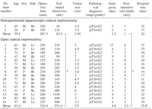
Outcome data were retrospectively com-pared between groups of patients treated bet-ween March 1998 and August 2001 at our de-partment clinic of Tottori University Hospital: the 2 initial patients undergoing laparoscopic radical nephrectomy versus 16 patients with lo-calized disease undergoing open radical ne-phrectomy (Table 1). Mean tumor size was 2.95 cm in the laparoscopic group and 3.50 cm in the open group (P = 0.52). Outcome analysis revealed that both groups were comparable in regard to operative time (207.5 min versus 210.4 min). However, the laparoscopic group showed lower blood loss (P < 0.05) and earlier postoperative oral intake (P < 0.05).
Although a long-term follow-up is neces-sary to evaluate the efficacy of this procedure, it
Table 1. Results of retroperitoneal laparoscopic versus open radical nephrectomy Pa- Age Sex Side Opera- Esti- Tumor Patholog- Anal- Post- Hospital
tient tive mated size ical gesia operative stay
number time blood loss (cm) diagnosis (day) oral intake (day)
(min) (mL) (stage/grade)† (day)
Retroperitoneal laparoscopic radical nephrectomy
1 65 M Rt 220 10 2.4 pT1a/G2 3 1 31
2 54 F Rt 195 115 3.5 pT1a/G2 2 1 17
Mean 59.5 207.5 62.5 2.95 2.5 1 24
Open radical nephrectomy
1 63 M Lt 220 235 3 pT1a/G2 17 4 27 2 59 F Lt 185 110 4.5 pT1b/G2 4 2 33 3 73 F Rt 185 260 3 pT1a/G2 7 3 18 4 43 M Rt 142 90 2 pT1a/G2 9 5 31 5 62 M Lt 235 530 3.3 pT1a/G2 2 8 24 6 71 M Lt 180 220 2.9 pT1a/G2 3 4 16 7 59 M Rt 205 385 3.5 pT1a/G2 3 3 56 8 47 F Rt 210 250 3.6 pT1a/G2 2 2 14 9 39 M Rt 180 190 3 pT1a/G2 3 4 17 10 77 F Rt 305 145 4.5 pT1b/G2 5 5 11 11 72 M Rt 200 220 4.4 pT1b/G2 1 2 16 12 63 F Rt 285 220 4 pT1b/G2 4 3 34 13 51 F Rt 180 680 6 pT1b/G2 3 3 35 14 66 F Lt 260 1150 3 pT1a/G2 4 2 32 15 83 M Lt 160 440 3 pT1a/G1 5 2 16 16 57 M Lt 235 500 2.3 pT3a/G2 4 3 33 Mean 61.6 210.4 351.6 3.5 4.8 3.4 25.8
F, female; Lt, left; M, male; Rt, right. * P < 0.05 compared by the Mann-Whitney test.
† According to the general rule for clinical and pathological studies on renal cell carcinoma (Japanese Urological Association, The Japanese Society of Pathology and Japanese Radiological Society, 1999).
will probably become a standard treatment modality for localized renal cell carcinoma.
Acknowledgments: Special thanks to Drs. Yoichi
Arai and Koji Yoshimura, Department of Urology, Kurashiki Central Hospital, for their kind advice on the surgical technique.
References
1 Abbou CC, Cicco A, Gasman D, Hoznek A, Antiphon P, Chopin DK, et al. Retroperitoneal lapa-roscopic versus open radical nephrectomy. J Urol 1999;161:1776–1780.
2 Cadeddu JA, Ono Y, Clayman RV, Barrett PH, Janetschek G, Fentie DD, et al. Laparoscopic
phrectomy for renal cell cancer: evaluation of ef-ficacy and safety: a multicenter experience. Urolo-gy 1998;52:773–777.
3 Cicco A, Salomon L, Hoznek A, Saint F, Alame W, Gasman D, et al. Results of retroperitoneal laparoscopic radical nephrectomy. J Endourol 2001;15:355–359.
4 Clayman RV, Kavoussi LR, Soper NJ, Dierks SM, Meretyk S, Darcy MD, et al. Laparoscopic nephrectomy: initial case report. J Urol 1991; 146:278–282.
5 Fornara P, Doehn C, Friedrich HJ, Jocham D. Nonrandomized comparison of open flank versus laparoscopic nephrectomy in 249 patients with benign renal disease. Eur Urol 2001;40:24–31. 6 Gaur DD. Laparoscopic operative
retroperito-neoscopy: use of a new device. J Urol 1992;148: 1137–1139.
7 Gill IS, Schweizer D, Hobart MG, Sung GT, Klein EA, Novick AC. Retroperitoneal laparo-scopic radical nephrectomy: the Cleveland clinic experience. J Urol 2000;163:1665–1670. 8 Hemal AK, Talwar M, Wadhwa SN, Gupta NP.
Retroperitoneoscopic nephrectomy for benign diseases of the kidney: prospective nonrandom-ized comparison with open surgical nephrectomy. J Endourol 1999;13:425–431.
9 Japanese Urological Association, The Japanese Society of Pathology and Japanese Radiological Society. General rule for clinical and pathologi-cal studies on renal cell carcinoma. The 3rd edi-tion. Tokyo: Kanehara; 1999.
10 Kadowaki H, Miyagawa I. Radical nephrectomy. Rinsho Hinyokika 2001;55:27–32.
11 Kavoussi LR, Kerbl K, Capelouto CC, McDougall EM, Clayman RV. Laparoscopic nephrectomy for renal neoplasms. Urology 1993;42:603–609. 12 Minervini A, Lilas L, Morelli G, Traversi C,
Battaglia S, Cristofani R, et al. Regional lymph node dissection in the treatment of renal cell carc-inoma: is it useful in patients with no suspected adenopathy before or during surgery? BJU Int 2001; 88:169–172.
13 Ono Y, Kinukawa T, Hattori R, Gotoh M, Kamihira O, Ohshima S. The long-term outcome of laparo-scopic radical nephrectomy for small renal cell carcinoma. J Urol 2001;165:1867–1870. 14 Paul R, Mordhorst J, Busch R, Leyh H, Hartung
R. Adrenal sparing surgery during radical ne-phrectomy in patients with renal cell cancer: a new algorithm. J Urol 2001;166:59–62. 15 Rassweiler J, Frede T, Henkel TO, Stock C,
Alken P. Nephrectomy: a comparative study be-tween the transperitoneal and retroperitoneal laparoscopic versus the open approach. Eur Urol 1998;33:489–496.
16 Robson CJ. Radical nephrectomy for renal cell carcinoma. J Urol 1963;89:37.
17 Savage SJ, Gill IS. Intact specimen extraction during renal laparoscopy: muscle-splitting ver-sus muscle-cutting incision. J Endourol 2001; 15:165–169.
Received December 19, 2001; accepted January 17, 2002 Corresponding author: Dr. Tadahiro Isoyama