J. Mamm. Ova Res. Vol. 28, 40-46, 2011 40
Coptis Rhizome and Phellodendron Extracts and Berberine Inhibit the Development of Mouse Embryos
Bark
Yukio Tsunoda* and Yoko Kato
Laboratory of Animal Reproduction, College of Agriculture, Kinki University,Nara 631-8505, Japan
Abstract: After screening 269 crude drugs for their ability to inhibit the development of mouse zygotes, we found Coptis rhizome and Phellodendron bark to have inhibitory effects. We examined the effects of both extracts and of berberine, a major component of these plants, on in vitro development of zygotes and on full- term fetal development in the mouse. Mouse zygotes were cultured in medium containing water-soluble extracts of Coptis rhizome or Phellodendron bark, or berberine at various concentrations for 5 days and the potential of zygotes to develop to blastocysts was examined. In addition, superovulated mice were intramuscularly injected with berberine and mated, and examined for the in vivo development of fertilized eggs to blastocysts and full-term fetuses. In vitro development of zygotes to blastocysts was almost completely inhibited when they were cultured in medium containing more than 0.1 ,ug/m1 Coptis rhizome, 10 ,ug/m1 Phellodendron bark, or 0.01 pg/ml berberine chloride or berberine sulfate. When superovulated and mated females received 100 pg berberine chloride once a day for 2 to 14 days, the proportions of recovered blastocysts and full-term fetuses were significantly decreased. The present study indicates the potential use of berberine as a contraceptive for animals.
Key words: Coptis rhizome, Phellodendron bark, Berberine, Antifertility
Introduction
Assisted reproductive technologies such as in vitro fertilization, intracytoplasmic sperm injection, freezing of embryos and gametes are now widely used in animal Received: July 26, 2010
Accepted: November 30, 2010
*To whom correspondence should be addressed . e-mail: [email protected]
husbandry, to rescue endangered species, as well as in human infertility therapy. Somatic cell nuclear transfer technology has also been developed to address these needs [1]. Fertility reduction and contraception in female animals are also important for avoiding unplanned reproduction in companion animals, wild animals and animals in zoos. Although effective contraceptive pills to control human fertility have been developed [2], practical non-invasive methods to inhibit fertility in female animals have not yet been established.
A few natural products are known to have an anti- fertility effect on animals. One example is gossypol, a natural component of cottonseed, which inhibits fertility [3]. Gossypol is a toxic factor indigenous to the cotton plant genus. In vitro treatment of gametes with gossypol or in vivo administration of gossypol disrupts estrous cycles, pregnancy, early embryonic development, sperm viability and sperm count [4, 5].
Gossypol administration in feed, especially to non- ruminant and immature ruminant animals, however, also produces toxic effects such as labored breathing, dyspnea, depressed growth rate, and anorexia [4].
In our preliminary studies, we examined the in vitro development of mouse zygotes treated with 269 crude drugs, as listed in Table 1, at a concentration of 10 ,ug/
ml. We found that 2 of the 269 crude drugs in the list, Coptis rhizome and Phellodendron bark, had inhibitory effects on the in vitro development of mouse zygotes to blastocysts. The roots of Coptis rhizome and Phellodendron bark are well known and widely used in traditional oriental herbal medicine. Coptis rhizome and
Phellodendron bark contain berberine, which has anti- malarial, anti-diarrheal, anti-inflammatory, and anti- microorganism effects, as well as inhibitory effect on morphine-induced locomotor sensitization [6, 7].
In the present study, we demonstrated for the first
Presented by Medical*Online
cD to CD
0 CD (1.) 0 CD
Agaricus subrufescens Lycium fluit Ginseng Ligustrum lucidum Safflower Siberiang ginseng Asparagus root Nuphar rhizome Polygonatum thizome Maca
Epimedium herb Japanese angelica root Chinese yam Cnidium rhizome Chinese knotweed Silvervine Cistanchis Harba Eucommia bark Mugwort Ganodermataceae Leonuri herbs Turmeric Peony root Rehmannia root Swertia herb(1) Saposhnikovia root Cassia Clove Fig
Sweet hydrangea leaf Aloe
Pine needle Shiitake mushroom Ginger
Garlic Green tee Chinese clematis root Uncaria hook Apricot Loquart leaf
Astragalus root Perilla herb
Artremisia capillaris flower(1 ) St John's wort
Medical evodia Moutan bark Amomum seed Hachimijio-gan
Artemisia capillaris flower(2) Fragrant wormwood Doku-dami Japanese pepper Comus fruit Nelumbo seed(1) Sinomenium stem(2) Leaf mustard Lonicera leaf and stem Plantago seed Enmeisou Anemarrhena rhizome Bupleurum root Greenbrier Coptis rhizome Scutellaria root Pueraria root Geranium herb Gardenia fruit Ophiopogon tuber Areca Atractylon Fennel Japanese gentian Chinese sage(1) Jujube fruit Ephedra herb Gambir Glehnia root Glycyrrhiza Unpolished rice Rehmannia root
Platycodon root Cyperus rhizome Mulberry bark
Atractylodes lancea rhizome Saffron
Phellodendron bark Alisma Rhizome Trichosanthes root Rhubarb
Keishi-bukuryo-gan-ryo Toki-shakuyaku-san-ryo
Boi-ogi-to Gorei-san-ryo Hannge-shashin-to Kakkon-to-ka-senkyu-shing Daio-kanzo-to
Shakuyaku-kanzo-to Bofu-tsusho-san
Seijo-bofu-to Hochu-ekki-to Chorei-to Kakkon-to Otsuj i-to Hachimi-jio-gan-ryo
Sho-saiko-to
Keishi-ka-ryukotsu-borei-to Mangnolia bark
Chuling Poria selerotium Cherry bark
Wood betony Pinellia tuber Cimicifuga rhizome
Pomegranate leaf Guava leaf Chu-sho-to
Epigallocatechin Epigallocatechin gallate Catechin
Epicatechin
Epicatechin gallate Yutan-gan Zuishi Zuishi-torio Smilax rhizome Saussurea root Achyranthes root(1 ) Tartary buckwheat Black soybean leaf Frankincense Jujube seed Forsythia Fruit Angelica dahurica root Burdock fruit Asiassarum root Mangnolia flower Japanese peppermint Corydalis tuber Eagle-wood Sinomenium stem(2) Polygonum root Sophora root Wax myrtle
Lesser galangal rhizome Jasmine tea
Pu-ear-tea Indian hemp Fragrant tea Citrus unshiu peel Oolong tea
Chrysanthemum flower Japanese honeysuckle bud Suwarowato
Tiger lily Lithospermum root Figuwato Lindera roo Schizonepeta spike Notopterygium rhizome Cnidium monnieri fruit
Processed aconite root Upland white Rangoon creeper fruit Dolichos seed Japanese rush stem Hawthon Gentiana Peucedani radix Ligusticum Sappn wood Gastrodia tuber
Rehmannia root steeped in sake Nelumbo seed(2)
Schisandra fruit Coltsfoot flower Roundleaf chastetree fruit Saiko-ka-ryukotsu-borei-to Sokei-katsu-ketsu-to Zokumei-to Resty Rumin A Langan Malt Hemp fruit Winter melon seed Fritillaria bulb China berry Cobra lily Digenea Chinese sage(2) Sawtooth oak Japanese pagoda tree Massa medicata fermentat Mallows bark Peach kernel Benzoin
Japanese quassia wood Bamboo shavings Polygonatum thizome Swertia herb(2)
Japanese angelica bark Japanese yew
Rose fruit Nandin fruit Nandin leaf Catalpa fruit Kaki persimmon leaf Lycium leaf Coffee senna Dandelion Japanese red elder Field balm
Common sage Tarragon Red pepper Anise seed Allspice Clove Cayenne pepper Bay laurel Solomon's seal Korean cherry Dwarf I ilytur
Cow herb Ume
Hedysarum polybotrys Mimosa
Immature orange Japanese torreya Fortune drynaria Soybean Scented rosewood Common rush Millettia Galls rhois Hairvein agrimony Variegated coral tree Trichosanthes kernel
Clove Powder Japanese varelian
Low striped bamboon Sweet hydrangea leaf Talc
Japanese climbing fern Zedoary
Cassia seed Rakkyo
Crested latesummer mint Sicklefruit fenugreek Processed ginger Ashitaba Achyranthes root(2) Pharbitis seed Cherokee rose Beefsteak geranium Polygala root Ginkgo leaf German chamomile Field balm Chinaberry Field horsetail Hikiokoshi Stellaria dichotoma Fern rhizome Valerian Chuling
Conandron ramondioides Prunella spike False daisy
z O Q O cr, CD
42 J. Mamm. Ova Res. Vol. 28, 2011
time that extracts of Coptis rhizome and Phellodendron bark, and berberine inhibit the development of zygotes in vitro and full-term fetal development in the mouse.
Materials and Methods
All experiments and protocols were performed in strict accordance with the Guiding Principles for the Care and Use of Research Animals adopted by the Kinki University Committee on Animal Research and Bioethics. All chemicals were purchased from Sigma- Aldrich Chemicals Co. (St. Louis, MO), unless otherwise
stated.
Extracts of crude drugs
Ten grams each of Coptis rhizome and Phellodendron bark (Uchida Wakennyaku Co.,Tokyo, Japan) were ground to powder using a blender, and each powder sample was then boiled in 50 ml distilled water for 20 min. The extracts were centrifuged at 900 g for 5 min at 4°C. The supernatant was decanted, freeze-dried, and stored at 4°C until use.
Embryo culture
Superovulation was induced in adult hybrid Fl female mice (C57BL/6x DBA) by the injection of 5 IU equine chorionic gonadotrophin (eCG) and 5 IU human chorionic gonadotropin (hCG) 48 h apart. The mice were then mated with Fl males. Mated females were sacrificed 20 h after hCG injection, and eggs with cumulus cells were collected and treated with 300 NFU/
ml hyaluronidase in M2 [8]. Denuded eggs with two pronuclei were used for the experiments. Five to ten zygotes were cultured in 10 pl KSOM/aa [9, 10]
containing 0.01 pg/ml to 1,000 pg /ml Coptis rhizome or Phellodendron bark extract, or 0.0001 pg/ml to 10 ,ug/m1 berberine chloride or berberine sulfate trihydrate (Wako Pure Chemical Industries, Ltd., Osaka, Japan) under 5% CO2 in air at 37°C for 5 days. The addition of Coptis rhizome or Phellodendron bark extracts, berberine chloride or berberine sulfate to the medium at 10 pg/m1
had no effect on the pH (6.76 to 6.87) or osmolarity (250 to 251 mOsm).
To examine the effect of the timing of berberine treatment on the potential of zygotes to develop into
blastocysts, we transferred 1-cell, 2-cell and 4-to 8-cell embryos and morulae cultured for 0, 22, 46 and 70 h, respectively, into the KSOM/aa supplemented with 0.1 pg/ml berberine chloride and cultured them for 118, 96,
72 and 48 h, respectively.
Effect of berberine administration on the in vivo development of embryos to blastocysts
Superovulation was induced in Fl females with eCG and hCG injections, and then the females were paired with Fl males and intramuscularly injected with 100 pg
berberine chloride according to the injection schedules described below. The day on which a female was paired with a male was designated as day 0. Day 1 was the afternoon of the day when a vaginal plug was found, and day -1 means the day before day 0. The females received 100 ,ug berberine once a day on day 1 (Group 1; 1 injection), days 1 and 2 (Group 2; 2 injections), days 1 to 3 (Group 3; 3 injections), days -2 to 3 (Group 4; 6 injections), or days -10 to 3 (Group 5; 14 injections). All females were sacrificed on day 4 to recover embryos. Recovered eggs/embryos were classified as morulae, blastocysts, or others according to their developmental stage. Eggs/embryos classified as "others" included unfertilized eggs, and degenerated and retarded embryos. Control females received 0.1 ml distilled water following the same schedule as that used for the berberine administration.
Effect of berberine administration on fetal development For experimental convenience, superovulation was induced in Fl females with injections of 2.5 IU eCG and 2.5 IU hCG 48 h apart, and the females were mated with Fl males and then received 100 pg berberine chloride for 6 days according to the schedule for Group 4 (6 injections, on days -2 to 3). Mated females were sacrificed to examine the numbers of live and dead fetuses on day 18.5 (day 0.5 was the morning of the day
on which a vaginal plug was observed). Control females received 0.1 ml distilled water. Preliminary studies demonstrated that the proportion of blastocysts that developed in vitro into young after transfer to
pseudopregnant recipients injected with 2.5 IU eCG and 2.5 IU hCG was not significantly different from that after transfer to naturally mated pseudopregnant recipients
(36% vs. 46%).
Statistical analysis
The developmental data were analyzed by the chi- square test and data on the number of fetuses were analyzed by t-test. A P-value of less than 0.05 was considered to be statistically significant.
Presented by Medical*Online
Table 2. Effect of the extracts of Coptis rhizome and Phellodendron bark on the in vitro development of mouse zygotes
Treatment Concentrations
(,ug/m1)
No. of zygotes cultured
No. (%) of zygotes developed to
2-cell blastocysts
Control Coptis rhizome
Phellodendron bark
0.01 0.1 1 10 100 1,000
0.01 0.1
1 10 100 1,000
94 61 71 73 71 30 30 43 36 20 19 26 30
92 (98)a 60 (98)a 65 (92)a 66 (90)a 14 (20)b
0 0 43 (100)a 36 (100)"
20 (100)a 18 (41)C
0 0
76 (81)a 24 (39)h 2 (3)C 0 0 0 0 37 (86)a 21 (58)h 10 (50)h 3 (7)C 0 0 a-c: Values with different superscripts in the same columm differ significantly (P < 0 .05).
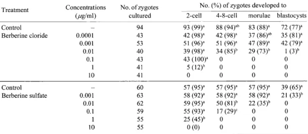
Table 3. Effect of berberine on the in vitro development of mouse zygotes
Treatment Concentrations
(pg/ml)
No. of zygotes cultured
No. (%) of zygotes developed to 2-cell 4-8-cell morulae blastocysts Control
Berberine cloride 0.0001 0.001 0.01 0.1 1
10
94 43 53 40 43 41 41
93 (99)a 42 (98)a 51 (96)a 39 (98)a 43 (100)a
5 (12)h 0
88 (94)ab 42 (98)a 51 (96)a 34 (85)b
0 0 0
83 (88)a 37 (86)ab 47 (89)a 29 (73)b
0 0 0
72 (77)a 35 (81)a 42 (79)a 1 (3)b 0 0 0 Control
Berberine sulfate 0.001 0.01 0.1 1
10
60 63 62 59 55 55
57 (95)a 58 (92)a 59 (95)a 55 (93)a 25 (45)h
0 (0)
57 (95)a 58 (92)a 50 (81)h 17 (29)c
0 0
57 (95)a 58 (92)a 22 (35)h
0 0 0
39 (65)a 21 (33)h
0 0 0 0 a-c: Values with different superscripts in the same columm differ significantly (P < 0 .05).
Results
Effect of Coptis rhizome, Phellodendron bark and berberine on the in vitro development of zygotes
Coptis rhizome at a concentration of more than 0.1 ,ug/m1 and Phellodendron bark extracts at a concentration of more than 10 ,ug/mIalmost completely inhibited the potential of zygotes to develop into blastocysts (Table 2). The development of zygotes to blastocysts was significantly reduced by Coptis rhizome at a concentration of 0.01 pg/ml and by Phellodendron bark at concentrations over 0.1 pg/ml.
The potential of zygotes to develop into blastocysts
was almost completely inhibited when embryos were treated with 0.01 ,ug/mlberberine chloride or berberine sulfate (Table 3). The inhibitory effect of berberine was dose-dependent.
The inhibitory effects of berberine chloride on in vitro development were observed in embryos treated at all stages from the 1-cell to the morula. Even morulae treated with 0.1 pg/ml berberine chroride did not develop into blastocysts (Table 4).
Effect of berberine chloride administration on early embryonic development in vivo
The administration of berberine did not affect the
Presented by Medical"Online
44 J. Mamm. Ova Res. Vol. 28, 2011
Table 4. Effect of the timing of berberine chloride treatment on the development of mouse embryos
Group Timing of treatment No. of embryos
cultured
No. (%) of embryos developed to
Developmental stage (h after culture) 2-cell 4-8-cell morulae blastocysts
Control Berberine
1-cell (—) 1-cell (0) 2-cell (22) 4-8-cell (46) morula (70)
70 48 29 47 47
70 (100) 44 (92)
70 (100)3 3 (6)b
1 (3)b
66 (94)a 0 0 8 (17)b
66 (94) 0 0 0 0 Berberine chloride was added to the culture medium at the concentration of 0.1 ,ug/ml.
in the same columm differ significantly (P < 0.05).
a-h: Values with different superscripts
Table 5. Effect of berberine chloride administration on the in vivo development of embryos
Group
Administration period
(day)
No. of females paired with
males
No. (%)ofmated females (with vaginal plug)
No. of eggs/
embryos recovered (average per mated female)
No. (%) of revovered embryos at rnorulae blastocysts others
G3: DW G1 : Berberine G2: Berberine G3: Berberine
3 1 2 3
14 9 8 14
10 (71) 6 (67) 6 (75) 13 (93)
334 (33.4) 173 (28.8) 126 (21.0) 366 (28.2)
124 (37)a 49 (28)h 37 (29)a 105 (29)b
145 (43)a 95 (55)h 41 (33)C 138 (38)C
65 (19)a 29 (17)a 48 (38)h 123 (34)b G6: DW
G6: Berberine
6 6
14*
25**
11(85)***
18 (82)***
131 (11.9) 123 (6.8)
54 (41) 50 (41)
46 (35)a 12 (10)h
31 (24)a 61 (50)b G14: DW
G14: Berberine
14 14
7*
11*
5 (83) 9 (90)
103 (20.6) 164 (18.2)
23 (22) 62 (38)
68 (66)a 68 (41)b
12 (12) 34 (21) a': Values with different superscripts in the same columm and the same experiment differ significantly (P < 0.05). *One female in
each group died before mating. **Three females died before mating. ***One female in each group died before embryo recovery.
Table 6. Effect of berberine chloride administration on fetal development
Treatment
No. of females paired with
males
No. of females mated (with vaginal plug)
No. (%) ofmated females with
fetuses
No. of fetuses live (average ± SD) dead DW
Berberine
16 16
15 ( 94) 16 (100)
9 (60) 10 (63)
156 (17.3 ± 6.7)a 121 (12.1 ± 3.7)b
4 5
a-h: Values with different superscripts differ significantly (P < 0 .05).
proportions of mated females (Table 5). When 100 dug berberine was intramuscularly injected for 2, 3, 6 or 14 days, the percentage of recovered embryos at the blastocyst stage in each group was significantly smaller than that in controls (10% to 41% vs. 35% to 66%, respectively). A few mice died following berberine injection, before or after mating (4 out of 25 and 1 out of
11 mice), or following distilled water injection (2 out of 14 and 1 out of 7 mice) in Group 6 and Group 14, respectively.
Table 6 shows the effect of berberine injection on the development to full-term. The proportions of females
mated (100% vs. 94% in the control group) and with fetuses (63% vs. 60% in the control group) on day 18.5 did not differ significantly from the control group. The average number of live fetuses in the berberine- administered females was significantly lower than that of the control females (12.1 vs. 17.3, respectively).
Discussion
Coptis rhizome and Phellodendron bark are widely used for the treatment of gastroenteritis, diarrhea, cholera, and human immunodeficiency virus (HIV) in
Presented by Medical"Online
traditional Chinese medicine [6, 7], and herbal medicines containing Coptis rhizome or Phellodendron bark are commercially available in Japan. Berberine is an alkaloid component of Coptis rhizome and Phellodendron bark [7] that is used to treat diseases such as hypotension, vasorelaxation, diarrhea, respiratory infection, HIV and human cancer cells [7, 11]. Although berberine administration to pregnant women is not recommended, because berberine displaces bilirubin from serum-binding proteins, causing jaundice, kernicterus, and brain damage in infants,
there are no reports on the toxicity of berberine at clinically relevant doses [7]. When berberine chloride dihydrate was administered in the feed to pregnant mice on days 6 to 17 of gestation, 33% of mice administered a high dosage of berberine (1,000 mg/kgBW/day) died from unknown causes, but prenatal mortality, average
litter size, and percentage of male fetuses were not affected [12].
To date, although there have been a number of pharmacologic and therapeutic studies of Coptis rhizome, Phellodendron bark and berberine [7, 13], their effects on fertility have not been reported. We found that water-soluble extracts of Coptis rhizome, and Phellodendron bark inhibited the development of mouse zygotes to blastocysts in vitro. We also found that berberine, a component of both natural products, inhibited the development of mouse zygotes. Moreover, intramuscular injection of berberine decreased the frequency of blastocysts and full-term fetuses. To our knowledge, this is the first report that Coptis rhizome, Phellodendron bark, and berberine inhibit the development of mouse zygotes.
The precise mechanisms by which both natural products and berberine inhibit the development of zygotes are not clear. Because the development of 1- cell, 2-cell, 4 to 8-cell, and morula-stage embryos to later stages was inhibited after the administration of berberine chloride, its inhibitory effect is likely related to cell division of blastomeres rather than embryonic genome activation, which occurs at the 2-cell stage in the mouse [14]. Berberine inhibits the growth of various types of cancer cells by inhibiting DNA topoisomerase I and by inducing cell-cycle arrest and apoptosis, mainly through the caspase-3 or Fas/FasL signaling pathway [13, 15, 16]. Serafim et al. [17] reported that berberine at low doses promotes G1 arrest but at higher doses results in G2 arrest. In the present study, we did not examine the cell cycle stage or apoptosis in development-arrested embryos after berberine treatment. Because most zygotes treated with 0.01 ,ug/
ml berberine chloride developed to the morula stage, but only a few berberine-treated embryos developed into blastocysts, and morulae treated with 0.1 ,ug/m1 berberine chloride did not develop to blastocysts, it is possible that berberine inhibits the cell differentiation in the morula.
The present study demonstrated that berberine administered to female mice once a day for 2 to 14 days after superovulation significantly decreased the proportion of recovered blastocysts and the fetal rate compared with those of control females. Although the precise mechanism is not clear, berberine administered intramuscularly may travel to oviducts and uteri through epithelial cells and inhibit embryonic development.
Unlike gossypol [4], the toxicity of berberine except at an unusually high dosage has not been reported. The findings of the present study indicate the potential use of berberine as a contraceptive for animals. However, effective procedures to enhance the transport of berberine to the genital tracts have to be developed, because the inhibition of fetal development is not complete. Follow-up in vivo studies are needed to demonstrate that fertility can be restored. Further studies should be conducted to reveal the mechanism of inhibitory action of berberine on the embryonic development.
Acknowledgements
This work was supported by the Ministry of Education, and Culture, Sports, Science and Technology, Japan (16200030, 20650063, 21028022).
References
1) Tsunoda, Y. and Kato, Y. (2006): Cloning in cattle. In:
Epigenetic Risks of Cloning (Inui, A., ed.), pp.33-57,
Taylor & Francis, Boca Raton.
2) Chang, M.C. (1978): Development of the oral contraceptives. Am. J. Obstet. Gynecol., 132,217-219.
3) Galvao, K.N., Sanntos, J.E.P., Coscioni, A.C., Juchem, S.O., Chebel, W.M., Sischo,W.M. and Villasenor, M.
(2006): Embryo survival from gossypol-fed heifers after
transfer to lactating cows treated with human chorionic
gonadotropin. J. Dairy Sci., 89,2056-2064.
4) Randel, R.D., Chase, C.C. Jr. and Wyse, S.J. (1992):
Effects of gossypol and cottonseed products on
reproduction of mammals. J. Anim. Sci., 70,1628-1638.
5) Brocas, C., Rivera, R.M., Paula-Lopes, F.F., McDowell, L.R., Calhoun, M.C., Staples, C.R., Wilkinson, N.S.,
Boning, A.J., Chenoweth, P.J. and Hansen, P.J. (1997):
Deleterious actions of gossypol on bovine spermatozoa,
oocytes, and embryos. Biol. Reprod., 57,901-907.
Presented by Medical*Online
46 J. Mamm. Ova Res. Vol. 28, 2011
6) Chan, C-0., Chu, C-C., Mok, D.K-W. and Chau, F-T.
(2007): Analysis of berberine and total alkaloid content in
Cortex Phellodendri by near infrared spectroscopy (NIRS)
compared with high-performance liquid chromatography
coupled with ultra-viable spectrometric detection. Analyt.
Chim. Acta., 592,121-131.
7) Imanshahidi, M. and Hosseinzadeh, H. (2008):
Pharmacological and therapeutic effects of Berberis
vulgaris and its active constituent, berberine. Phyt. Res., 22,999-1012.
8) Fulton, B.P. and Whittingham, D.G. (1978): Activation of mammalian oocytes by intacellular injection of calcium.
Nature, 273,149-151.
9) Erbach, G.T., Lawittsm, J.A., Papaioannou, V.E. and Biggers, J.D. (1994): Differential growth of the mouse
preimplantation embryo in chemically defined media. Biol.
Reprod., 50,1027-1033.
10) Kato, Y. and Tsunoda, Y. (1994): Effect of culture density of mouse zygotes on the development in vitro and in vivo.
Theriogenology, 41,1315-1322.
11) Yu, F-S., Yang, J-S., Lin, H-J., Yu, C-S., Tan, T-W., Lin, Y-T., Lin, C-C., Lu, H-F. and Chung, J-G. (2007):
Berberine inhibits WEHI-3 leukemia cells in vivo. In Vivo, 21,407-412.
12) Jahnke, G.D., Price, C.J., Marr, M.C., Myers, C.B. and
George, J.D. (2006): Developmental toxicity evaluation of berberine in rats and mice. Birth Defects Res. (Part B), 77,
195-206.
13) Tang, J., Feng, Y., Tsao, S., Wang, N., Curtain, R. and Wang, Y. (2009): Berberine and Coptidis Rhizoma as
novel antineoplastic agents: A review of traditional use and
biomedical investigations. J. Ethnopharm., 126,5-17.
14) Kidder, G.M. (1992): The genetic program for preimplantation development. Develop. Genetics, 13,319—
325.
15) Li, X.K., Motwani, M.T., William, B., William, S. and Gary, K.S. (2000): Huanglian, a Chinese herbal extract,
inhibits cell growth by suppressing the expression of cyclin B1 and inhibiting CD2 kinase activity in human cancer
cells. Mol. Pharm., 58,1287-1293.
16) Kuo, C.L., Chi, C.W. and Liu, T.Y. (2005): Modulation of apoptosis by berberine through inhibition of cyclooxygenase-2 and Mc1-1 expression in oral cancer
cells. In Vivo, 19,247-252.
17) Serafim, T.L., Olivera, P.J., Sardao, V.A., Perkins, E., Parke, D. and Holy, J. (2008): Different concentrations of
berberine result in distinct cellular localization patterns and
cell cycle effects in a melanoma cell line. Cancer Chem.
Pharm., 61,1007-1018.
ハム ス ター凍 結乾 燥精 子 の顕 微 授精 に よ る産 仔作 出
宗 藤 朋 美 ・堀 内 俊 孝 県 立 広 島 大 学 大 学 院 総 合 学 術 研 究 科,庄 原 市 〒727‑0023
32‑39
ハ ム ス ター凍 結 乾 燥精 子 は顕微 授 精 に よ って前 核 を形 成 す るが, この前 核 期卵 子 の発 生 能 は不明 であ る.こ れ までに,凍 結乾 燥 精子 の顕 微 授 精 か ら産 仔作 出 に成 功 した動 物 はマ ウ ス,ウ サ ギ,ラ ッ トの み で あ る,我 々 は,今 回,ハ ム ス タ ー凍 結 乾 燥精 子 の顕 微 授 精 に よ って は じめ て産 仔 作 出 に成 功 した.凍 結 乾 燥 ・再 水 和 後 の 精 子DNAの 正 常性 は,凍 結 乾 燥精 子 の 顕微 授精 に よ る胚 発 生 に重 要 で あ る.本 研 究 で は,TUNEL法 に よ っ て,M2培 地 ま た は50 mMEGTA添 加Tris‑HCI溶 液(EGTA溶 液)を 用 いて凍 結 乾 燥 し たハ ム ス ター精子 のDNA断 片化 と,精 子 染色 体 の正 常性 を調 べ た.
EGTA溶 液 で 凍 結 乾 燥 し た ハ ム ス タ ー 精 子 のDNA断 片 化 の 割 合 は M2培 地 よ り も 有 意 に 低 か っ た(P〈0。05,4.3%vs.41.4%).正
常 な 精 子 染 色 体 の 割 合 も,EGTA溶 液 区 がM2培 地 区 よ り も 有 意 に 高 く(P<0.05,8t1%vs。41.0%),顕 微 授 精 後 の 桑 実 胚 ・胚 盤 胞 へ の 発 生 率 も,EGTA溶 液 区 が 有 意 に 高 か っ た(P<0.05, 62.2%vs.12.5%).23個 の 桑 実 胚 と胚 盤 胞 を 移 植 し た 結 果,3匹 の 産 仔 が 誕 生 し た.
キ ー ワ ー ド:DNA断 片 化,凍 結 乾 燥 卵 細 胞 質 内 精 子 注 入,ハ ム ス タ ー 精 子
オ ウ レ ン.オ ウバ ク な ら び に そ れ ら の 主 要 成 分 ベ ル ベ リ ン は マ ウ ス 受 精 卵 の
発 生 能 を阻害 す る
角 田 幸 雄 ・加 藤 容 子 近 畿 大 学 農 学 部,奈 良 市 〒631‑8505
40‑46
生 薬269種 の 水 抽 出 物 に つ い て,マ ウ ス 前 核 期 受 精 卵 の 発 生 能 に 及 ぼ す 阻 害 効 果 を検 討 し た.そ の 結 果,オ ウ レ ン と オ ウ バ ク 抽 出 物 な ら び に そ れ ら の 主 要 成 分 で あ る ベ ル ベ リ ン に,マ ウ ス 前 核 期 受 精 卵 の 発 生 能 を阻 害 す る作 用 が あ る こ と が 判 明 し た.オ ウ レ ン で は 0.1μglml,オ ウ バ ク で は10μg!ml,ベ ル ベ リ ン で は0.01μglmlを 添 加 し た 培 地 で 培 養 さ れ た マ ウ ス 前 核 期 受 精 卵 は,胚 盤 胞 へ の 発 生 が ほ と ん ど完 全 に 阻 害 さ れ た.ま た,100μgベ ル ベ リ ン を1日1 回,交 配 の 前 後 に2〜14日 間 雌 マ ウ ス に 筋 肉 内 投 与 す る と,胚 盤
胞 へ の発 生率 が有 意 に低 下 す る と ともに,妊 娠満 期 に お ける胎子 数 が有意 に減 少 した.ベ ルベ リン は,人 や動 物 医薬 品 の成 分 と して配 合 され てい る、 ベル ベ リン投与 雌 に お ける受 胎 阻害 作用 は完 全 で は な かっ たが,さ らに投 与方 法 を検 討 す るこ とに よ り,雌 動物 へ のベ ル ベ リン投与 は動物 の 非侵 蝕的 な受胎 阻 害法 とな る可能 性 が示 唆 さ れ た.
キ ーワー ド:オ ウ レン,オ ウバ ク,ベ ル ベ リン,受 胎 阻害
Cryotopを 用 い た 単 精 子 凍 結 保 存 の 検 討47‑52
遠 藤 雄 史1・ 藤 井 好 孝1・ 新 谷 香 澄 ㌧ 瀬 尾 百 百 代[・ 本 山 洋 明1・ 舟 橋 弘 晃21倉 敷 成 人 病 ク リニ ッ ク体 外 受精 セ ン ター,倉 敷 市 〒710・0824,2岡 山 大学 大 学 院 自 然 科 学 研 究 科,岡 山市 〒700‑8530
非 生 体 由 来 で 取 り扱 い が 容 易 なCryotopを 用 い て 単 精 子 凍 結 保 存 を試 み た.Cryotop先 端 ス トリ ッ プ 部 分 に 置 い た 凍 結 液 中 に 精 子 を 入 れ,液 体 窒 素 蒸 気 で 凍 結 保 存 し た.そ の 結 果,Cryotopを 用 い た 精 子 凍 結 は 可 能 で あ り,融 解 後 は 透 明 帯 と 同 等 の 回 収 率(98.0%
vs.88.0%),運 動 率(30.6%vs.23.9%)だ っ た.ま た,Cryotop を 用 い て 精 巣 上 体 由 来 精 子 を 凍 結 保 存 し て も 融 解 後 の 生 存 率 は 射 出 精 子 と比 べ 差 を 認 め な か っ た(44.4%vs.42.1%).さ らに,異
な る 凍 結 保 護 物 質 で 精 子 を 凍 結 保 存 し た と こ ろ,融 解 後 の 生 存 率 は シ ョ 糖 の み の 方 がSpermFreezeに 比 べ 有 意 に 高 か っ た(65.3%
vs.37.3%,P<0.01).Cryotopは 単 精 子 凍 結 保 存 に お い て 有 効 な 保 存 容 器 で あ り,シ ョ 糖 は 効 果 的 な凍 結 保 護 物 質 で あ る こ と が 明 ら か と な っ た.
キ ー ワ ー ド:凍 結 保 存,Cryotop,透 明 帯,単 精 子,シ ョ糖
体外 成 熟培 養 に おけ る培 養 液量 と供 試 卵母 細胞 数 の違 い によ る初 期発 生 への影 響
西 尾 愛 美 ・星 野 由 美 ・佐 藤 英 明
53‑60 東 北 大 学 大 学 院 農 学 研 究 科 動 物 生 殖 科 学 分 野,仙 台 市 〒981‑8555
培 養 液 量 と供 試 卵 子 数 が マ ウ ス 卵 子 の 体 外 成 熟 と そ の 後 の 発 生 に 及 ぼ す 影 響 を調 べ 最 適 な 条 件 を 選 定 す る こ と を 目 的 と した.培 養 液 量 は20,50,100,200μ1,供 試 卵 子 数 は1,5,10,20,50 個 と し た.体 外 成 熟 培 養(IVM)後18時 間 で 成 熟 率 と卵 丘 膨 化 を 調 べ た.受 精 お よ び 胚 発 生 へ の 影 響 を 調 べ る た め,成 熟 率 が75%
以 上 の 区 に お い て は体 外 受 精 ・体 外 培 養 を 行 い,前 核 形 成 率 お よ び 初 期 胚 発 生 率 を算 出 し た.さ ら に 初 期 胚 の 質 を 評 価 す る た め,胚 の 細 胞 数 お よ びg'αoose飴 ηsρo/feFプ とdesmoco〃'η 〃'のmRNA発
現 を解析 した.現 行 条 件 に最 も近 い20個/100μ1区 は,発 生 率 や 胚 の質 が体 内成 熟卵 と有意差 が なか った こ とか ら,よ り適 したIVM 条 件 であ る こ とが裏 付 け られ た.さ らに,5個 区 で も胚 を得 られ た
こ とか ら,現 行 条件 よ り少数 で も培 養可 能 で あ るこ とが明 らか とな っ た。
キ ーワ ー ド:卵母細 胞 の質,体 外 成熟 培養,初 期 胚発 生,培 養 条件, マ ウス
PresentedbyMedical★Online