37
AfatEkiNf* 7 : 37-46 (1999)
Bull. Inst. Compr. Agr. Sci. Kinki Univ. 7 : 37-46 (1999)
Structural Characterization of Glycolipids in Edible Mushroom by FAB/MS"
Akiyoshi SAWABE*, Masanori MORITA**, Seiji OUCHI***, and Tadashi OKAMOTO***
Synopsis
The structures of glycolipids isolated from mushrooms, Hypsizigus marmoreus (Bunashimeji) and Pleurotus citrinopileatus (Nireouma) were determined to be (4E,8E)-N-2-hydroxyhexadecanoy1-1-0- - glucopyranosy1-9-methyl-C,g-sphinga-4,8-dienine (1), phosphodihexose N-(2-hydroxyoctadecanoy1)-4- hydroxy-C18-sphinganine (2), phosphodihexose N-(2-hydroxyhexadecanoy1)-4-hydroxy-Ci8-sphinganine (3), phosphodihexose N-(2-hydroxytetracosanoy1)-4-hydroxy-C,1-sphinganine (4), and phosphodihexose N-(2-hydroxydocosanoy1)-4-hydroxy-C18-sphinganine (5). In particular, location of the double bonds in the long-chain base of 1 was clearly determined by the B/E constant linked scan method. The structure of cerebroside having a long-chain base of 9-methyl-C18-sphinga-4,8-dienine could be determined in general by the presence of characteristic fragment ions of [C,9-sphingadienine + H - H20]+
at m/z 276 and [C19-sphingadienine + H]+ at m/z 294, and the fatty acid carbon number could be calculated from the characteristic fragment ion of [ceramide - 180]+ ([MH - GlcOH - 180]+) in positive ion mode FAB mass spectrometry. In the structural determination of 2-5, the ions of m/z 421 and 720 in the negative ion mode analyses are assigned to be characteristic peaks of phosphodihexose and phytosphingosine containing phosphodihexose, respectively.
This method proved to be useful for the structural determination of unstable natural products such as lipids.
Introduction
During the course of our search for functional molecules in edible fungi, we have been studying the characterization and structural determination of glycolipids in edible mushrooms. In this report applications of B/E constant linked scan fast atom bombardment (FAB) mass spectrometry for glycolipids (1 - 5, Fig. 1), which were found in Hypsizigus marmoreus (Bunashimeji) and Pleurotus citrinopileatus (Nireouma) 2-"are reviewed.
Cerebrosides having a 9-methyl-C,8-sphingadienine unit were discovered independently by two groups almost at the same time in 1979 (Ballio et al. ') and Karlsson et al. 9)) from a Deuteromycetes fungus, Fusicoccum amygdali, and from a sea anemone, Metridium senile, respectively. Recently, Kawai and Ikeda found a cerebroside having 9-methyl-C,9-sphinga-4,8-dienine in Schizophyllum commune (Suehirotake, a mushroom) and elucidated the complete structure. The long-chain base moiety with a 9-methyl branch of the cerebroside was suggested to constitute an essential part of the fruit-inducing activity in some fungi 10-13).
* Institute for Comprehensive Agricultural Sciences
, Kinki University, Nakamachi 3327-204, Nara 631-8505, Japan
** Joint Research Center , Kinki University, Kowakae 3-4-1, Higashi-Osaka 577-8502, Japan
***Faculty of Agriculture
, Kinki University, Nakamachi 3327-204, Nara 631-8505, Japan
38 Bull. Inst. Compr. Agr. Sci. Kinki Univ. No. 7 (1999)
OH
3 5 7 9
HOCH2 .- 0 18
HO OH NH
OH o-C 16 OH
1
O OH
. 1 3 5 7
Hex-Hex-O—P-0
OH NH OH
0' .0-`4''(CH2)x-CH3
2
OH
2 3 4 5
x=1 5 x=1 3 x=21 x=19
18
Fig. 1
Structures of glycolipids (1 - 5).
Recently, glycosphingolipids and sphingomyelin were postulated to be modulators of cell growth and cell differentiation 14,15).
Although structures of these glycolipids have been analyzed in the past by gas chromatography-mass spectrometry (GC-MS) of their derivatives such as trimethylsilyl (TMS) ethers 16-18), we have characterized in this work, the fatty acid composition and the location of double bonds in the long-chain base (C,9-sphingadienine), by B/E linked scan FAB mass spectrometry of the glycolipids itself.
Results and Discussion
Structural elucidation of glycolipid 1
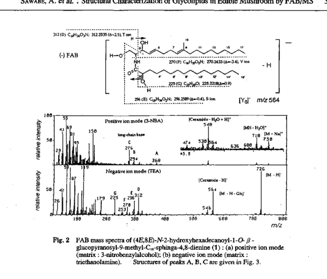
Positive and negative ion FAB mass spectra of cerebroside (1) are shown in Fig. 2. The positive ion mode FAB mass spectrum can be divided into three regions as reported in Refs. 19 and 20; (i) the molecular ion region, (ii) the ceramide ion region, and (iii) the long-chain base ion region. In region i, the molecular weight is successfully determined from m/z 750 [M + Na]' and m/z 710 [MH - 1120]+
ions. In region ii, the ceramide ion peak was observed clearly at m/z 548 [MH GlcOH]+. In region iii, the peaks derived from the long-chain base of C,9-sphingadienine were found at m/z 294 [C,9- sphingadienine + H]+ and m/z 276 [C,9-sphingadienine + H H2O].+
The B/E constant linked scan spectra of the ceramide ion and the long-chain base ion in positive ion FAB mass spectra are shown in Figs. 3 and 4, respectively. The B/E constant linked scan spectrum of the ceramide ion indicated the fragmentation of ion m/z 548 at the doublly allylic position of two double bonds to give fragment A at m/z 368 and at the amide position to generate fragment B at m/z 294, and the following dehydration of fragment B to generate fragment C at m/z 276, as shown in Fig.
3. High-resolution mass spectra of the peaks A and C at m/z 368 and 276, respectively, produced in the positive ion mode FAB mass spectrum of 1, are summarized in Table 1. The possible elemental formulas calculated on the basis of the exact mass are also listed in Table 1. The B/E constant linked scan spectrum of the long-chain base ion in region iii gave an informative result shown in Fig. 4, which indicates the location of the double bonds in the long-chain. From the results of high resolution mass spectra, the fragment ions of high intensities in Fig. 3 at m/z 94 21) and 148 are assigned to C61-18N+
produced by ring formation between C-6 and C-1, and CioH,,N+, respectively. On the basis of the 'H- 'H COSY analysis of 1 , all the 'H chemical shifts could be unambiguously determined, which indicated
SAWABE, A. et al. Structural Characterization of Glycolipids in Edible Mushroom by FAB/MS 39
312 (I)) C18H3403N;
(-) FAB
312.2535 T ion H TY
OH
[H-0"
/9
13
NH o' ,c H
0
270 (F) C16H3202N; 270.2433 (A=-3.4), V ion
4. 8' 8' ID' 12' 14' 18'
225 (G) C15H220; 225.2218.9) H
296 (E) C181-13402N; 296.2589 (0=-0.4), S ion
-H
[Yor m/z 564
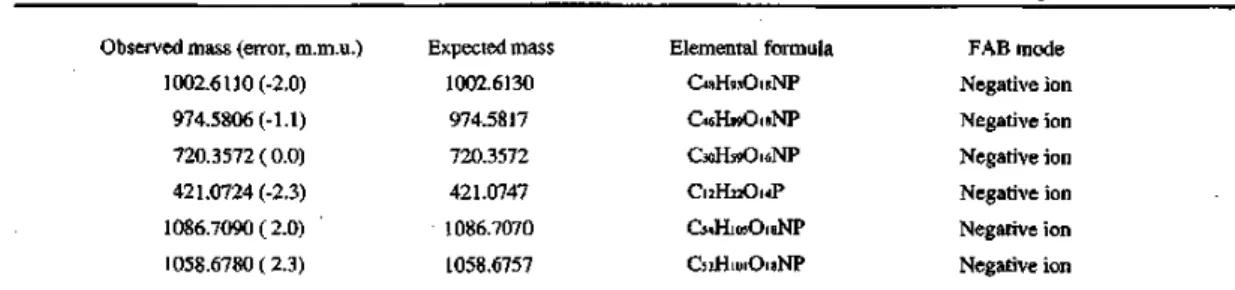
Table I. Determination of the exact mass of molecular ion and fragment ions of compound 1 Observed mass (error, m.m.u.)
368.3165 (-0.5)
276.2697 ( 0.6) 726.5521 ( 0.1) 312.2539 ( 2.1)
296.2589 (-0.4)
270.2433 (-3.4)
225.2218 (-0.9)
Expected mass 368.3160 276.2691 726.5520 312.2560 296.2585 270.2399 225.2209
Elemental formula C22H4203N CI9FNN CA-17609N Cid-13403N CisH3402N Cio.H.:02N Cl5H290
FAB mode Positive ion Positive ion Negative ion Negative ion Negative ion Negative ion Negative ion
Fragment*
A C
D E F G *Assignments of fragment ions in Fig . 2,
the presence of a vinylmethyl group at 8 1.50 (3H, s, C-19), two methylene groups of allylic positions at 1.97 (4H, m, C-6 and C-7), three olefinic protons at 5.20 (1H, br. s, C-8), 5.39 (1H, dd, J=15.5, 7.1Hz, C-4), and 5.66 (1H, br. d, J=15.5Hz, C-5), and an anomeric proton of a fl -glucopyranoside moiety at 4.17 (1H, d, J=7.6Hz, Glc-1). The location of the double bonds in the fragment ion at m/z 276 were then assigned at C2-C3, C4-05, and C5-C9 on the basis of the above-mentioned results. The structure of the long-chain base was thus found to be 9-methyl-C18-sphinga-4,8-dienine by B/E constant linked scan method. The characteristic fragment ions indicating the structure are determined to be [C19-sphingadienine + H - H20]+ at m/z 276 and [ceramide - 180]+ ([MH - G1cOH - 18(]') at m/z 368 in the positive ion mode FAB mass spectrum.
40 Bull. Inst. Compr. Agr. Sci. Kinki Univ. No. 7 (1999)
Gic-O
548 (ceramide), Zo ion 368 (A) 294 (13)
OH
1 3 s
NH C 0' OH
4 6 8
11
10 13 _15
12 14
OH -NNNN,
CH2 NH
2 4 6 8 10 12 14 16
OH
m/z 368 (A) C22H4203N; 368.3165 (A=-0 5)
17
16
OH
H2C 35 79 11 13 15 17
NH2 m/z 294 (B), W' ion
-H20
H2N
m/z 276 (C) C19H34N; 27626970=0.6)
3 H2N
55 81
_
94
7 9
•.****"..
-H
_
148
11 13 15 17
-H
(r)
• a.) cts
100
50
Fig. 4
a 50 100 150 260 250
m/z B/E constant linked scan spectrum of [C19-sphingadienine + H - H2O]+ ion at m/z 276 in positive
ion mode FAB mass spectrum of 1.
SAWABE, A. et al. : Structural Characterization of Glycolipids in Edible Mushroom by FAB/MS 41
The molecular ion structure was not accessible from the positive ion mode FAB mass spectrum, but was more clearly elucidated by the negative ion mode experiments. The negative ion mode FAB mass spectrum was also divided into three regions 19,20) ; (i) the molecular ion region, (ii) the ceramide ion region, and (iii) the fatty acid region. The molecular weight can be determined from the intense peak at m/z 726 [M - The exact mass of the ion at m/z 726 in the negative ion mode is also included in Table 1, which clearly indicates the elemental formula in accordance with the proposed structure.
The spectra in the ceramide ion region showed the presence of two ions at m/z 564 [M - Glc] and m/z 546 [M - H - G1c01-1]-. The spectra in the fatty acid ion region showed fragment ions due to [C14H29- CH(OH)-CONH-(CH-CH-2)0 - Hj- at m/z 312, [C,,H„-CH(OH)-CONH-CH=CH, - HI at m/z 296, [C14- H29-CH(OH)-CONH2 - HI at m/z 270 and [C13H2,-(CH-CH2)0 - H]- at m/z 225, which correspond to the fragmentations D, E, F, and G, respectively, in Fig. 2. The exact mass of fragments D, E, F, and G in the negative ion mode are also listed in Table 1. Consequently, the structure of fatty acid residue was found unequivocally to be 2-hydroxyhexadecanoic acid, which is supported by the result of positive ion mode analysis.
Based on the evidence of positive and negative ion mode FAB/MS, the structure of cerebroside (1) was determined to be (4E,8E)-N-2-hydroxyhexadecanoy1-1-0- j9 -glucopyranosy1-9-methyl-C10- sphinga-4,8-dienine (Fig. 1), which had been isolated from Schizophyllum commune by Kawai and Ikeda 10) as a fungal fruiting body inducer.
For glucosylceramides (cerebrosides) having the long-chain base of 9-methyl-C,8-sphinga-4,8- dienine, it might be concluded that the presence of characteristic fragment ion peaks at m/z 276 and 294 are accepted as the diagnosis of the long-chain base, and the fatty acid carbon number can be calculated from the characteristic fragment ion of [ceramide - 180]+ ([MH - G1cOH - 180]1 in positive ion mode FAB mass spectrum.
These data might contribute to the determination of structures of cerebrosides having the long-chain base of 9-methyl-C,0-sphinga-4,8-dienine, which are widely distributed among fungi.
Structural elucidation of glycolipids 2 - 5
The molecular ion structures were not accessible from the positive ion mode FAB mass spectra, but were more clearly elucidated by the negative ion mode.
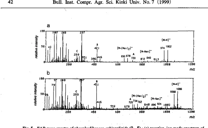
The negative ion FAB mass spectra of fraction II and fraction III are shown in Fig. 5. The molecular weights can be determined from intense peaks of [M - H]- in Figs. 5a and 5b, at m/z 1002 and 974, and 1086 and 1058, respectively. The difference of 28 a.m.u. suggests that these fractions are mixtures containing two molecular species. The exact mass of these molecular ions determined by the negative ion mode are listed in Table 2. The methanolysis of these fractions gave mixtures containing two FAMEs with the difference of carbon numbers as indicated above.
Table 2 Determination of the exact mass of molecular ion and fragment ions of compound 2-5 Observed mass (error, m.m.u.)
1002.6110 (-2.0) 974.5806 (-1.1)
720.3572 ( 0.0)
421.0724 (-2.3)
1086.7090 ( 2.0) 1058.6780 ( 2.3)
Expected mass 1002.6130 974.5817 720.3572 421.0747 1086.7070 1058.6757
Elemental formula C481-1930,8NP Ca6H890,8NP C3oHs90,6NP C,21-1220,4P Cg4H105018NP C52H,,,,018NP
FAB mode Negative ion Negative ion Negative ion Negative ion Negative ion Negative ion
42 Bull. Inst. Compr. Agr. Sci. Kinki Univ. No. 7 (1999)
a
100
c73 z Se
a)
0 79 I 48 2 97
[M—H
974 100242.1[M—(Hex)2]—
[M—Hex 259
U./
3II 396-
A 650446678720
812840913
. 111
0 ^r•
zee 400 sea 800 1.000 1200
m/z
b
100
50 ci)
8
9 7 1 48 2 37
79 421 [M-H)-
1086
C 1058
8-26
259 [M-(Hex)2]-
ii]IJ .1
A [M-Hex]-
446 720734762
940 896 924 100255B678
0 • I I
200 400 GOO BOO 1000 1290
m/z
Fig. 5 FAB mass spectra of phosphodihexose-sphingolipids (2 - 5) : (a) negative ion mode spectrum of fraction II (matrix : triethanolamine) ; (b) negative ion mode spectrum of fraction III (matrix : triethanolamine). Structures of peaks A, B, and C are given in Fig. 7.
Structural elucidation of glycosyl phosphosphingolipids 2 and 3 in FAB mass spectrum of fraction II The peaks at m/z 924 and 896 in Fig. 5a were assigned to fragment ions due to [M - Hex]- and peaks at m/z 678 and 650 to fragment ions due to [M - (Hex)2]-.
The B/E constant linked scan spectra of the molecular ions m/z 1002 and 974 indicated that fragmentations occurred at the amide position to generate fragment A at m/z 720, and at the polar groups to generate fragment B at m/z 421. Subsequent loss of a hexose from fragment B generated fragment C at m/z 259, and then loss of a phosphate group took place giving rise to ions of m/z 97 and 79, as shown in Fig. 6. The results of high-resolution mass spectra of peaks A and B at m/z 720 and 421, respectively, produced in the negative ion mode FAB mass spectra (Fig. 5a), are summarized in Table 2. The possible elemental formula calculated on the basis of the exact masses are also listed in Table 2. From the results of high-resolution mass spectra, the fragment ions in Fig. 6 at m/z 259, 97 and 79 are assigned to C6I-11209P-, 1-1204P-, and 03P-, respectively. The characteristic fragment ion at m/z 259 closely resembled the phosphoinositol part of phosphoinositol N-stearoyl-sphinganine which had been previously isolated from Leishmania by Singh et al. '9,22). Moreover, the long-chain base can be elucidated as a phytosphingosine with a eighteen carbon chain (4-hydroxy-Cwsphinganine) on the basis of a characteristic fragment ion at m/z 720. Consequently, the fatty acid carbon numbers can be calculated from the difference between the molecular ions and the characteristic ion at m/z 720, which indicated the presence of 2-hydroxyoctadecanoic acid for the compound of m/z 1002 and 2- hydroxyhexadecanoic acid for the compound of m/z 974. The structures of these compounds having peaks at m/z 1002 and 974, respectively, in the negative ion spectrum (Fig. 5a) were thus found to be phosphodihexose N-(2-hydroxyoctadecanoy1)-4-hydroxy-C,8-sphinganine (2) and phosphodihexose N- (2-hydroxyhexadecanoy1)-4-hydroxy-C,rsphinganine (3) as shown in Fig. 1.
SAWABE, A. et : Structural Characterization of Glycolipids in Edible Mushroom by FAB/MS 43
m/z 744 H
—,--o-- (C)771/2 259 OH 0 I ; He>;.-Hex-O—P-0 ;
I ;
OH ; NH OH
m/z 1002 C48H93018NP; 1002.6130 .6,=-2.0) m/2 974 C46H89018NP; 974.5817 (A=-1.1)
I 2 (B) m/z 421 C12l-122014P; 421.0747 (L1=-2.3) : OH
0' 'T"2"
OH
4 6
8 10 12 14
4
10 12 14
16 18 (C18h)
100
16 (C16h)
59
_515
79
197 259
.5 .
4 I (-) FAB-B/E constant linked scan spectrum of m/z 1002 1 A
728 678
744 8I
2
Structural elucidation of glycosyl phosphosphingolipids 4 and 5 in FAB mass spectrum of fraction III The structures of 4 and 5 were similarly determined as those of 2 and 3, as follows. The two peaks at m/z 421 and 720 in the negative ion spectrum (Fig. 5b) indicated the presence of a phosphodihexose and a phytosphingosine containing phosphodihexose, respectively. The ions at m/z 259, 97, and 79 also support the structures of the phosphodihexoses. Consequently, the structures of fatty acids are determined to be 2-hydroxytetracosanoic acid for the compound of m/z 1086 and 2-hydroxydocosanoic acid for the compound of m/z 1058 from the characteristic fragment ion at m/z 720. The B/E constant linked scan spectra of the molecular ions m/z 1086 and 1058 (Fig. 7) supported the above-mentioned postulation. The structures of these compounds having peaks at m/z 1086 and 1058, respectively, in the negative ion spectrum (Fig. 5b) were thus found to be phosphodihexose N-(2- hydroxytetracosanoy1)-4-hydroxy-Cirsphinganine (4) and phosphodihexose N-(2-hydroxydocosanoy1)- 4-hydroxy-C,8-sphinganine (5) as shown in Fig. 1.
Thus, the ions of m/z 421 and 720 in the negative ion mode are found to be very useful for the structural determination of glycosyl phosphosphingolipids (phosphodihexose-sphingolipids) in the analyses by FAB mass spectrometry, because these ion peaks are characteristic of a phosphodihexose and a phytosphingosine (4-hydroxy-C,8-sphinganine) containing phosphodihexose, respectively.
FAB mass spectrometry is a powerful tool for analyzing the structure of glycolipids. Especially in combination with a linked scan at constant B/E, it provides unambiguous information on the fatty acid composition, the location of double bonds in the long-chain base (C19-sphingadienine), or the presence of phosphodihexose.
44 Bull. Inst. Compr. Agr. Sci. Kinki Univ. No. 7 (1999)
100
z z 50 a.)—
-r ts
9- 79 97
259
LL4o-L-
4 1
B
462 529
,
A 726
762
• .
924
19 6
5.0.
6 260 460 662 lobe
Fig. 7 B/E constant linked scan spectra of [M - H]- ions at m/z 1086 and 1058 in negative ion mode FAB mass spectrum of fraction III.
Experimental
Isolation of Glycosyl Phosphosphingolipids. Fresh fruiting bodies (1.4 kg) of Hypsizigus marmoreus were kept in hot H2O at 96°C for 20 min, chopped by a commercial blender, and homogenized after making up the total volume to 1.5 L with hot H2O. Cold EtOH (3.5 L) was added to the hot H2O homogenate, and the mixture was allowed to stand overnight in the dark. The 1120- Et0H extract (68.5 g) was extracted with n-hexane and n-BuOH, successively. The n-BuOH extract (4.7 g) was chromatographed over an Amberlite XAD-2 column (Japan Organo Co. Ltd., 2.3 x 24 cm).
The loaded column was washed with 500 mL of H2O, and successively eluted with 20% Me0H-H20, 50% Me0H-H20, and Me0H (500 mL each). The solvent-removed H2O eluate (2.7 g) was chromatographed over silica gel (Wako gel C-300, Wako Pure Chemical Industries Ltd.) with CHC13- Me0H-H20 (60:29:6) as an eluent to obtain fraction I (1335 mg), fraction II (22 mg), and fraction III (218 mg). The fraction I was rechromatographed over silica gel (Wako gel C-300) with CHC13-MeOH (5:1) as an eluent, and 85 mg of a pure cerebroside (1) was obtained. The fractions II and III were analyzed by FAB/MS to have negative ion spectra shown in Figs. 5a and 5b, respectively.
Methanolysis of Fractions II and ITE Three mg of each fraction was heated at reflux with 0.9M HC1 in 82% Me0H (1 mL) for 18 hr 23), respectively. The reaction mixture was extracted with n- hexane, the n-hexane layer was concentrated in vacuo, and the residue was analyzed by GC-MS to give the fatty acid methyl esters (FAMEs); methyl 2-hydroxyoctadecanoate: m/z 314 [M]+, 255 [M - 59 (COOCH3)] and methyl 2-hydroxyhexadecanoate: m/z 286 [M]+, 227 [M-59] for fraction II; methyl 2- hydroxytetracosanoate: m/z 398 [M]+, 339 [M-59] and methyl 2-hydroxydocosanoate: m/z 370 [M]+, 311 [M - 59] for fraction III, respectively.
Conditions of FAB mass spectrometry. MS was carried out with a JEOL JMS-HX 100 double- focusing mass spectrometer of EB geometry that was connected to a JEOL DA 5000 data system. The mass spectrometer was fitted with a high-field magnet, a FAB ion source, and a post-accelerating detector. The sample was dissolved in chloroform-methanol (1:1 ; 1 u g/ 1u1) for glycolipid 1 or water-methanol (5:1 ; 1 ,u g/ii 1) for glycolipids 2 - 5 and 1 ,u 1 of the solution was added to the matrix
SAWABE, A. et al. : Structural Characterization of Glycolipids in Edible Mushroom by FAB/MS 45
(triethanolamine for the negative ion mode) on the stainless-steel FAB probe target. The sample was bombarded with a 6 KeV xenon atom beam. The exact mass measurement by FAB mass spectrometry was carried out using a mixture of cesium iodide, sodium iodide, and glycerol (5:1:25) as a mass calibrant. For linked scan analysis with constant B/E, collisional activation was performed in a collision chamber in the first field-free region using helium as the collision gas.
References
1) Sawabe, A., Morita, M., Ouchi, S., and Okamoto, T., Proceedings of International Symposium on Plant Glycosides 1997.
2) Sawabe, A., Morita, M., Okamoto, T. and Ouchi, S., Biological Mass Spectrometry, 1994, 23, 660.
3) Sawabe, A., Morita, M., Ouchi, S. and Okamoto, T., Journal of Mass Spectrometry Society of Japan, 1995, 43, 115.
4) Sawabe, A., Morita, M., Inaba, K., Ouchi, S. and Okamoto, T., Mushroom Science and Biotechnology,
1995, 2, 166.
5) Sawabe, A., Morita, M., Ouchi, S. and Okamoto, T., Journal of Mass Spectrometry, 1996, 31, 921.
6) Sawabe, A. and Okamoto, T., New Food Industry, 1996, 38, 45.
7) Morita, M. and Sawabe, A., Journal of Mass Spectrometry Society of Japan, 1998, 46, 204.
8) Ballio, A., Casinovi, C.G., Framondino, M., Marino, G., Nota, G. and Santurbano, B., Biochimica et Biophysica Acta, 1979, 573, 51.
9) Karlsson, K., Leffler, H. and Samuelsson, B.E., Biochimica et Biophysica Acta, 1979, 574, 79.
10) Kawai, G. and Ikeda, Y., Biochimica et Biophysica Acta, 1983, 754, 243.
11) Kawai, G. and Ikeda, Y., Journal of Lipid Research, 1985, 26, 338.
12) Funaki, Y., Kawai, G. and Mori, K., Agricultural and Biological Chemistry, 1986, 50, 615.
13) Kawai, G., Nippon Nogeikagaku Kaishi, 1986, 60, 1027.
14) Okazaki, T., Bielawska, A., Bell, R.M. and Hannun, Y.A., Journal of Biological Chemistry, 1990, 265, 15823.
15) Bielawska, A., Livardic, C.M. and Hannun, Y.A., Journal of Biological Chemistry, 1992, 267, 18493.
16) Kawai, G. and Ikeda, Y., Journal of Lipid Research, 1985, 26, 338.
17) Yasugi, E., Kasama, T. and Seyama, Y., Journal of Biochemistry, 1991, 110, 202.
18) Garg, H.S., Sharama-Pandey, M., Bhakuni, D.S., Pramanik, B.N. and Bose, A.K., Tetrahedron
Letters, 1992, 33, 1641.
19) Castello, C.E. and Vath, J.E., in Methods in Enzymology, 193, ed. McCloskey, J.A., Academic Press, San Diego, 1990, p 738.
20) Adams, J. and Ann, Q., Mass Spectrometry Reviews, 1993, 12, 51.
21) Ohashi, Y. and Nagai, Y., Carbohydrate Research, 1991, 221, 235.
22) Singh, B.N., Castello, C.E., Beach, D.H. and Holz, G.G., Biochemical and Biophysical Research Communication, 1988, 157, 1239.
23) Higuchi, R., Kagoshima, M. and Komori, T., Liebigs American Chemistry, 1990, 659.
Bull. Inst. Compr. Agr. Sci. Kinki Univ. No. 7 (1999)
FABIMSに よ る食 用 キ ノ コ に含 まれ る糖 脂 質 の 構 造 特 性
沢 辺 昭 義 ・森 田 全 律 ・大 内 成 志 ・岡 本 忠
摘 要
機 能 性 天 然 物 分 子 探 索 の 一 環 と し て,食 用 キ ノ コ に 含 ま れ る 糖 脂 質 の 構 造 解 析 に つ い て 研 究 を 行 っ て い る 。 本 稿 で は,ブ ナ シ メ ジ (耳 γP5fzfgり5π1armoreロ5)お よ び 楡 黄 麻 (pleHro加5cffアfηopflea∫ ロ5)か ら 得 た 糖 脂 質 の FABIMSの 構 造 特 性 に つ い て 紹 介 す る 。 得 ら れ た 糖 脂 質 の 構 造 は,(4E,8E)‑N‑2‑
hydroxyhexadecanoyl‑1一 σ β 一glucopyranosy1‑9‑methyl‑
Cl8‑sphinga‑4,8‑dienine(1),phosphodihexoseN(2‑
hydroxyoctadecanoyl)‑4‑hydroxy‑C18‑sphinganine(2),
phosphodihexoseハ 弄一(2‑hydroxyhexadecanoyl)‑4‑
hydroxy‑C18‑sphinganine(3),phosphodihexose坪(2‑
hy(廿oxytetracosanoyl)‑4‑hydroxy‑Cl8‑sphinganine(4)お よ びphosphodihexose1Ψ(2‑hydroxydocosanoy1)‑4‑
hydroxy‑C18‑sphinganine(5)と 決 定 し た 。 特 に, こ れ ら の 構 造 特 性 に お い て,長 鎖 ア ル キ ル の 不 飽 和 結 合 の 位 置,極 性 部 お よ び 脂 肪 酸 の 種 類 はB/E‑一 定 リ ン ク ド ス キ ャ ン 法 を 用 い た FABIMSを 測 定 す る こ と に よ り 容 易 に 決 定 さ れ た 。
![Fig. 7 B/E constant linked scan spectra of [M - H]- ions at m/z 1086 and 1058 in negative ion mode FAB mass spectrum of fraction III.](https://thumb-ap.123doks.com/thumbv2/123deta/9937289.1390008/8.892.113.778.91.594/fig-constant-linked-scan-spectra-negative-spectrum-fraction.webp)
