Doctoral Thesis
Non-genomic Actions of Diterpenoid Acids:
with Special Reference to Differentiation Induction of Human Neuroblastoma and Hepatoma Cells
Chiharu SAKANE
Molecular and Cellular Biology
Graduate School of Human Health Science
University of Nagasaki
CONTENTS
GENERAL INTRODUCTION ... 1
I. 1. Terpenoids biosynthesis ... 2
I. 2. Retinoid effects; genomic and non-genomic regulations of gene expression ... 11
I. 3. Acyclic retinoids ... 14
I. 4. Brief outline of the thesis ... 16
RAPID DOWNREGULATION OF CYCLIN D1 ... 18
II. 1. Abstract ... 19
II. 2. Introduction ... 20
II. 2. 1. Cell cycle and its related proteins ... 20
II. 2. 2. Cyclin D1 as a potent chemoprevention target ... 24
II. 2. 3. Non-genomic actions of retinoids, posttranslational downregulation of cyclin D1 ... 25
II. 2. 4. Aim of the study... 27
II. 3. Results ... 28
II. 3. 1. Rapid decrease in cyclin D1 content after GGA treatment in HuH-7 cells ... 28
II. 3. 2. GGA-induced downregulation of RB protein in HuH-7 cells ... 33
II. 3. 3. GGA-induced downregulation of E2F1 expression in HuH-7 cells ... 37
II. 3. 4. Mode of action for GGA to downregulate cyclin D1 levels in HuH-7 cells ... 39
II. 4. Discussion ... 44
II. 5. Conclusion ... 50
UPREGULATION OF NEUROTROPHIC TYROSINE KINASE, RECEPTOR, TYPE 2 ... 52
III. 1. Abstract ... 53
III. 2. Introduction ... 54
III. 2. 1. Neurotrophins and their receptors ... 54
III. 2. 2. BDNF ... 57
III. 2. 3. Differentiation-inducing effect of ATRA on neuroblastoma SH-SY5Y cells ... 60
III. 2. 4. Metabolites of the mevalonate pathway in neurons ... 60
III. 2. 5. Aims of the study ... 63
III. 3. Results ... 64
III. 3. 1. Proliferation profile of SH-SY5Y cells with GGA ... 64
III. 3. 2. Suppression of growth-related gene expression by GGA in SH-SY5Y cells ... 66
III. 3. 3. Morphologic alterations of SH-SY5Y cells by GGA ... 68
III. 3. 4. Downregulation of hexokinase-2 (HK2) with GGA in SH-SY5Y cells ... 71
III. 3. 5. Expression of neurotransmitter synthesizing enzymes tyrosine hydroxylase (TYH) and choline acetyltransferase (ChAT) in SH-SY5Y cells ... 73
III. 3. 6. Expression of NTRK2 in SH-SY5Y cells ... 75
III. 3. 7. Expression of nuclear retinoid receptors in SH-SY5Y cells ... 80
III. 4. Discussion ... 87
III. 5. Conclusion ... 91
INHIBITION OF LYSINE-SPECIFIC DEMETHYLASE 1A ... 92
IV. 1. Abstract ... 93
IV. 2. Introduction ... 94
IV. 2. 1. Epigenetic regulatory mechanisms ... 94
IV. 2. 2. Epigenetic regulation in cancer ... 101
IV. 2. 3. KDM1A as a candidate target of isoprenoids ... 102
IV. 2. 4. Aim of the study ... 104
IV. 3. Results ... 105
IV. 3. 1. Inhibition of KDM1A activity by farnesol ... 105
IV. 3. 2. Effects of isoprenoid chain length on KDM1A inhibitory activity ... 108
IV. 3. 3. Effect of dihydrogenation on KDM1A-inhibitory activity of GGA ... 111
IV. 3. 4. Upregulation of H3K4me2 bound to the promoter region of the NTRK2 gene by GGA ... 114
IV. 4. Discussion ... 119
IV. 5. Conclusion ... 122
RET KINASE SIGNAL TRANSDUCTION ... 123
V. 1. Abstract ... 124
V. 2. Introduction ... 125
V. 2. 2. MeCP2 is required for regulation of RET transcription ... 126
V. 2. 3. Aims of the study ... 132
V. 3. Results ... 133
V. 3. 1. Phosphorylation of MeCP2 by GGA treatment ... 133
V. 3. 2. Upregulation of RET expression by GGA treatment ... 135
V. 3. 3. Induction of lysine-4 methylation of histone H3 in upstream region of the RET gene ... 137
V. 3. 4. Knockdown of RET attenuated GGA-induced upregulation of NTRK2 expression ... 139
V. 3. 5. Effect of RET kinase inhibitor on GGA-induced NTRK2 upregulation ... 141
V. 4. Discussion ... 144
V. 5. Conclusions ... 149
GENERAL DISCUSSION ... 150
VI. 1. Genomic actions ... 151
VI. 1. 1. Retinoid receptors... 151
VI. 1. 2. Orphan receptors ... 154
VI. 2. Non-genomic actions ... 156
VI. 2. 1. Histone modification ... 156
VI. 2. 2. Signal transduction effects ... 158
VI. 3. Implications ... 160
VI. 4. Conclusions ... 161
VII. 1. Materials ... 163
VII. 1. 1. Chemical compounds ... 163
VII. 1. 2. qPCR primers and siRNAs ... 164
VII. 1. 3. Antibodies ... 166
VII. 2. Methods... 167
VII. 2. 1. Cell culture ... 167
VII. 2. 2. Reverse transcription and quantitative polymerase chain reaction (RT-qPCR) ... 168
VII. 2. 3. Chromation immunoprecipitation assay (ChIP) ... 169
VII. 2. 4. SDS-PAGE and immunoblotting ... 169
VII. 2. 5. Immunofluorescence ... 170
VII. 2. 6. Monoamine oxidase (MAO) activity and lysine-specific demethylase 1A (KDM1A) inhibitory analysis ... 171
VII. 2. 7. Statistical analysis ... 172
ACKNOWLEDGMENTS ... 173
REFERENCES ... 175
ABBREVIATIONS
2-PCPA trans-2-phenylcyclopropylamine 4EGI inhibitor of eIF4E/eIF4G interaction 9CRA 9-cis retinoic acid
ACR acyclic retinoid
AOF amine oxidase (flavin-containig) APP amyloid-β precursor protein ATCC American Type Culture Collection ATRA all-trans retinoic acid
BDNF brain-derived neurotrophic factor
BINAP 2,2'-bis(diphenylphosphino)-1,1'-binaphthyl BLAST Basic Local Alignment Search Tool
BLBP brain lipid binding protein
bp base pairs
CCD charge coupled device
CCND1 cyclin D1
CDK cyclin-dependent kinase
cDNA complementary DNA
ChAT choline acetyltransferase ChIP chromatin immunoprecipitation
ChIP-seq chromatin immunoprecipitation sequencing
CHX cycloheximide
CoA coenzyme A
COUP-TF chicken ovalbumin upstream promoter-transcription factor CRABP cellular retinoic acid binding protein
DMAPP dimethylallyl diphosphate
DME Dulbecco's modified Eagle's
DMSO dimethyl sulfoxide
DNA deoxyribonucleic acid
DP DNA-binding partner of E2Fs
DR direct repeat
eIF eukaryotic translation initiation factor
ES embryonic stem
FA farnesoic acid
FABP fatty acid binding protein FAD flavin adenine dinucleotide
FBS fetal bovine serum
FBXO F-box protein for other domain
FPP farnesyl diphosphate
FPPS farnesyl diphosphate synthase
G1 gap phase 1 in cell cycle
G2 gap phase 2 in cell cycle
GAPDH glyceraldehyde-3-phosphate dehydrogenase GDNF glial cell derived neurotrophic factor
GGA geranylgeranoic acid
GGal geranylgeranial or geranylgeranyl aldehyde
GGOH geranylgeraniol
GGPP geranylgeranyl diphosphate
GGPPS geranylgeranyl diphosphate synthase
GluR glutamate receptor
GPP geranyl diphosphate
GSK glycogen synthase kinase
H3K4 histone H3 lysine-4
HDAC histone deacetylase
HK hexokinase
HMG-CoA 3-hydroxy-3-methyglutaryl-CoA
HMT histone methyltransferase
HOX homeobox
IC50 inhibitory concentration at a half maximum
IPP isopentenyl diphosphate
JARID Jumonji, AT rich interactive domain
JMJD Jumonji domain
kDa kilodalton
KDM lysine (K)-specific demethylase Ki inhibitory constant for enzyme reaction
M mitosis phase in cell cycle
MAO monoamine oxidase
MeCP methyl CpG binding protein MEP methylerythritol phosphate
miRNA microRNA
mRNA messenger RNA
MVA mevalonic acid
NR nuclear receptor
NT neurotrophin
NTF neurotrophic factor
NTR neurotrophin receptor
NTRK neurotrophic tyrosine kinase, receptor
PBS phosphate buffered saline
PBS-T phosphate buffered saline with Tween 20
PCR polymerase chain reaction
PKC protein kinase C
PLC phospholipase C
PPAR peroxisome proliferator-activated receptor PPRE peroxisome proliferator response element PTB polypyrimidine tract binding protein PTEN phosphatase and tensin homolog PTM post-translational modification
Pu purine
PVDF polyvinylidene difluoride RAR retinoic acid receptor
RARE retinoic acid response element
RB retinoblastoma protein, ret proto-oncogene
RET ret (rearranged during transfection) proto-oncogene RIPA radio immunoprecipitation assay
RNA ribonucleic acid
RNAi RNA interference
ROR retinoic acid receptor-related orphan receptor
RORE ROR-response element
RPI-1 ret protein inhibitor-1
rRNA ribosomal RNA
RT-qPCR reverse transcription and quantitative polymerase chain reaction
RTT Rett syndrome
RXR retinoid X receptor
RXRE retinoid X response element
S DNA synthesis phase
SD standard deviation
SDS-PAGE sodium dodecyl sulfate-polyacrylamide gel electrophoresis Shc Src homology 2 domain containing
siRNA small interfering RNA
SMCY selected mouse cDNA on Y
SUMO small ubiquitin-related modifier SWI/SNF switch/sucrose nonfermentable
T1/2 half-life
TFDP transcription factor DP-1
TLX trophoblast-lymphocyte cross-reaction
TNF tumor necrosis factor
Tirs tris(hydroxymethyl)aminomethane
TR testicular receptor
TYH tyrosine hydroxylase
UTR untranslated region
UV ultraviolet
ZF-Jmj zinc finger Jumonji
LIST OF TABLES AND FIGURES
Tables
Table IV-1. Tentative classification of histone lysine demethylases (KDMs) in NCBI gene database...100
Table VII-1. Primers for RT-PCR………164
Table VII-2. Primers for ChIP analysis……….………..165
Table VII-3. List of antibodies used in the present study……….…………...166
Figures Fig. I-1. The mevalonate (MVA) pathway. ... 4
Fig. I-2. Schematic diagram of the isoprenoid production. ... 7
Fig. I-3. Enzymatic biosynthesis of GGA from GGPP. ... 10
Fig. I-4. Vitamin A production and its metabolites. ... 13
Fig. I-5. Schematic diagram for brief outline of the thesis. ... 17
Fig. II-1. Cell cycle. ... 21
Fig. II-2. Genes related G1/S transition. ... 22
Fig. II-3. Downregulation of the cellular cyclin D1 level in hepatoma-derived cell lines by treatment with GGA. ... 29
Fig. II-4. Rapid decrease in cellular cyclin D1 levels after GGA treatment in HuH-7 cells. ... 31
Fig. II-5. Dose effects of GGA on cyclins in HuH-7 cells... 32
Fig. II-6. Effects of GGA on the phosphorylation of retinoblastoma protein (RB). ... 34
Fig. II-7. Effects of GGA on subcellular localization of RB. ... 36
Fig. II-8. Effects of GGA on cellular expression of E2F1. ... 38
Fig. II-9. Suppression of cyclin D1 synthesis by GGA treatment. ... 41
Fig. II-10. Effect of GGA removal on expression of cyclin D1 in HuH-7 cells. ... 43
Fig. II-11. Hypothesis for molecular mechanism of GGA effect on cyclin D1. ... 51
Fig. III-1. Schematic diagram of neurotrophins and their cell-surface membrane receptors. ... 55
Fig. III-2. Crosstalk between NTRK2 and p75NTR with NTs. ... 56
Fig. III-3. BDNF/NTRK2 signaling pathways. ... 59
Fig. III-4. Isoprenoid metabolism in mammalian brain cells including steroid and nonsterol isoprenoids. ... 62
Fig. III-5. Effect of GGA on proliferation of SH-SY5Y cells. ... 65
Fig. III-6. Downregulation of cell cycle-related gene expression by GGA. ... 67
Fig. III-7. Effect of GGA on morphology of SH-SY5Y cells. ... 69
Fig. III-8. Embossment images. ... 70
Fig. III-9. Effects of GGA on hexokinases (HKs) expression. ... 72
Fig. III-10. Effects of GGA on TYH and ChAT expression. ... 74
Fig. III-11. Effects of GGA on NTRK2 expression in SH-SY5Y cells. ... 76
Fig. III-12. Effects of GGA on NTRK2 splice variant expression in SH-SY5Y cells. ... 77
Fig. III-14. Effects of GGA on retinoid receptors expression in SH-SY5Y cells. ... 81
Fig. III-15. Effects of GGA on RARB expression in SH-SY5Y cells. ... 82
Fig. III-16. Effects of RARB knockdown on NTRK2 gene expression in SH-SY5Y cells. ... 84
Fig. III-17. Effects of RARB gene dosage on NTRK2 gene expression in SH-SY5Y cells. ... 86
Fig. IV-1. 3D-structure of KDM1A and MAOB. ... 103
Fig. IV-2. Inhibition of recombinant human MAOB and KDM1A activities with farnesol. ... 106
Fig. IV-3. KDM1A inhibitory effects of farnesol derivatives. ... 107
Fig. IV-4. Isoprenoid chain length-dependent inhibition of KDM1A activity by polyprenoic acids. ... 109
Fig. IV-5. GGA inhibits KDM1A in a non-competitive fashion. ... 110
Fig. IV-6. Effect of dehydrogenation on the KDM1A-inhibitory activity of GGA. ... 112
Fig. IV-7. Concentration dependence of the KDM1A-inhibitory activity of 14,15-dihydroGGA. ... 113
Fig. IV-8. Time-dependent upregulation of the NTRK2 gene by GGA treatment. ... 115
Fig. IV-9. GGA induced dimethylated H3K4 bound to promoter regions of the NTRK2 gene. ... 116
Fig. IV-10. Correlation between KDM1A inhibition and NTRK2 expression levels. ... 118
Fig. V-1. MeCP2-mediated regulation of gene expression. ... 128
Fig. V-2. Potential posttranslational modifications in MeCP2. ... 131
Fig. V-3. Phosphorylation of MeCP2 by GGA treatment. ... 134
Fig. V-4. Time-dependent upregulation of RET expression with GGA. ... 136
Fig. V-5. Induction of lysine-4-methylations of histone H3 at upstream of the RET gene by GGA. .... 138
Fig. V-6. Knockdown of the RET gene attenuated NTRK2 mRNA level in the presence of GGA. ... 140 Fig. V-7. Effect of RET kinase inhibitor RPI-1 on GGA-induced NTRK2 upregulation. ... 142 Fig. V-8. Effect of RET kinase inhibitor RPI-1 on GGA-induced RET upregulation. ... 143 Fig. VI-1. Working hypothesis for molecular mechanism how GGA act in NTRK2 gene expression. . 159
Chapter I
GENERAL INTRODUCTION
Despite a plethora of studies establish “genomic actions” of diterpenoid acids, the actions partially explain the mechanisms of broader biological effects of diterpenoid acids. Hence, “non-genomic actions” become prospective research topics during the last decade. In this thesis, we describe differentiation-inducing effects of a diterpenoid geranylgeranoic acid, and discuss novel “non-genomic actions” of the diterpenoid acid in human neuroblastoma and hepatoma cells in comparison with another diterpenoid acid, retinoic acid.
I. 1. Terpenoids biosynthesis
Terpenoids, also known as isoprenoids, a class of natural organic compounds consisting of the five-carbon unit called “isoprene”, are synthesized ubiquitously and play essential roles among eubacteria, archaebacteria and eukaryotes including human. They are derived through condensations of isoprene compound isopentenyl diphosphate (IPP) and its isomer dimethylallyl diphosphate (DMAPP). Two distinct and independent pathways exist to biosynthesize IPP: the classical mevalonate (MVA) pathway (Fig. I-1) [Chaykin et al, 1958; Spurgeon & Porter, 1981] and a mevalonate-independent methylerythritol phosphate (MEP) pathway [Surmacz & Swiezewska, 2011]. Although MEP pathway is limited to photosynthetic plants and bacteria, the MVA pathway is an important cellular metabolic pathway present ubiquitously in all organisms. Acetyl-CoA is generated in a cell, which is added another acetyl-CoA and becomes acetoacetyl-CoA in a reaction catalyzed by thiolase. Produced and pooled IPP are converted to DMAPP by an IPP isomerase. The enzymes of the MVA pathway have been studied from a number of organisms, including humans.
the cholesterol synthetic pathway, is the target of the statin class of cholesterol-lowering drugs [Alberts et al, 1980], the treatment of cardiovascular disease, and inflammatory processes [Liao & Laufs, 2005]. It is important for the production of DMAPP and IPP, which serve as the basis for the biosynthesis of molecules used in processes as diverse as isoprenoid synthesis, protein prenylation, cell membrane maintenance and N-glycosylation. It is also a part of steroid biosynthesis.
Beginning with the C5 molecule DMAPP, a series of C10 GPP (geranyl diphosphate), C15 FPP (farnesyl diphosphate), C20 GGPP (geranylgeranyl diphosphate), and higher-molecular-weight isoprenoid diphosphates are formed by addition of C5 IPP to the growing chain (Fig. I-2). GPP and FPP are produced through a
“tail-to-head” addition of IPP by farnesyl diphosphate synthase (FPPS). The metabolic pathway from FPP
branches into two, which is decided by FPPS and its interacting membrane enzymes. The major pathway is steroidogenesis. FPPS interacts with enzyme squalene synthase that produces squalene by a “tail-to-tail”
condensation of 2 molecules of FPP. Squalene is a hydrocarbon and a triterpene, and is a natural and vital part of the synthesis of cholesterol, steroid hormones, and vitamin D in the human body. Another pathway is polyprenoid synthesis or non-steroidogenesis, where FPPS interacts with geranylgeranyl diphosphate synthase (GGPPS) that produces GGPP by prenyltransfer reaction from IPP to FPP. When described in more detail, there are two alternative pathways to produce GGPP [Bansal & Vaidya, 1994]. One pathway produces all-trans GGPP by trans-prenyltransferase interacting with FPPS. Such a course that produces all-trans GGPP is called “non-steroid” pathway. In plants, all-trans GGPP is also the precursor of carotenoids,
gibberellins, tocopherols, and chlorophylls. Bimolecular condensation of GGPP in a tail-to-tail manner composes phytoene, a tetraterpene starting material for hundreds of carotenoids in plant cells but no phytoene synthase has been found in animal cells. In other words, the end products of all-trans GGPP include ubiquinone, chlorophyll or carotenoids. Contrastively in animal cells, all-trans GGPP is discovered with almost all organ tissues but no phytoene synthase has been found. Another one is cis-polyprenol pathway, where FPPS interacts with cis-prenyltransferase that produces 2-cis GGPP. The end products of
2-cis GGPP include polyprenols and their 2,3-dihydroderivatives, dolichols (C80-100). Although significant amounts of polyprenols and dolichols are produced and detected in mammalian tissues, biological implication of these metabolites is scarcely understood, except for glycosyl-carrier function in glycoprotein and glycolipid synthesis [Surmacz & Swiezewska, 2011].
Endogenous isoprenoid metabolites, including monoterpenes (C10), sesquiterpenes (C15), diterpenes conteinig retinoids (C20), sterols (C30), carotenoids (C40), ubiquinones (C50), and dolichols (C80-100) have physiological roles in plants and animals. Specifically, isoprenoids are involved in various steps of biological processes such as compartmentation, enzyme reaction, signal transduction, and even social activity.
Furthermore exogenous terpenoids play as nutrient and/or medicinal compounds. Vitamin A is chemically a part of retinoids (C20) and its metabolite all-trans retinoic acid has high activity of biological effects in particular (see later). In addition to providing one or two molecules of retinoids by β-carotene monooxygenase, some carotenoids (C40) indicate associations between human health and alleviating metabolic diseases. Various other terpenoids are on the list of agents have the potential to treat disease, or some of them have entered clinical trial or therapy.
GGPP is one of the key isoprenoid intermediates to be allocated to the synthesis of various end products necessary for plant growth and defense. Recent observations have led to the identification of new biologically active compounds including farnesol and geranylgeraniol (GGOH), which can be derived by dephosphorylation of FPP and GGPP, respectively. Mitake and Shidoji suggested that a diterpenoid acid, GGA (geranylgeranoic acid) could be enzymatically derived from GGOH in mammal cells (Fig. I-3) [Mitake
& Shidoji, 2012]. Inasmuch as the chemically-synthesized GGA shows antitumor and cell-differentiation inducing effects, a future detailed study may warrant novel biological roles of the intermediates of MVA pathway. In other words, GGPP can be exo- and/or endogenous source of physiologic active diterpenoid
In conclusion, metabolites of isoprenoid pathway are crucially important for health promotion. However, the detailed metabolic pathway map and the molecular based evidence of these effects still remain unclear.
Or clarification of the molecular mechanism is requisite.
Fig. I-3. Enzymatic biosynthesis of GGA from GGPP.
I. 2. Retinoid effects; genomic and non-genomic regulations of gene expression
Most widely and deeply recognized diterpenoid are retinoids including 11-cis retinal and all-trans retinoic acid (ATRA), which has multiple biological activities in development, maintenance of epithelial tissues, vision, immune response and carcinogenesis (Fig. I-4). According to analysis of previous studies, retinoic acid binds to nuclear receptor proteins in the steroid and thyroid hormone receptor superfamily, so-called retinoic acid receptors (RARs). Three types of RARs gene (RARA, RARB and RARG) have been cloned [Ruberte, 1994], moreover, several isoforms generated by differential promoter usage and alternative splicing have been identified. RARs form heterodimers with retinoid X receptors (RXRs), which also belong to the superfamily and consist of three subtypes RXRα (coded by RXRA), RXRβ (coded by RXRB) and RXRγ (coded by RXRG), and these dimers act as transcriptional trans-regulatory factors. Recent studies have shown that retinoic acid binds to not only RARs but also peroxisome proliferator-activated receptors (PPAR) β/δ (but not the other PPARα and γ) [Al Tanoury et al, 2013], which a member of the nuclear receptor superfamily. Moreover, some in vitro studies suggest that retinoic acid signaling could be mediated by other receptors such as RORα, COUP-TFII or TR2/4 [Al Tanoury et al, 2013].
Nuclear retinoid receptors bind genome wide retinoid response elements through contact with coregulator, which are common for RARs and PPARs. The classical retinoid response elements are composed of a direct repeat of the motif 5’-PuG (G/T) TCA spaced by 0 (DR0), 1 (DR1), 2 (DR2), 5 (DR5), or 8 (DR8) base pairs [Al Tanoury et al, 2013]. Therefore, retinoic acid is considered to regulate gene expression via ligand-dependent nuclear receptor activation, which we call genomic action of retinoic acid in this thesis.
However, during the last decade, this scenario became more complicated with the discovery that retinoic acid also has extranuclear and nontranscriptional effects which influences expression of target genes, so-called nongenomic effects [Al Tanoury et al, 2013]. Indeed, several laboratories reported that retinoic acid activates rapidly the p38 mitogen-activated protein kinase, the p42/p44 extracellular signal-regulated kinase and the Janus kinase / signal transducer and activator of transcription 5 signaling cascade [Al Tanoury et al, 2013]. Moreover, it was demonstrated that, in response to retinoic acid, unconventional cytosolic located RARα triggers rapid local translation of the postsynaptic glutamate receptor GluR1 and subsequently an increase in synaptic strength [Al Tanoury et al, 2013]. Today, it is admitted that, in addition to the above classical genomic effects, retinoic acid also has number of nongenomic effects [Al Tanoury et al, 2013].
I. 3. Acyclic retinoids
Today, several compounds that do not fit with the chemical atructure of retinoic acid but are much more active in several assays have been synthesized, and now, retinol, retinoic acid, other active metabolites and active synthetic compounds are grouped as “retinoids” [Al Tanoury et al, 2013]. Retinoids including ATRA, 9-cis retinoic acid (9CRA) and other retinoic acid derivatives are clinically utilized as chemotherapeutic agents for acute promyelocytic leukemia, but their side effects are sometimes so serious that it becomes difficult to continue administration of the retinoids [Yob & Pochi, 1987]. Therefore, synthetic retinoids without serious side effects are now certainly desirable especially in cancer prevention field. In this point of view, a promising synthetic retinoid with few side effects has been developed in Japan.
It has been shown that hepatocytes and neurons are both targets for retinoic acid in terms of the gene expression of intracellular retinoic acid binding protein (CRABP), RARs, and RXRs. For as much as GGA is a potent ligand for CRABP, RAR, and RXR and it acts as a potential agonist of natural retinoids [Muto et al, 1981; Araki et al, 1995; Yamada et al, 1994], GGA and its biological active derivatives have been called as
“acyclic retinoids”. However, the acid shows some different properties from those of retinoic acid. For
example, GGA induced cell death in human hepatoma-derived cells, whereas ATRA and 9CRA did not [Nakamura et al, 1995]. In addition, although like natural retinoids, acyclic retinoids show a strong antitumor activity, their toxicity is much less than that of retinoic acid [Muto & Moriwaki, 1984; Moriwaki et al, 1988].
Furthermore, recent studies on GGA have reported several cytological actions such as an incomplete
preventable cell death [Shidoji et al, 2006], both of which are impossible to be explained by nuclear retinoid receptor-mediated pathways or genomic actions of retinoids.
I. 4. Brief outline of the thesis
In this thesis, we describe some biological effects of diterpenoid acids, particularly GGA and ATRA (Chapters I and VI). Their biological actions described here are categorized into either genomic (Chapter III) or non-genomic actions. The genomic actions described in this thesis consist of 2 categories, which are conveyed through nuclear retinoid receptors or other orphan receptors. The non-genomic actions are further subdivided to structural changes of chromatin through histone modification, or so-called “epigenetic effects”
(Chapter IV) and transcriptional regulation through signal transduction including phosphorylation cascade (Chapter II, V). A schematic diagram for brief outline for the thesis is illustrated in Fig. I-5.
Fig. I-5. Schematic diagram for brief outline of the thesis.
Chapter II
RAPID DOWNREGULATION OF CYCLIN D1
Shohei Shimonishi Takashi Muraguchi Maiko Mitake Chiharu Sakane Kyoko Okamoto Yoshihiro Shidoji
Rapid downregulation of cyclin D1 induced by geranylgeranoic acid in human hepatoma cells.
Nutrition and Cancer (2012) 64: 473-480
Molecular and Cellular Biology, Graduate School of Human Health Science,
II. 1. Abstract
Geranylgeranoic acid (GGA) and its derivatives are currently under development as chemopreventive agents against second primary hepatoma in Japan. We aimed to evaluate chemoprevention targets of GGA and a surrogate marker of chemopreventive response to clarify the molecular mechanism of hepatoma chemoprevention with GGA. Human hepatoma-derived cell lines such as HuH-7, PLC/PRF/5, and HepG-2, were treated with GGA and its derivatives. Cellular dynamics of several cell-cycle-related proteins were assessed by either immunoblotting or immunofluorescence method.
The cellular expression of cyclin D1 protein was suppressed immediately after GGA treatment. This reduction was partially blocked by pretreatment with 26S proteasome inhibitor MG-132, indicating that proteasomal degradation was involved in GGA-induced disappearance of cyclin D1. A phosphorylation of retinoblastoma protein (RB) at serine 780, a target site of cyclin D1-dependent kinase 4, was rapidly decreased in GGA-treated HuH-7 cells. Furthermore, subcellular fractionation, immunoblotting, and immunofluorescence revealed GGA-induced nuclear accumulation of RB. These results strongly suggest that cyclin D1 may be a target of chemopreventive GGA in human hepatoma cells. GGA-induced rapid repression of cyclin D1, and a consequent dephosphorylation and nuclear translocation of RB, may influence cell cycle progression and may be relevant to GGA-induced cell death mechanisms.
II. 2. Introduction
II. 2. 1. Cell cycle and its related proteins
Normal cells as they are proliferating and renewing are going through different phase, in a process referred as the cell cycle (Fig. II-1). From the G1 phase where the cell has 2 sets of its genome, it goes through the DNA-synthesis, S-phase, over to the G2 phase where its DNA content becomes doubled. Finally the cell enters the mitosis, M-phase to give two daughter cells with identical genome. During its life a cell can be exposed to various DNA damaging agents such as UV irradiation. Interestingly it has been observed that following DNA damage the cell is able to arrest either at the G1/S transition or at the G2/M in order to initiate DNA repair before the cell goes on [Hartwell & Weinert, 1989]. The role of these 2 checkpoints is to avoid the propagation of mutagenic lesions to the daughter cells.
E2F
Regulation of cell cycle results in complex interaction with cell-cycle related proteins. Cell-cycle related proteins that regulate G1/S transition are shown in Fig. II-2. The E2F transcription factor family members are the key regulators of cell proliferation. For example, E2Fs control the cell cycle by regulating the expression of number of genes, whose products are required for the S-phase entry and cell cycle progression [Helin et al, 1998]. E2F1 promotes cell cycle by regulating critical regulator genes involved in the DNA replication and G1/S transition [Bracken et al, 2004].
Fig. II-1. Cell cycle.
Fig. II-2. Genes related G1/S transition.
The transcriptionally active forms of E2F are a collection of heterodimeric protein complexes [Girling et al, 1993], each composed of one E2F protein family subunit and one DP (a DNA-binding partner of E2Fs) protein family subunit. The DP family contains two well-characterized members, TFDP1 and -2. These two proteins share high homology in the DNA binding/hetero-dimerization domain but diverge from each other in the C-terminus [Girling et al, 1993]. Due to the lack of a transactivation domain, DP proteins themselves have no transcriptional activity. Instead, they exert a regulatory function by dimerizing with E2F proteins. In fact, the hetero-dimerization of E2F-DP is essential for both high affinities of DNA binding and efficient transcriptional regulation by E2Fs [Helin et al, 1993]. As heterodimers, the E2F-DP complexes bind to the consensus E2F DNA recognition site TTT(C/G)GCGC(C/G) identified in a large number of cellular promoters. This could lead to either activation or repression of the target genes, depending on the specific E2F members involved. E2F1 to -3, for example, usually lead to the activation of genes critical for DNA synthesis and cell cycle progression. E2F4 and -5, on the other hand, recruit RB and related proteins to E2F-regulated promoters and actively repress gene expression [Dimova & Dyson, 2005].
RB
The initial functional characterization of the retinoblastoma protein (RB) following the seminal discovery of the RB gene as first tumor suppressor focused on its role as a central regulator of cell cycle progression.
RB tumor suppressor function was originally thought to be largely due to its capacity to arrest cells in G1 by inhibiting the activity of E2F transcriptional factors [Riley et al, 1994]. It is now believed that RB has many
cellular roles in addition to serving as a G1 checkpoint, including control of cellular differentiation during embryogenesis and in adult tissue, regulation of apoptotic cell death, maintenance of permanent cell-cycle arrest and preservation of chromosomal stability [Zheng & Lee, 2001].
Cyclins
Cyclins are key molecules in cell-cycle control because of their specific and periodic expression during cell cycle progression. The D-type cyclins (cyclin D1, D2, and D3) complex with CDK (cyclin-dependent kinase) 4 and CDK6 and thereby regulate transition from the G1 phase into the S phase by phosphorylation and inactivation of RB [Holnthoner et al, 2002]. The activity of the cyclin D/CDK complexes is negatively regulated by the CDK inhibitors [Holnthoner et al, 2002]. Cyclin E binds to CDK2, which is required for the transition from G1 to S phase of the cell cycle that determines cell division. As the cell passes from G1 into S phase, cyclin A in turn associates with CDK2, replacing cyclin E. Cyclin B is necessary for the progression of the cells into and out of M phase of the cell cycle by a formation of the cyclin B/CDK1 complex.
II. 2. 2. Cyclin D1 as a potent chemoprevention target
Recently it has been demonstrated that nuclear cyclin D1 could be a potential oncogene product because it may promote tumorigenic growth [Kim & Diehl, 2009]. The early onset of deregulation of the cyclin D1 (CCND1) gene, or aberrant cyclin D1 expression in premalignant tissues relative to normal tissues, implies
years ago that high intra-lesional cyclin D1 protein expression was linked to shorter cancer-free survival [Papadimitrakopoulou, Izzo et al, 2009]. Furthermore, a specific polymorphism of CCND1 (G/A870) located at the splice junction of exon 4/5, which is proposed to influence the relative amounts of this spliced shorter isoform, is reported to be associated with the aggressive phenotype to esophageal adenocarcinoma [Izzo et al, 2007]. CCND1 genotype and cyclin D1 protein expression are now expected to be important risk markers for laryngeal cancer [Papadimitrakopoulou, Izzo et al, 2009].
II. 2. 3. Non-genomic actions of retinoids, posttranslational downregulation of cyclin D1
Successful clinical cancer chemoprevention studies have so far been conducted with retinoid [Lippman et al, 1990], although it has been found in randomized phase III intergroup chemoprevention trials that it did not reduce second primary tumors [Papadimitrakopoulou, Lee et al, 2009]. Pioneering work by Dmitrovsky’s group proposed that posttranslational downregulation of cyclin D1 by ATRA, a natural retinoid, may be a chemoprevention mechanism [Langenfeld et al, 1997]. In this decade, an important concept has been established, namely that ATRA is a promising compound playing a key role in the rapid downregulation of cyclin D1 by inducing enhanced proteasomal proteolysis [Dragnev et al, 2007]. Another successful example of clinical cancer chemoprevention has been provided by unique acyclic retinoid (ACR or Peretinoin) [Muto et al, 1996; 1999]. Serious side effects are often an unavoidable problem with any cancer therapeutic agent, including retinoids. During retinoid therapy for promyelocytic leukemia, some of the patients will suffer from ATRA syndrome, such as weight gain, dyspnea, fever, respiratory distress, pulmonary infiltrates,
episodic hypotension, and acute renal failure, which sometimes may cause lethal events [Larson et al, 2003].
Therefore, prior to becoming retinoid resistant, many patients have to withdraw from the drug or stop taking it [Ortega et al, 2005]. In comparison with a natural retinoid such as ATRA, it has been established that ACR or Peretinoin is a far safer retinoid. Muto’s group has developed acyclic retinoid as safe chemicals that have been proven efficient for cancer chemoprevention [Muto et al, 1996; 1999]. In fact, the efficacy of acyclic retinoid in the prevention of second primary hepatoma has been demonstrated in a placebo-controlled double-blinded and randomized phase II clinical trial involving postoperative hepatoma patients with few side effects [Muto et al, 1996, 1999]. Subsequently, it was revealed that oral administration of Peretinoin (600 mg/d) for 12 mo significantly increased the 5-yr survival rate in those patients, without any side effects after radical therapy for primary hepatoma [Muto et al, 1996, 1999]. Downregulation of cyclin D1 has also been repeatedly reported with the use of this novel synthetic retinoid in several cellular systems [Shimizu et al, 2004a; 2004b; Suzui et al, 2006]. However, the molecular mechanism underlying the downregulation of cyclin D1 is thought to be quite different from ATRA-induced posttranslational suppression of cyclin D1. It has been demonstrated that proteasomal inhibitor did not rescue an ACR-induced decrease in the cellular level of cyclin D1 protein, in contrast to its preventive effect on ATRA-induced downregulation [Suzui et al, 2006]. Furthermore, ACR took more than 2 d to induce downregulation of cyclin D1 gene expression at its transcriptional level. A natural mother compound of Peretinoin is GGA, which consists of 4-isoprene units in a straight chain and has a carboxylic group at its tail terminus [Shidoji & Ogawa, 2004]. In the past 2
animals [Moriwaki et al, 1988] and has been demonstrated to be an efficient chemical in the prevention of second primary hepatoma in a phase II clinical trial [Muto et al, 1998]. GGA and 4,5-didehydroGGA were initially screened as acyclic retinoid to bind to the cellular retinoic acid-binding protein [Muto et al, 1981].
Furthermore, both compounds possess ligand activities for the retinoid receptor (retinoic acid receptor and retinoid X receptor) in the reporter assay [Yamada et al, 1994] and also show efficient activity in the induction of cell death in human hepatoma-derived cell lines [Shidoji et al, 2006].
II. 2. 4. Aim of the study
In the present study, we aimed to determine whether GGA is able to downregulate the cellular level of cyclin D1 in comparison with ATRA-induced proteasomal degradation of cyclin D1 protein.
II. 3. Results
II. 3. 1. Rapid decrease in cyclin D1 content after GGA treatment in HuH-7 cells
To observe the changes of cyclin D1 expression by GGA treatments, immunoblotting analysis was performed.
In HuH-7 cells, cyclin D1 was clearly detected before GGA treatment. Cyclin D1 rapidly disappeared over a period of 2 h after the addition of GGA, and the GGA -treated cells did not accumulate any detectable cyclin D1 protein during the experiment (Fig. II-3A). A rapid reduction in cyclin D1 content induced by GGA also occurred in the other hepatoma-derived cell lines, PLC/PRF/5 and HepG2 (Fig. II-3A). Fig. II-3B shows that the downregulation of cyclin D1 is specific for GGA, in other words neither geranylgeraniol (a terminal carboxyl group of GGA reduced to an alcohol group) nor farnesoic acid (the number of isoprenoids units in GGA is reduced from 4 to 3) decreased cyclin D1 level in these 3 cell lines. ATRA was active in reducing the cyclin D1 contents but was less active than GGA.
Fig. II-3. Downregulation of the cellular cyclin D1 level in hepatoma-derived cell lines by treatment with GGA.
A: Total cell lysates from HuH-7, PLC/PRF/5, or HepG2 cells treated with 10 μM GGA for the indicated times were analyzed by immunoblot with anti-cyclin D1, and the same membrane was reprobed with anti-β-actin as loading control. B: Cyclin D1 was measured by immunoblot with the lysates from HuH-7, PLC/PRF/5, or HepG2 cells treated for 2 h with ethanol (C), 10 μM GGA, 10 μM geranylgeraniol (GGOH), 10 μM farnesoic acid (FA), or 10 μM all-trans retinoic acid (ATRA) in medium. Actin-β was reprobed as a loading control.
In HuH-7 cells, the reduction of the cyclin D1 contents had already started at 30 min after the addition of GGA and exhibited a half-life T1/2 of 22 min (Fig. II-4). GGA downregulated the cellular level of cyclin D1 in a dose-dependent manner with an IC50 of 6.8 μM (Fig. II-5A). The inhibitory effect of GGA was specific for cyclin D1 protein, due to the fact that the cellular levels of other cyclins such as cyclin B1 and cyclin E were not significantly changed after GGA treatment, at any concentrations tested herein (Fig. II-5B).
Fig. II-4. Rapid decrease in cellular cyclin D1 levels after GGA treatment in HuH-7 cells.
Total cell lysates from cells treated with 10 μM GGA for the indicated times were analyzed on
immunoblot with anti-cyclin D1 antibody. The calculated density (mean ± SD, n = 3) of each band was plotted against sampling time.
Fig. II-5. Dose effects of GGA on cyclins in HuH-7 cells.
A: Cyclin D1 was measured using immunoblot with the lysates from the cells treated for 1 h with the indicated concentrations of GGA in medium. The same membrane was reprobed with anti-β3-tubulin as loading control. The calculated density of each band was plotted against final concentrations of GGA. B:
Cyclin B1 and E were measured with loading control of β3-tubulin.
II. 3. 2. GGA-induced downregulation of RB protein in HuH-7 cells
To examine whether or not GGA had an effect on RB expression, immunoblotting was performed. As shown in Fig. II-6 (upper panel), the doublet bands for RB were detected at the positions around 105 kDa in control HuH-7 cells. The upper band, possibly phosphorylated forms of RB, was continuously decreased until 6 h and had disappeared at 16 h after GGA treatment. In contrast, the lower band, unphosphorylated RB, was gradually decreased after GGA treatment with a transient increase at 2h. In the literature, it is well established that RB is phosphorylated at serine-780 (Ser708) by protein kinase CDK4 whose activity is dependent on cyclin D1. Therefore, to examine whether or not the cellular level of RB phosphorylated at Ser780 was changed after GGA treatment, phosphorylated-Ser780-RB specific antibody was employed. As shown in Fig. II-6 (lower panel), the cellular level of the phospho-RB was immediately downregulated by GGA treatment.
Fig. II-6. Effects of GGA on the phosphorylation of retinoblastoma protein (RB).
RB in the total cell lysates from HuH-7 cells treated with 10 μM GGA for the indicated times was measured using immunoblot with anti-RB or anti-phosphoRB (serine-780). pRB; phosphorylated RB.
In some cancer cell lines, RB proteins have been reported to aberrantly accumulate in their cytoplasmic space. In HuH-7 cells, RB was detected both in the nuclear and cytoplasmic fractions prior to the addition of GGA. After GGA treatment, the density of the nuclear RB band was increased at 6 h and the cytoplasmic RB was gradually and continuously decreased (Fig. II-7A). Nucleocytoplasmic localization was demonstrated using an immunofluorescence technique, which revealed both cytoplasmic and nuclear localization of RB in untreated cells (Fig. II-7B). The fluorescence intensity of the nuclear RB was increased, and the cytoplasmic RB fluorescence was gradually decreased after GGA treatment.
Fig. II-7. Effects of GGA on subcellular localization of RB.
A: RB was measured using immunoblot with the cytoplasmic extracts or nuclear extracts from HuH-7 cells treated with 10 μM GGA for the indicated times. B: Immunofluorescence images of RB with HuH-7 cells treated with 10 μM GGA for the indicated times. PC; phase-contrast image, Merge;
immunofluorescence image + phase-contrast image.
II. 3. 3. GGA-induced downregulation of E2F1 expression in HuH-7 cells
Two molecular species of E2F1 were found on the immunoblot. Both bands were continuously decreased during GGA addition in a time-dependent manner (Fig. II-8A). However, a small change in E2F1 mRNA expression level was observed (Fig. II-8B).
Fig. II-8. Effects of GGA on cellular expression of E2F1.
A: E2F1 in the tonal cell lysates from HuH-7 cells treated with 10μM GGA for the indicated times was measured using immunoblot with anti-E2F1, and the same membrane was reprobed with anti-β-actin as loading control. B: Semi-quantitative RT-PCR was performed to measure E2F1 mRNA levels of HuH-7 cells treated with 10 μM GGA for the indicated times. GAPDH mRNA was used as a loading control.
II. 3. 4. Mode of action for GGA to downregulate cyclin D1 levels in HuH-7 cells
Weinstein’s group have reported that ACR (Peretinoin or 4,5-didehydroGGA) downregulates cyclin D1 (CCND1) gene expression at the transcriptional level [Shimizu et al, 2004a; 2004b; Suzuki et al, 2006].
Since GGA is one of the acyclic retinoids, we first examined the cellular level of CCND1 mRNA. Fig. II-9A clearly shows that no downregulation of the CCND1 mRNA level was induced by GGA treatment. In the literature, Dmitrovsky’s group have established that ATRA is active in inducing proteasomal degradation of cyclin D1, resulting in its downregulation [Dragnev et al, 2007; Feng et al, 2007; Freemantle et al, 2007;
2009; Langenfeid et al, 1997]. Since ATRA was also active in decreasing the cyclin D1 level to some extent in our system (Fig. II-3B), the preventive effect of MG132, a proteasome inhibitor, on GGA-induced dowregulation of cyclin D1 was examined. As a result, 30-min preincubation with MG132 definitely protected the GGA-induced downregulation of cyclin D1, but its effect was apparently partial (Fig. II-9B).
In the presence of MG132 alone at 1 h, as expected HuH-7 cells time-dependently accumulated cyclin D1, at the cellular level (Fig. II-9C, upper panel). At the same time, ATRA did not provide any additional effect on MG132-induced accumulation of cyclin D1 (Fig. II-9C, lower panel), whereas GGA evidently inhibited MG132-induced accumulation of cyclin D1 (Fig. II-9C, middle panel). Next, the inhibitory effect of GGA on translation of the CCND1 gene was tested. When translation of the CCND1 gene was stopped with cycloheximide, the intracellular half-life of cyclin D1 protein was calculated to be 20 min (Fig. II-9D, upper panel). No additional effect of GGA was detected on the cycloheximide-induced downregulation of cellular cyclin D1 (Fig. 2-9D middle panel), whereas ATRA further enhanced the downregulation of cellular cyclin
D1 protein level in the presence of cycloheximide (Fig. II-9D, lower panel).
Fig. II-9. Suppression of cyclin D1 synthesis by GGA treatment.
A: Reverse transcription and quantitative real-time polymerase chain reactions was performed for cyclin D1 mRNA in total RNA from HuH-7 cells treated for 1 h with ethanol or 10 μM GGA, and the relative abundance was calculated against the amounts of 28S rRNA. B: Cyclin D1 levels were measured using immunoblot, with total cell lysates from 30-min MG132-pretreated or nontreated HuH-7 cells, treated for 2 h with ethanol or 10 μM GGA. C: Cyclin D1 levels were analyzed using immunoblots with total cell lysates from HuH-7 cells treated with 20 μM MG132, in the presence or absence of 10 μM GGA or 10 μM all-trans retinoic acid (ATRA) for the indicated times. D: Cyclin D1 levels were analyzed using immunoblots, with total cell lysates from HuH-7 cells treated with 50 μg/ml cycloheximide (CHX), in the presence or absence of 10 μM GGA (+ GGA) or 10 μM ATRA (+ ATRA) for the indicated times.
II. 3. 5. Reversibility of GGA-induced downregulation of cyclin D1 in HuH-7 cells
To examine whether or not GGA-induced downregulation could be reversed after removing GGA from culture medium, immunoblotting was performed with anti-cyclin D1 antibody. At 1h after removal of GGA in medium, cyclin D1 band, which had disappeared after 1-h pretreatment with GGA, was back, and the density of the bands even exceeded over the original level at 6 h after removal of GGA (Fig. II-10).
Fig. II-10. Effect of GGA removal on expression of cyclin D1 in HuH-7 cells.
Cells were treated with 10 μM GGA for 1 h and consecutively incubated in culture medium alone for 1 or 6 h. Cyclin D1 levels were analyzed using immunoblots with total cell lysates. The same membrane was reprobed with anti-β3-tubulin as loading control.
II. 4. Discussion
In the present study, we found for the first time that a diterpenoid acid, GGA induced a rapid downregulation of cyclin D1 in human hepatoma-derived cell lines. And it was also found that the phosphorylated RB (at Ser780) immediately disappeared and a gradual decrease in E2F1 level occurred in HuH-7 cells after GGA treatment. In addition, it was unexpectedly shown that GGA induced a subsequent nuclear translocation of the cytoplasmic RB protein. These findings are all consistent with previous findings that GGA induced growth suppression in hepatoma cells.
Among the GGA-induced changes found in the present study, we focused on the rapid downregulation of cyclin D1, because cyclin D1 is upstream signal of the RB/E2F1 signal transduction pathway for cell growth.
At the cellular level, cyclin D1 may be directly modulated by GGA. In other words, cyclin D1 may be a non-genomic target of GGA when it exerts a cancer chemopreventive action. It is well established that cyclin D1 is one of the important regulatory proteins that promotes G1-to-S phase progression in many different cell types [Alao et al, 2007]. In fact, most tumor cells show a higher expression level of the CCND1 gene, and overexpression of cyclin D1 is known to correlate with the early onset of cancer and the risk of tumor progression and metastasis of clinical cancer cells, including hepatoma [Zhang et al, 2002].
In considering the molecular mechanism of GGA-induced downregulation of cyclin D1 (Fig. II-11), it is noteworthy that cyclin D1 is, in general, a highly labile or rapid-turnover protein with a half-life of 20-30 min. In addition, proteolysis of cyclin D1 requires polyubiqutination by F-box protein FBXO31 [Santra et al,
therapeutic agents have been observed to induce cyclin D1 degradation in vitro through proteasome degradation. Among them, ATRA, a diterpenoid acid similar to GGA, is well known to induce the enhanced proteasomal degradation of cyclin D1 [Dragnev et al, 2007; Feng et al, 2007; Freemantle et al, 2007; 2009;
Langenfeld et al, 1997] as well as in the literature the first described posttranscriptional downregulation of fibronection gene expression by ATRA treatment [Scita et al, 1996]. Therefore, we were very much interested in whether GGA was able to enhance the proteasomal degradation of cyclin D1. Although, as expected, ATRA in the presence of a proteasome inhibitor, MG132, was totally inactive in decreasing the cyclin D1 level, the inhibitor was completely unable to facilitate the accumulation of cellular cyclin D1 in the presence of GGA. This result indicated that GGA may induce a putative protease degradation of cyclin D1 through an ubiquitin-independent or antizyme-dependent process [Newman et al, 2004], although the present study failed to show any enhancement of cyclin D1 proteolysis after GGA treatment (Fig. II-9C). In other words, the MG132 experiment demonstrated that GGA maintained or slightly decrease the initial cellular level of cyclin D1, even while proteasomal degradation was blocked by MG132.
GGA is one of the acyclic retinoids that possess no cyclic structure in their chemical formula and show ligand activity for nuclear retinoid receptors [Araki et al, 1995]. According to Weinstein’s group, the most scrutinized acyclic retinoid so far is 4,5-didehydroGGA (ACR or Peretinoin), which downregulates CCND1 gene expression at the transcriptional level with cells in culture, including hepatoma-derived cell lines [Shimizu et al, 2004a, 2004b, Suzuki et al, 2006]. Recently, the suppressive effect of 4,5-didehydroGGA on CCND1 gene expression was shown in vivo in diethylnitrosoamine-induced liver tumor of obese and
diabetic mice [Shimizu et al, 2011]. As compared with GGA tested in the present study, 4,5-didehydroGGA downregulates the CCND1 transcript and protein levels more slowly over a period of 24 to 48 h even in the similar in vitro systems [Shimizu et al, 2004a, 2004b, Suzuki et al, 2006]. After taking into consideration that 4,5-didehydroGGA or Peretinoin required such a long time to reduce the CCND1 transcript level, GGA-induced rapid downregulation of cyclin D1 protein may not be transcriptional but posttranscriptional.
Indeed, the cellular level of CCND1 mRNA still remained at the initial level at 1 h after GGA treatment (Fig.
II-9A). Furthermore, as mentioned earlier, the proteasomal degradation of cyclin D1 was not enhanced by GGA in the presence of cycloheximide (Fig. II-10D).
In this context, we finally speculated that the apparent synthetic rate of cyclin D1 protein should be reduced after GGA treatment. When the cells were incubated with MG132, with the assumption that cyclin D1 is degraded solely through proteasomal proteolysis, the accumulation rate of cellular cyclin D1 should reflect the synthetic rate. Interestingly, after blocking of the proteolysis of cyclin D1 with MG132 treatment, a time-dependent accumulation of cyclin D1 was completely prevented by co-treatment with GGA (Fig.
II-10D). This result strongly suggests that the impaired translational synthesis of cyclin D1 protein occurred immediately after the addition of GGA. In sharp contrast, ATRA treatment had no significant effect on MG132-induced accumulation of cyclin D1 protein, indicating that ATRA may not be involved in translational control of the CCND1 gene (Fig. II-11).
It could easily be considered that a rapid decrease in cyclin D1 content after GGA addition may be
translation of the CCND1 gene have so far been reported to be miR-34a, miR-16, miR-193, miR-200b, miR-17, and miR-20 [Yu et al, 2010; Xia et al, 2010; Qin et al, 2010; Chen et al, 2010; Jiang et al, 2009; Sun et al, 2008]. These microRNAs potentially bind to the 3’-untranslated region (UTR) of CCND1 mRNA and repress translation of the CCND1 gene. Among these miRNAs, we were interested in looking at the cellular levels of miR-17, consisting of a miR 17-92 polycistronic cluster of 7 miRNAs, some of which are known to knock down E2F1 gene expression. In the present experiments, the cellular levels of E2F1 protein were gradually reduced after GGA treatment (Fig. II-8). However, both matured form and pri-miR 17 were unexpectedly downregulated by GGA (unpublished result). To our knowledge, transcription and maturation of cellular miRNA will take around 1 h [Davies et al, 2008], so that it may be difficult for any miRNAs to downregulate cyclin D1 over a period of 15 min.
Another possible mechanism for rapid downregulation of cyclin D1 is the potential blocking of a specific nuclear export of CCND1 mRNA by GGA due to inhibition of eukaryotic translation initiation factor eIF4E bound to the 3’ UTR of CCND1 mRNA. A recent report on the small molecule 4EGI-1 supports the idea of reducing the level of cyclin D by inhibiting eIF4e/eIF4G interaction in human cancer cells [Fan et al, 2010].
Taken together, GGA might regulate the subcellular distribution of CCND1 mRNA (Fig. II-11).
We also have to mention some cell biological consequences from GGA-induced downregulation of cyclin D1. It is well established that cyclin D1 forms holoenzymes as an oncogenic component with CDK4 and CDK6, which phosphorylates nuclear localized tumor suppressor RB protein to release the E2F1 transcription factor. This factor can then activate some of the genes essential for the G1-S transition [Jiao et
al, 2008]. In the present study, as expected the phosphorylated RB (at serine 780) had disappeared by 2 h after GGA treatment, probably because of the downregulation of cyclin D1.
In the present study, the cytoplasmic RB was detected in HuH-7 and GGA reduced the cytoplasmic RB level but increased the nuclear content of RB. In other words, GGA produced nuclear translocation of the cytoplasmic RB protein. In recent studies, it has been demonstrated that deregulated CDK activity, often associated with hyperphosphorylation of RB, might alter RB subcellular localization and thereby compromise its tumor suppressor function [Jiao et al, 2008]. These studies presented evidence that the pharmacological inhibition of CDK activity allowed the cytoplasmically mislocalized RB to accumulate in the nucleus, providing a reasonable explanation for GGA-induced relocalization of RB to the nucleus in HuH-7 cells. Another possible mechanism for GGA-induced nuclear translocation of the cytoplasmic RB is CDK independent. Most recently, Iwao and Shidoji reported that GGA induced translocation of tumor-suppressive protein p53 accumulated in cytoplasm to nuclei through de-sequestration of the cytoplasmic p53 [Iwao & Shidoji, 2014]. It suggests that GGA induced nuclear transport of RB protein by cyclin D1 dependent/independent manner. However, the influence on tumor repressive effects of GGA-induced reduction of the cytoplasmic RB level and accumulation of the nuclear RB were unknown.
Finally, we would like to discuss the biological significance of GGA-induced rapid downregulation of cyclin D1 in cancer chemoprevention, especially in the prevention of hepatoma. Amplification, polymorphism, and/or overexpression of the CCND1 gene have been found in human hepatomas [Nishida et
[Huang et al, 2010]. Besides hepatoma, cyclin D1 is also frequently overexpressed in a variety of other human carcinomas [Kim et al, 2009]. These findings suggest that aberrant expression of cyclin D1 protein may play an important role in the development of human hepatoma and other carcinomas. Indeed, overexpression of cyclin D1 is sufficient to initiate hepatocarcinogenesis in transgenic mice [Deane et al, 2001]. Thus, cyclin D1 can function as an oncogene product in the liver, and is therefore a potential target for hepatoma prevention and therapy.
The concept of oncogene addiction, a term coined by Bernard Weinstein, refers to the dependence of a cancer cell on one overactive gene or pathway for cell survival and growth. The source of Weinstein’s theory was his observation that only partial blocking of cyclin D1, a key component of the Rb/E2F1 pathway, was sufficient to arrest the growth of cancer cells that were overexpressing the protein [Arber et al, 1997, Weinstein et al, 1997]. Over the last decade, researchers have accumulated further evidence for “oncogene addiction”. They are now studying how such addiction takes place and identifying the candidate genes in the different tumors.
II. 5. Conclusion
We were able to demonstrate that GGA induced rapid downregulation of cyclin D1 in several human hepatoma cell lines. This may be conveyed by non-genomic actions of GGA, and may be a plausible mechanism involved in hepatoma chemoprevention.
Fig. II-11. Hypothesis for molecular mechanism of GGA effect on cyclin D1.
Chapter III
UPREGULATION OF NEUROTROPHIC TYROSINE KINASE, RECEPTOR, TYPE 2
Chiharu Sakane Yoshihiro Shidoji
Reversible upregulation of tropomyosin-related kinase receptor B by geranylgeranoic acid in human neuroblastoma SH-SY5Y cells.
Journal of Neuro-Oncology (2011) 104:705-713
III. 1. Abstract
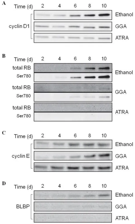
All-trans retinoic acid (ATRA) plays crucial roles in cell survival and differentiation of neuroblastoma cells. In the present study, we investigated the effects of geranylgeranoic acid (GGA), an acyclic retinoid, on differentiation and neurotrophic tyrosine kinase, receptor, type 2 (NTRK2) gene expression in SH-SY5Y human neuroblastoma cells in comparison with ATRA. GGA induced growth suppression and neural differentiation to the same extent as ATRA. Two variants (145 and 95 kDa) of the NTRK2 protein were dramatically increased by GGA treatment, comparable to the effect of ATRA. Following 6- to 8-d GGA treatment, the effect of GGA on NTRK2 was reversed after 2–4 d of its removal, whereas the effect of ATRA was irreversible under the same conditions. Both GGA and ATRA upregulated the cellular levels of three major NTRK2 messenger RNA splice variants in a time-dependent manner. Time-dependent induction of cell cycle-related genes, such as cyclin D1 and retinoblastoma protein, and amplification of the neural progenitor cell marker, brain lipid binding protein, were suppressed by GGA treatment and were completely abolished by ATRA. ATRA and GGA induced retinoic acid receptor β (RARβ) expression, whereas the time-dependent expression of both RARα and RARγ was abolished by ATRA, but not by GGA. Our results suggest that GGA may be able to restore neuronal properties of SH-SY5Y human neuroblastoma cells in a similar but not identical way to ATRA.
III. 2. Introduction
III. 2. 1. Neurotrophins and their receptors
Neurotrophins (NTs) play crucial roles in the development of the central nervous system, influencing proliferation, differentiation, survival and death of neuronal and non-neuronal cells. NTs include nerve growth factor (NGF), brain-derived neurotrophic factor (BDNF), neurotrophin 3 (NTF3) and neurotrophin 4 (NTF4). NTs mediate their cellular effects through the actions of two different receptors, the neurotrophic tyrosine kinase, receptor (NTRK, previously known as Trk; tropomyosin-related kinase receptor) and p75 neurotrophin receptor (p75NTR), a member of the TNF (tumor necrosis factor) receptor superfamily (Fig.
III-1). NGF binds most specifically to NTRK1, BDNF and NTF4 to NTRK2, and NTF3 to NTRK3. All NTs can bind to p75NTR with low affinity, enhancing the activation of NTRK receptors [Bothwell et al, 1995]
(Fig. III-2).
Fig. III-1. Schematic diagram of neurotrophins and their cell-surface membrane receptors.
[modified from Chao, 2003]
Fig. III-2. Crosstalk between NTRK2 and p75NTR with NTs.
[modified from Chao, 2003]
JNK; mitogen-activated protein kinase 8, MAPK; mitogen-activated protein kinase 14, NF-κB; nuclear factor of kappa-light polypeptide gene enhancer in B-cells, PI3K;
phosphatidylinositol-4,5-bisphosphate 3- kinase, PLCγ; phospholipase C gamma
III. 2. 2. BDNF
In the pathophysiology of depression, BDNF and neurogenesis have an important function [Nibuya et al, 1995; Santarelli et al, 2003; Monteggia et al, 2007; Adachi et al, 2008]. In addition, electroconvulsive seizures [Altar et al, 2004] and administration of antidepressant drugs can increase hippocampal BDNF levels [Nibuya et al, 1995]. Moreover, Taliaz et al. reported that reduced BDNF protein levels, in hippocampal sub-regions, could reduce neurogenesis in vivo and affected behaviors associated with depression [Taliaz et al, 2010]. Furthermore, several studies show that BDNF regulates synaptic transmission, synaptic plasticity and synaptic growth [Lu et al, 2013]. Since the synaptic dysfunction is a key pathophysiological hallmark in neurodegenerative disorders, BDNF may provide the synaptic repair therapies for neurodegenerative disorders, including Alzheimer’s disease [Lu et al, 2013].
NTRK2 is expressed in three major splice variants [Middlemas et al, 1991]. Full-length receptors (NTRK2-FL) possess an intracellular tyrosine kinase domain as well as an extracellular ligand-binding domain. The two truncated receptors, NTRK2-T1 and NTRK2-Shc, lack tyrosine kinase activity [Klein et al, 1990] (Fig. III-2). NTRK2-T1 has a direct functional role in mediating calcium release from intracellular stores [Rose et al, 2003]. NTRK2-Shc contains an SHC (Src homology 2 domain containing) transforming protein binding domain and is predominantly expressed in the brain, probably as a negative regulator of NTRK2 signaling [Zuccato et al, 2008]. Consequently, impairment of the BDNF/NTRK2 signaling system (Fig. III-3) is considered to associate with neurodevelopmental and neurodegenerative diseases [Yoshii &
Constantine-Paton, 2010]. In other words, proper maintenance of the BDNF/NTRK2 signaling system may
be important in prevention of neurodevelopmental/ neurodegenerative disorders, or neuroblastoma.
Fig. III-3. BDNF/NTRK2 signaling pathways.
AKT; RAC-alpha serin-threonine-protein kinase, CaMK; calcium/calmodulin-dependent protein kinase, CREB; cyclic AMP-responsive element-binding protein, ERK; mitogen-activated protein kinase 1, GAB2; GRB2-associated binding protein 2, GRB2; growth factor receptor-bound protein 2, IRS; insulin receptor substrate, MEK; mitogen-activated protein kinase kinase, p38MAPK; mitogen-activated protein kinase 14, PI3K, phosphatidylinositol-4,5-bisphosphate 3-kinase, RAF; RAF proto-oncogene
serin/threonin-protein kinase, RAS; rat sarcoma viral oncogen homolog, SHC; SHC-transforming protein 1, SOS; son of sevenless homolog, TK; tyrosine kinase
III. 2. 3. Differentiation-inducing effect of ATRA on neuroblastoma SH-SY5Y cells
It is well known that all-trans retinoic acid (ATRA) predisposes undifferentiated SH-SY5Y neuroblastoma cells to BDNF treatment; for example, ATRA-treated SH-SY5Y cells produce and secrete β-amyloid precursor protein (APP) upon BDNF treatment; however, naive SH-SY5Y cells do not produce APP after exposure to BDNF alone [Ruiz-Leon & Pascual, 2003]. These results indicate that ATRA shifts BDNF-insensitive SH-SY5Y cells to BDNF-responsive SH-SY5Y cells. In fact, many researchers have reported that NTRK2 gene expression is upregulated by ATRA treatment in SH-SY5Y cells [Kaplan et al, 1993; Hu & Koo, 1998; Edsjo et al, 2003; Ruiz-Leon & Pascual, 2003; Hecht et al, 2005; Holback et al, 2005; Kou et al, 2008; Nishida et al, 2008], however, a couple of exceptional papers demonstrated that ATRA decreased NTRK2 mRNA levels in SH-SY5Y cells [Enrhard et al, 1993; Chen et al, 2010].
III. 2. 4. Metabolites of the mevalonate pathway in neurons
Neuron is one of the second most active cells that are producing cholesterol from the mevalonate pathway after hepatocyte. In neurons, a fraction of cholesterol is metabolized and eliminated by the enzyme cholesterol 24-hydroxylase, as shown in Fig. III-4. The mevalonate pathway in neurons produces neuronal steroids as well as nonsterol isoprenoids including geranylgeraniol (GGOH). Recently, Kotti’s group have shown that constant production of GGOH in neuronal cells is required for long-term potentiation but not via protein geranylgeranylation [Kotti et al, 2006; 2008; Russell et al, 2009]. GGOH may be metabolically