• 検索結果がありません。

A Specimen of the Ammodytid Genus Ammodytoides (Teleostei, Perciformes) from off Southern Shikoku Island, Japan

N/A
N/A
Protected

Academic year: 2021

シェア "A Specimen of the Ammodytid Genus Ammodytoides (Teleostei, Perciformes) from off Southern Shikoku Island, Japan"

Copied!
8
0
0

読み込み中.... (全文を見る)

全文

(1)

A Specimen of the Ammodytid Genus Ammodytoides (Teleostei, Perciformes) from off Southern Shikoku Island, Japan

Eri Katayama

1

, Hiromitsu Endo

2

and Gento Shinohara

1,3

1

National Museum of Nature and Science, 4–1–1 Amakubo, Tsukuba, Ibaraki 305–0005, Japan

E-mail: ekata@kahaku.go.jp

2

Laboratory of Marine Biology, Faculty of Science, Kochi University, 2–5–1 Akebono-cho, Kochi 780–8520, Japan

3

Hokkaido University Museum, 3–1–1 Minato-cho, Hakodate, Hokkaido 041–8611, Japan (Received 18 September 2015; accepted 23 March 2016)

Abstract A juvenile specimen, 59.2 mm standard length (SL), of the ammodytid genus Ammody- toides was collected with a midwater trawl off Cape Ashizuri-misaki, Kochi Prefecture, Shikoku Island, Japan. This specimen has the following combination of characters: dorsal fin rays 48; anal fin rays 22; pectoral fin rays 15; pored lateral line scales ca. 95; gill rakers 5+22=27; vertebrae 33+24=57; head length 25.3% SL; predorsal length 26.7% SL; caudal peduncle depth 5.7% SL;

2 supratemporal pores; pale orange-red body with blackish dorsal and anal fins posteriorly when fresh. Among the 10 valid congeners, this specimen is similar to A. gilli (Bean, 1895) (tropical eastern Pacific), A. kimurai Ida and Randall, 1993 (Ogasawara Islands) and A. kanazawai Shibu- kawa and Ida, 2013 (Ogasawara Islands) in most counts and proportions, but differs from them by the combination of coloration when fresh, and counts of lateral line scales and supratemporal pores. The validity of A. kanazawai described from a juvenile is also discussed.

Key words: Ammodytidae, Ammodytoides sp., A. gilli, A. kanazawai, A. kimurai, Kochi, Japan.

Introduction

The ammodytid genus Ammodytoides Duncker and Mohr, 1939 is composed of 10 valid species (Shibukawa and Ida, 2013): A. gilli (Bean, 1895), A. vagus (McCulloch and Waite, 1916), A. ren- niei (Smith, 1957), A. kimurai Ida and Randall, 1993, A. pylei Randall, Ida and Earle, 1994, A.

leptus Collette and Randall, 2000, A. idai Ran- dall and Earle, 2008, A. praematura Randall and Earle, 2008, A. xanthops Randall and Earle, 2008, and A. kanazawai Shibukawa and Ida, 2013. One nominal species, A. lucasanus (Beebe and Tee- Van, 1938), is regarded as a junior synonym of A.

gilli (Collette and Robertson, 2001; Shibukawa and Ida, 2013). The genus is distinguished from other ammodytid genera in having the following combination of characters: 45–50 dorsal fin rays,

21–25 anal fin rays, no teeth in the jaws, no developed ventro-lateral skin fold on the body, no pelvic fin, and expanded haemal and neural spines on the caudal vertebrae (for more detail, see Ida et al., 1994; Shibukawa and Ida, 2013).

During midwater-trawl surveys carried out by the R/V Soyo-maru (National Research Institute of Fisheries Science, Fisheries Research Agency, Yokohama) in the western North Pacific off Kyusyu and Shikoku islands of Japan, an ammo- dytid specimen with numerous fine black dots on the dorsal and anal fins was collected off Cape Ashizuri-misaki in southern Shikoku Island (Fig.

1). The specimen is very close to three species of

Ammodytoides, A. gilli distributed in the tropical

eastern Pacific, and A. kimurai and A. kanazawai,

both known only from types collected off the

Ogasawara Islands, but different from them by

(2)

some counts, proportions, and coloration includ- ing fin pigmentation. We herein report it as Ammodytoides sp. and provide comments on the validity of A. kanazawai.

Materials and Methods

Methods for counts and measurements follow Ida and Randall (1993) and Randall et al. (1994).

Vertical fin rays and vertebrae were counted from radiographs. Standard length is abbreviated as SL. Scales were observed with the aid of a scan- ning electron microscope (SEM; JEOL JSM- 6380LV, JEOL Ltd., Tokyo). Lateral line systems were temporarily stained with cyanine blue (Saruwatari et al., 1997). The skin for SEM was dehydrated with a graded series of ethanol and t-butyl alcohol, dried in a freeze dryer (JEOL JFD-300), and coated by a JEOL JFC-1600 with mixed platinum and palladium. Terminology of cephalic sensory canals and pores follows Shibu- kawa and Ida (2013). Counts for paired struc- tures are the same on both sides unless noted.

The specimens examined here are deposited in the following institutions: Laboratory of Marine Biology, Faculty of Science, Kochi University, Kochi (BSKU); National Museum of Nature and Science, Tsuku ba (NSMT); National Museum of Natural History, Smithsonian Institution, Wash- ington DC (USNM).

Ammodytoides sp.

(Figs. 1–3, Tables 1, 2)

Material examined. BSKU 71207, 59.2 mm SL, 32°40.0′N, 133°34.2′E, off Cape Ashizuri- misaki, Kochi Pref., Japan, midwater trawl, depth unknown (bottom depth: 912 m), R/V Soyo-maru, 25 April 2004, coll. by K. Kameda.

Description. Proportional measurements and counts are provided in Tables 1, 2. Dorsal fin rays 48; anal fin rays 22; pectoral fin rays 15; princi- pal caudal fin rays 9+9 (uppermost and lower- most rays unbranched); tubed (pored) lateral line scales ca. 95, followed by 5 unpored scales in

left side, right side damaged; scales above lateral line to origin of dorsal fin 2; scales below lateral line to origin of anal fin ca. 11; predorsal scales 13; gill rakers 5+22; pseudobranchial filaments 14; branchiostegal rays 7; vertebrae 33+24=57;

predorsal vertebrae 4; first two dorsal pterygio- phores in space between 4th and 5th neural spines; neural and haemal spines of vertebrae of caudal peduncle expanded distally; postdorsal vertebrae 11; vertebrae posterior to anal fin 9.

Body elongate, slightly compressed. A longitu- dinal ventro-lateral fold not developed. Dorsal fin origin behind a vertical line through pectoral fin base. Anus just before anal fin origin. Caudal peduncle deep, its length more than half of body depth. Broad gap between second and third sub- orbitals. Posterior corner of dorsal fin not form- ing lobe. All dorsal and anal fin rays unbranched, except last rays branched at base. Pelvic fins absent. Snout pointed, compressed, its length longer than orbit diameter. Interorbital space wide, its width equal to orbit diameter. Jaws extending slightly behind vertical through ante- rior margin of eye. Lower jaw projecting, sharply pointed. No teeth on jaws. Gill opening broad, posterior edge of operculum slightly pointed.

Gill rakers slender, longest at middle of 1st gill arch, slightly shorter than longest gill filament.

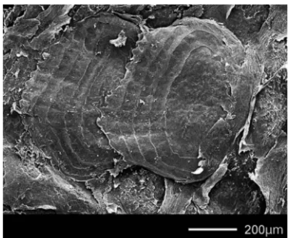
Scales small and weak, cycloid and arranged in diagonal straight rows. Four semi-circular ridges on scale, posterior edge of each scale ser- rated (Fig. 2). Ventrolateral skin fold undevel- oped. Head naked. V-shaped patterns of scale rows on nape. Caudal fin base covered with small scales. Lateral line scales tubed (pored), except on caudal peduncle. Lateral line posi- tioned dorsally on body, continuing to caudal fin.

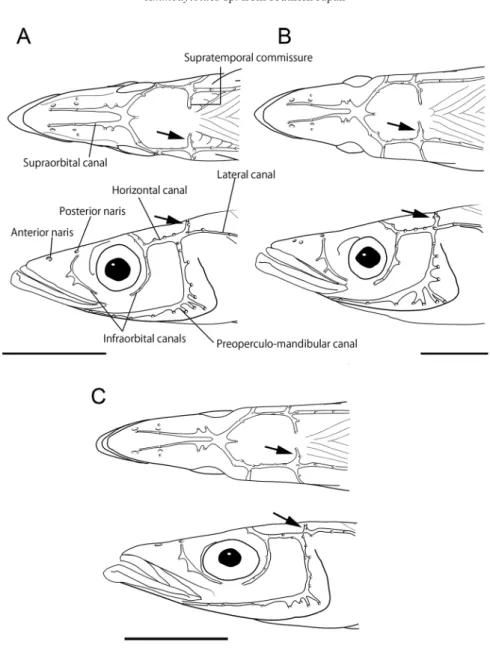
Cephalic lateral line system as shown in Fig.

3. Nostrils small, located dorsally on snout; ante- rior nostril below anterior supraorbital pore, pos- terior nostril above anterior infraorbital pore;

infraorbital canal broadly interrupted below eye;

supratemporal commissure with 2 pores.

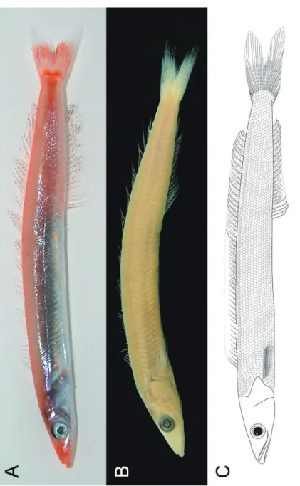
Color when fresh (Fig. 1A). Overall color of

head and body pale orange-red. Snout, pectoral

fin base, dorsal profile of body posteriorly, mid-

(3)

Fig. 1. Ammodytoides sp. collected of f Cape Ashizuri-misaki, BSKU 71207, 59.2 mm SL. — A, fresh condition (photographed by K. Kameda); B, preserved condition; C, schematic illustration (drawn by E. Katayama).

(4)

lateral caudal peduncle, caudal fin base, and mid- dle of caudal fin deep orange-red. Occipital area, preopercle, upper part of opercle, and mid-lateral trunk blackish to brownish internally. A broad silvery stripe mid-lateraly on body. Suborbital and opercular region, underside of head, and ventro-lateral portion of abdomen silver. Mem- branes of dorsal, anal, and paired fins transparent, with numerous fine black dots scattered posterior to 18th and 3rd rays of dorsal and anal fins, respectively. Dorsal and anal fin rays pale orange- red. Anterior third of caudal fin pale orange-red with numerous black dots, broadly edged with deep orange-red posteriorly; posterior third of upper and lower lobes of caudal fin translucent.

Color in alcohol (Fig. 2B). Head and body uniformly pale cream yellow. Occiput and upper region of opercle somewhat darkish. Numerous fine black dots on vertical fins membranes;

paired fins translucent. Fin rays whitish.

Remarks. The present specimen is clearly a species of Ammodytoides in having the following combination of characters: 48 dorsal fin rays (45–50 in the genus), 22 anal fin rays (21–25), no teeth in the jaws, no pelvic fins, undeveloped ventro-lateral skin fold, expanded haemal and neural spines of caudal vertebrae, and a discon- tinuous subocular canal below eye (see Ida et al., 1994; Shibukawa and Ida, 2013). Among the 10 valid species in the genus, this specimen can be distinguished from all species except A. gilli by the following counts: dorsal fin rays 48 (vs.

50–53 in A. leptus and A. renniei), vertebrae 57 (vs. 61–63 in A. leptus), predorsal vertebrae 4 (vs. 6 in A. idai, A. praematura, and A. xan- thops), gill rakers on upper limb 5 (vs. 10 in A.

renniei), and pored lateral line scales ca. 95 (vs.

103–118 in the congeners except for A. gilli) [Table 2; data of nine species are from Collette and Randall (2000), Collette and Robertson (2001), Ida and Randall (1993), McCulloch and Waite (1916), Randall and Earle (2008), Randall et al. (1994), Randall and Heemstra (2008) and Smith(1957)]. All counts for fin rays, scales, gill rakers, and vertebrae in the present specimen however are included in or extensions of ranges

of A. gilli known from the tropical eastern Pacific. Furthermore, two pores on the supratem- poral commissure are common to both species (Fig. 3A, C). Its pale orange-red coloration when fresh, however, is clearly different from the

“translucent gray-white” color of A. gilli (Fig. 1;

Collette and Robertson, 2001).

Collette and Robertson (2001) re-described A.

gilli from 50 specimens ranging in length from 42.3 to 115 mm SL (including the lectotype), and provided ranges for counts (Table 2) and described two ontogenetic changes, a reduction of the proportional length of the expanded poste- rior dorsal fin lobe and an increase in number of branched dorsal and anal fin rays with growth.

Furthermore, they observed that A. gilli has a yellow head and 1–5 large black blotches on the dorsal fin that appear in larger specimens (69.5–

84.4 mm SL), but not in smaller ones (42.3–

62.2 mm SL). The dorsal fin characters are there- fore not useful for identifying the present specimen of 59.2 mm SL.

In the western North Pacific, this specimen is similar to A. kimurai and A. kanazawai known from the Ogasawara Islands in counts, with the exception of pored lateral line scales as well as, its fin pigmentation (Fig. 1, Table 2; Shibukawa and Ida, 2013: fig. 2). The present specimen dif- fers from A. kimurai (98.3–116 mm SL) in three morphometric characters: a narrower body width

Fig. 2. SEM image of scales on lateral side of body

(posterior to right pectoral fin) of Ammodytoi-

des sp., BSKU 71207.

(5)

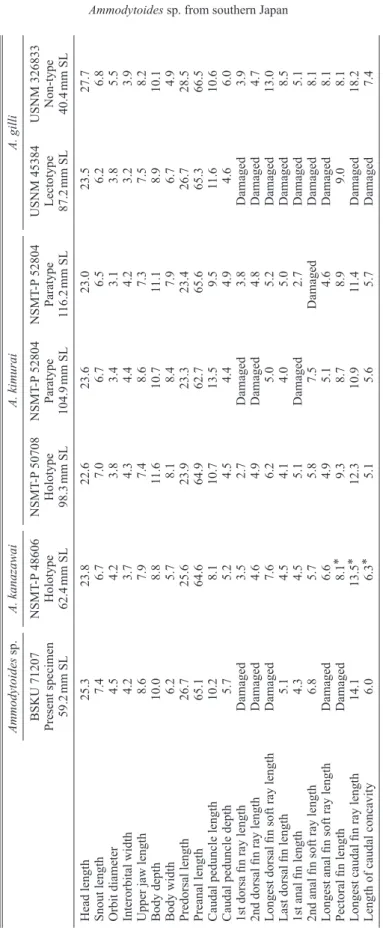
Table 1. Proportional measurements (% SL) for the present specimen and three species of Ammodytoides . Ammodytoides sp. A. kanazawai A. kimurai A. gilli BSKU 71207 Present specimen 59.2 mm SL

NSMT -P 48606 Holotype 62.4 mm SL

NSMT -P 50708 Holotype 98.3 mm SL

NSMT -P 52804 Paratype 104.9 mm SL

NSMT -P 52804 Paratype 116.2 mm SL

USNM 45384 Lectotype 87.2 mm SL

USNM 326833 Non-type 40.4 mm SL Head length 25.3 23.8 22.6 23.6 23.0 23.5 27.7 Snout length 7.4 6.7 7.0 6.7 6.5 6.2 6.8 Orbit diameter 4.5 4.2 3.8 3.4 3.1 3.8 5.5 Interorbital width 4.2 3.7 4.3 4.4 4.2 3.2 3.9 Upper jaw length 8.6 7.9 7.4 8.6 7.3 7.5 8.2 Body depth 10.0 8.8 11.6 10.7 11.1 8.9 10.1 Body width 6.2 5.7 8.1 8.4 7.9 6.7 4.9 Predorsal length 26.7 25.6 23.9 23.3 23.4 26.7 28.5 Preanal length 65.1 64.6 64.9 62.7 65.6 65.3 66.5 Caudal peduncle length 10.2 8.1 10.7 13.5 9.5 11.6 10.6 Caudal peduncle depth 5.7 5.2 4.5 4.4 4.9 4.6 6.0 1st dorsa fin ray length Damaged 3.5 2.7 Damaged 3.8 Damaged 3.9 2nd dorsal fin ray length Damaged 4.6 4.9 Damaged 4.8 Damaged 4.7 Longest dorsal fin soft ray length Damaged 7.6 6.2 5.0 5.2 Damaged 13.0 Last dorsal fin length 5.1 4.5 4.1 4.0 5.0 Damaged 8.5 1st anal fin length 4.3 4.5 5.1 Damaged 2.7 Damaged 5.1 2nd anal fin soft ray length 6.8 5.7 5.8 7.5 Damaged Damaged 8.1 Longest anal fin soft ray length Damaged 6.6 4.9 5.1 4.6 Damaged 8.1 Pectoral fin length Damaged 8.1 * 9.3 8.7 8.9 9.0 8.1 Longest caudal fin ray length 14.1 13.5 * 12.3 10.9 11.4 Damaged 18.2 Length of caudal concavity 6.0 6.3 * 5.1 5.6 5.7 Damaged 7.4

* Data from Shibukawa and Ida (2013)

(6)

Table 2. Counts for the present specimen and 10 valid species of Ammodytoides . ― CP , central Pacific; CWP , central western Pacific; EP , eastern Pacific; SP , South Pacific; WSI, western South Indian Ocean; WNP , western North Pacific; WSP , western South Pacific. Ammodytoides sp. A. gilli A. idai A. kanazawai A. kimurai A. leptus Present specimen Collette & Robertson (2001) Randall & Earle (2008) Present study Present study , Ida & Randall (1993) Collette & Randall (2000) n = 1 n = 50 n = 10 n = 1 n = 3 n = 23 Dorsal fin rays 48 44–47 44–46 50 48–49 50–53 Anal fin rays 22 22–24 21–22 23 23–24 24–25 Pectoral fin rays 15 14–15 14–16 16 14–15 16–17 Lateral line scales (tubed + unpored) ca. 95 + 5 85–94 + 2–5 103–107 + 4–6 ca.107 or 108 + 5 104–109 + 6–8 114–1 18 + 4–6 Predorsal scales 11 10–12 11 13 12 10–12 Gill rakers on first arch 5 + 22 5–7 + 21–25 5–6 + 21–23 5 + 21 6 + 21–23 6–7 + 22–25 Vertebrae 33 + 24 = 57 31–33 + 23–26 = 55–58 55–58 34 + 26 = 60 33–34 + 25–27 = 59–61 34–36 + 26–27 = 61–63 Predoral vertebrae 4 4 6 4 4 No data Postdorsal vertebrae 11 11 10 11 12 No data Distribution Shikoku I., Japan, WNP Tropical EP Papua New Guinea, CWP

Ogasawara Is., Japan, WNP Ogasawara Is., Japan, WNP Pitcairn I., SP A. pylei A. praematura A. r enniei A. vagus A. xanthops Randall et al. (1994) Randall & Earle (2008) Smith (1957) McCulloch & W aite (1916) Randall & Heemstra (2008) n = 17 n = 1 n = 3 n = 1 n = 19 Dorsal fin rays 48–52 48 50–51 48 48 Anal fin rays 22–25 24 23–24 22 23 Pectoral fin rays 15–17 14 No data 16 16 Lateral line scales (tubed + unpored) 109–1 16 (total) 103–106 + 4 113–1 16 (tubed) 107 + 5 106–1 12 + 4 Predorsal scales 10–13 No data 12 No data 11 Gill rakers on first arch 5–7 + 23–25 5 + 23 10 + 1 + 20–21 6 + 21 5–6 + 22–25 Vertebrae 34 + 25–26 = 59–60 58 32 + 26 = 57 No data 57–59 Predoral vertebrae 4 6 4* 4* 6 Postdorsal vertebrae 12 10 11 * 12 * 10 Distribution Hawaiian Is., CP Chagos Arch., WSI South Africa, WSI Lord Howe I., WSP Mozambique, WSI

* Data from Ida

et al. (1994)

(7)

(6.2% SL vs. 7.9–8.4), a longer predorsal (26.7%

SL vs. 23.3–23.9), and deeper caudal peduncle (5.7% SL vs. 4.4–4.9) (Table 1). These differ- ences, however, need further study to determine if proportional changes related with growth are involved as this specimen is smaller than avail- able specimens identified as A. kimurai, with regard to coloration when fresh, the pale orange- red of this specimen readily differs from the

“bluish gray” described for A. kimurai (Fig. 1;

Ida and Randall, 1993). The present specimen also resembles A. kanazawai known only from the holotype in the proportions, but differs from it in having 2 pores on the supratemporal com- missure (vs. 3; Fig. 3).

The original descriptions of A. kimurai and A.

kanazawai state they have 2 or 3 pores on the supratemporal commissure, respectively (Ida and Randall, 1993: fig. 2; Shibukawa and Ida, 2013:

fig. 3), but our examination revealed that the holo-

Fig. 3. Dorsal (above) and lateral (below) views of head of the present specimen and two species of Ammodytoi-

des. — A: A. sp., BSKU 71207, 59.2 mm SL, B: A. kimurai, paratype, 105 mm SL, C: A. gilli, USNM 326833,

40.4 mm SL. Bars indicate 5 mm. Arrows indicate pores of supratemporal commissure.

(8)

type and two paratypes of A. kimurai have 3 pores (Fig. 4B). Hence, these two species are not sepa- rable by the character. The lack of branched dor- sal fin rays in the holotype of A. kanazawai may be a juvenile feature as mentioned above for ontogenetic changes in A. gilli. In addition, dif- ferences of pigmentation of the dorsal fin in the two species, a series of black spots on the outer edge (absent in A. kanazawai at 63.2 mm SL vs.

present in A. kimurai at 99.4–120.6 mm SL; Ida and Randall, 1993) and numerous fine black dots (present vs. absent), may be attributable to growth as in A. gilli. Consequently, additional specimens for the full ranges of size are required to elucidate the variation of these characters and confirm the validity of A. kanazawai.

Comparative materials. Ammodytoides gilli:

USNM 45384, lectotype, 87.2 mm SL, “Dr. Stimp- sonʼs collections from the Pacific, date unknown;

USNM 326833, 40.4 mm SL, Panama, Azuero Peninsula, Playa Venao, small lagoon near Tuna Commission Laboratory, in rocky and sandy bot- tom 2–3 m depth, 29 Apr. 1990. Ammodytoides kanazawai: NSMT-P 48606, holotype, 63.2 mm SL, off southern Chichi-jima Island, Ogasawara Islands, Japan, 95–99 m depth, coll. by T.

Kanazawa, 19 June 1995. Ammodytoides kimurai:

NSMT-P 50708, holotype, 101.0 mm SL, off Minami-shima Island, Ogasawara Islands, Japan, coll. by H. Ida and R. L. Pyle, 1 June 1992;

NSMT-P 52804 (formerly 2 of FSKU 920601, transferred from the School of Fisheries Sciences, Kitasato University, Iwate), paratypes, same col- lecting data as holotype, 104.9–116.2 mm SL.

Acknowledgements

We are grateful to Kazunari Kameda (Sea Tur- tle Association of Japan) for supplying the pres- ent specimen and the picture when fresh. We also thank Koichi Shibukawa (Museum of Natural and Environmental History, Shizuoka) for valu- able suggestions and critical comments to early manuscript. Our special thanks go to Martin Gomon (Museum Victoria, Melbourne) for edit- ing the English text. This study was partly sup-

ported by a grant from the Fujiwara Natural His- tory Foundation, Tokyo (EK), a Grant-in-Aid for Scientific Research (B) from the Japan Society for the Promotion of Science, Tokyo (24370041) (HE), a Grant-in-Aid of the “Marine Science Project” of the Natural Science Cluster, Science Unit, Kochi University (HE), the “Kuroshio Sou- gou Project” of the National Museum of Nature and Science, Tsukuba (HE) and the MEXT KAKENHI Grant number 24120001 (GS).

References

Collette, B. B. and J. E. Randall 2000. Ammodytoides lep- tus, a new species of sand lance (Teleostei: Ammody- toidae) from Pitcairn Island. Proceedings of the Biolog- ical Society of Washington, 113: 397–400.

Collette, B. B. and D. R. Robertson 2001. Redescription of Ammodytoides gilli, the tropical eastern Pacific sand lance (Perciformes: Ammodytidae). Revista de Biolo- gia Tropical, 49, Supplement 1: 111–115.

Ida, H. and J. E. Randall 1993. Ammodytoides kimurai, a new species of sand lance (Ammodytidae) from the Ogasawara Islands. Japanese Journal of Ichthyology, 40: 147–151.

Ida, H., P. Sirimontaporn and S. Monkolprasit 1994.

Comparative morphology of the fishes of the family Ammodytidae, with a description of two new genera and two new species. Zoological Studies, 33: 251–277.

McCulloch, A. R. and E. R. Waite 1916. Additions to the fish-fauna of Lord Howe Island. No. 5. Transactions Royal Society of South Australia, 40: 437–451.

Randall, J. E. and J. L. Earle 2008. Two new Indo-Pacific sand lances of the genus Ammodytoides (Perciformes:

Ammodytidae). Pacific Science, 62: 603–612.

Randall, J. E. and P. C. Heemstra 2008. Ammodytoides xanthops, a new species of sandlance (Perciformes:

Ammodytidae) from Mozambique. Smithiana, Publica- tions in Aquatic Biodiversity, Bulletin, 9: 21–25.

Randall, J. E., H. Ida and J. L. Earle 1994. Ammodytoides pylei, a new species of sand lance (Ammodytidae) from the Hawaiian Islands. Pacific Science, 48: 80–89.

Saruwatari, T., J. A. López and T. W. Pietsch 1997. Cya- nine blue: a versatile and harmless stain for specimen observation. Copeia, 1997: 840–841.

Shibukawa, K. and H. Ida 2013. Ammodytoides kana- zawai, a new species of sand lance (Perciformes:

Ammodytidae) from the Ogasawara Islands, Japan.

Bulletin of the National Museum of Nature and Sci- ence, Series A, Supplement 7: 25–30.

Smith, J. L. B 1957. Four interesting new fishes from South

Africa. South African Journal of Science, 53: 219–222.

Fig. 1. Ammodytoides sp. collected off Cape Ashizuri-misaki, BSKU 71207, 59.2 mm SL.—A, fresh condition (photographed by K
Fig. 2.  SEM image of scales on lateral side of body  (posterior  to  right  pectoral  fin)  of   Ammodytoi-des sp., BSKU 71207.
Table 1. Proportional measurements (% SL) for the present specimen and three species of Ammodytoides
Table 2. Counts for the present specimen and 10 valid species of Ammodytoides. ― CP, central Pacific; CWP, central western Pacific; EP, eastern Pacific; SP, South  Pacific; WSI, western South Indian Ocean; WNP, western North Pacific; WSP, western South Pacific
+2

参照

関連したドキュメント

The input specification of the process of generating db schema of one appli- cation system, supported by IIS*Case, is the union of sets of form types of a chosen application system

Laplacian on circle packing fractals invariant with respect to certain Kleinian groups (i.e., discrete groups of M¨ obius transformations on the Riemann sphere C b = C ∪ {∞}),

The only thing left to observe that (−) ∨ is a functor from the ordinary category of cartesian (respectively, cocartesian) fibrations to the ordinary category of cocartesian

Eskandani, “Stability of a mixed additive and cubic functional equation in quasi- Banach spaces,” Journal of Mathematical Analysis and Applications, vol.. Eshaghi Gordji, “Stability

Finally, we give an example to show how the generalized zeta function can be applied to graphs to distinguish non-isomorphic graphs with the same Ihara-Selberg zeta

We use these to show that a segmentation approach to the EIT inverse problem has a unique solution in a suitable space using a fixed point

The main problem upon which most of the geometric topology is based is that of classifying and comparing the various supplementary structures that can be imposed on a

It turns out that the symbol which is defined in a probabilistic way coincides with the analytic (in the sense of pseudo-differential operators) symbol for the class of Feller