GUIDELINE
Clinical practice guide for the treatment of perforating dermatosis
Tamihiro KAWAKAMI,
1Masashi AKIYAMA,
2Akemi ISHIDA-YAMAMOTO,
3Hajime NAKANO,
4Chikage MITOMA,
5Kozo YONEDA,
6Yasushi SUGA
71
Division of Dermatology, Tohoku Medical and Pharmaceutical University, Sendai,
2Department of Dermatology, Nagoya University Graduate School of Medicine, Hagoya,
3Department of Dermatology, Asahikawa Medical University, Asahikawa,
4Department of Dermatology, Hirosaki University Graduate School of Medicine, Hirosaki,
5Ai Dermatology Clinic, Fukuoka,
6Department of Clinical Pharmacology, Faculty of Pharmaceutical Sciences, Osaka Ohtani University, Osaka,
7Department of Dermatology, Juntendo University Urayasu Hospital, Urayasu, Japan
ABSTRACT
Perforating dermatoses are a heterogeneous skin disease group defined by transepidermal elimination of various skin materials. Four classical forms of primary perforating dermatosis have been described, where the transepider- mal elimination mechanism represents the hallmark of the disease: acquired reactive perforating collagenosis, elas- tosis perforans serpiginosa, Kyrle’s disease and perforating folliculitis. Acquired reactive perforating collagenosis presents with transepidermal elimination of collagen fibers. Elastosis perforans serpiginosum presents with the elimination of elastic fibers. Kyrle’s disease presents with transepidermal elimination of abnormal keratin. In perfo- rating folliculitis, it is the content of the follicle. We established diagnostic criteria and severity classification. In addi- tion, the Japanese guideline for treatment of perforating dermatoses was updated using the Medical Information Network Distribution Service (MINDS) methodology. The guideline is based on a systematic published work review completed from 1989 to 2019, and on a formal consensus and approval process. Most medical published work on the treatment is limited to individual case reports and small series of patients. The guideline covers treatment options considered relevant by the expert panel and approved in Japan at the time of the consensus conference.
Key words: acquired reactive perforating collagenosis, elastosis perforans serpiginosa, Kyrle’s disease, perforating dermatosis, perforating folliculitis.
PREPARATION OF CLINICAL PRACTICE GUIDE
Features of clinical practice guide
Guidelines are “systematic documents designed to assist clinicians and patients in making appropriate decisions in specific clinical situations”. The clinical practice guide is based on the Medical Information Network Distribution Service (MINDS) clinical practice guideline. Its purpose is to present recommendations of evidence-based medicine on medical actions of high clinical importance, that we consider to be optimal for supporting decisions of the patient and the medical staff, while considering the bal- ance between benefits and harms. Each item was created in the clinical question (CQ) format, with the aim of creat- ing highly practical guidelines that general clinicians imme- diately understand and practice in the field, and disseminate thereafter.
Perforating dermatosis has been reported in a small number of patients and has no globally uniform diagnostic criteria;
therefore, clinical trials with a high level of evidence such as randomized controlled trials (RCT) and prospective cohort studies are difficult to perform. Sufficient clinical data cannot be accumulated and scientific evidence (clinical studies and articles) with a high level of evidence cannot be obtained. We did not perform a systematic review in this clinical practice guide. Instead, we decided to collect and evaluate the degree of consent to the recommendation by dermatologists to pro- vide the scientific basis with a low evidence level.
When we see an actual patient, there are various individual background factors such as differences in underlying diseases and degrees of symptoms and complications. Although this clinical practice guide shows the present standard medical care in Japan, in the general medical care, the physician should decide the treatment policy based on the consultation with the patient.
Correspondence: Tamihiro Kawakami, M.D., Ph.D., Division of Dermatology, Tohoku Medical and Pharmaceutical University, 1-15-1 Fukumuro, Miyagino-ku, Sendai, Miyagi 983-8536, Japan. Email: tami@tohoku-mpu.ac.jp
This is the secondary English version of the original Japanese manuscript for “Clinical practice guide for the treatment of perforating dermatosis”, published in theJapanese Journal of Dermatology2020; 130(9): 2007–2016, 2020.
Received 13 September 2020; accepted 16 September 2020.
Evidence level and article extraction
The evaluation and classification of the evidence level was based on the guideline for the MINDS clinical practice guideli- nes 20071and 2014 (Table 1)2. In the extraction of articles, we searched for published documents from January 1989 to Octo- ber 2019. In addition, the latest important articles were added as appropriate. The databases used were Medline, PubMed and the Japan Medical Abstracts Society Web, each of which had their own hand search. In the selection criteria, RCT papers were given priority. When not available, papers from cohort studies, case–control studies, non-control studies, case reports and case series were included. Published work of basic experiments was excluded.
Adaptation of consent level
After extracting articles, due to the lack of RCT papers, the low level of evidence became a problem. On the other hand, even if there is no scientific basis with a high level of evidence, there are many items that are generally recognized in dermato- logical practice. Based on the Behcßet’s disease clinical prac- tice guideline 2020,3we decided to create a five-tier consent level classification to supplement the recommendation level (Table 2). That is, if the recommendation is followed nine times or more in 10 clinical opportunities, the level of consent is 5 (strongly agree), the level of consent is 4 or more if the recom- mendation is followed seven times or more (agree), and the level of consent is 3 or more if the recommendation is followed five times or more (agree with conditions). This scoring task was performed by 19 dermatologists with mutual consultation prohibited. The aggregated results are shown as average val- ues. CQ and recommendations that did not obtain consent levels of 4 or higher were considered as not generally used in actual clinical settings, even if there was a certain level of evi- dence. The CQ itself was deleted and it was decided to not be included in this clinical practice guide.
Criteria for the degree of recommendation and recommendation sentence
The degree of recommendation is determined based on the level of evidence. CQ based on clinical trials and academic articles with a high level of evidence has a high degree of recommenda- tion. However, the strength of the evidence does not directly
indicate the strength of the recommendation. There is also a CQ that can be performed in the actual clinical setting even if the evi- dence level is not high. Because perforating dermatosis has a small number of patients reported and no globally uniform diag- nostic criteria, sufficient evidence cannot be proved in RCT arti- cles. Based on the Behcßet’s disease clinical practice guideline 2020,3the criteria for recommendation levels and recommenda- tion sentences were determined (Table 3). Furthermore, a con- sensus building meeting was held, and the level of recommendation was determined by an unbiased method.
Source of funding and conflict of interest
Regarding perforating dermatosis, the editorial board is not supported by any specific organization/company or pharma- ceutical company. In addition, the members involved in the clinical practice guide and the members involved in the verifi- cation have no relationship with any organization that may cause conflicts of interest. In other words, there is no conflict of interest to clarify.
DIAGNOSTIC CRITERIA AND SEVERITY CLASSIFICATION OF PERFORATING DERMATOSIS
Diagnostic criteria for perforating dermatosis
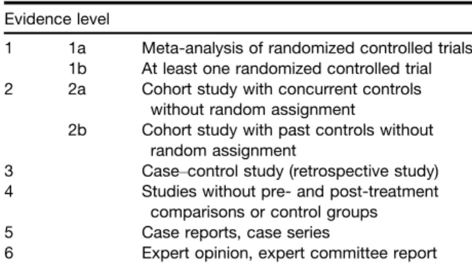
Each disease was reported for the first time by the following:Kyrle’s disease in 1916 by Kyrle,4elastosis perforans serpigi- nosa in 1953 by Lutz,5and perforating folliculitis and acquired reactive perforating collagenosis in 1968 by Mehregan and Coskey (Table 4).6 These diseases are characterized by the phenomenon of transepidermal elimination of denatured dermis as a histopathological finding. Rapiniet al.7collectively referred Table 1. Evidence level classification
Evidence level
1 1a Meta-analysis of randomized controlled trials 1b At least one randomized controlled trial 2 2a Cohort study with concurrent controls
without random assignment
2b Cohort study with past controls without random assignment
3 Case–control study (retrospective study) 4 Studies without pre- and post-treatment
comparisons or control groups
5 Case reports, case series
6 Expert opinion, expert committee report
Table 2. Degree of consent
Consent level
Frequency to follow recommendations in 10 clinical settings
5 Strongly agree ≥9 times
4 Agree ≥7 times
3 Agree with conditions ≥5 times
2 Moderately disagree ≥4 times
1 Disagree ≤1 time
Table 3. Determination of recommendation sentences Recommendation
sentence
Evidence level
Consent level
Recommendation level
Highly recommend Mainly 1 ≥4.8 A
Recommend Mainly
2 or 3
≥4.5 B
Suggest Mainly 4–6 ≥4.0 C1
Do not recommend due to no basis
No evidence
C2 Recommend
not to do
Invalid or harmful evidence
D
to them as acquired perforating dermatosis in 1989. At pre- sent, given that there are cases of early-onset disease that are not necessarily acquired, they are often collectively referred to as perforating dermatosis. A pathological tissue finding of transepidermal elimination image in which denatured skin com- ponents are excreted outside the skin is essential as the diag- nostic criteria for perforating dermatosis.
We first set up histopathological findings of transepidermal elimination of degenerated cutaneous components as basic findings and set it as item A. Item B was set to include the transepidermally eliminated substances: acquired reactive per- forated collagenosis that eliminates collagen tissue from the epidermis, elastosis perforans serpiginosa that eliminates elas- tic fibers, Kyrle’s disease that eliminates keratins and perforat- ing folliculitis that eliminates collagen tissue from hair follicles.
Because the substances that are transepidermally eliminated are not clearly classified according to each disease, the term
“mainly” was added. In addition, regarding acquired reactive perforating collagenosis, for histopathology, Faveret al.8pub- lished the diagnostic criteria as meeting all of the following three criteria: (i) histopathological findings of elimination of necrotic basophilic collagen tissue into a cup-shaped epider- mal depression; (ii) clinical presentation of umbilicated papules or nodules with a central adherent keratotic plug; and (iii) onset of skin lesions after the age of 18 years. The diagnostic criteria have been cited in many articles related to acquired reactive perforating collagenosis, and some consent has been obtained. Item C was set, and umbilicated papules or nodules with a central adherent keratotic plug, and the onset at 18 years or older were listed as clinical findings that lead to the diagnosis of acquired reactive perforating collagenosis.
Kim et al.9 verified 30 cases of acquired perforating der- matosis and observed pruritus in 83.3% and Koebner’s phe- nomenon in 31.8%. In some articles regarding acquired reactive perforating collagenosis that are most commonly encountered in clinical practice, confirmation of pruritus and Koebner’s phenomenon is helpful for diagnosis. It was deter- mined that the diagnostic value was also high, and reference diseases include prurigo nodularis, prurigo simplex, folliculitis, insect hypersensitivity and multiple keratoacanthoma.
Perforating dermatosis severity classification
We adopted the infiltration/papulation and excoriations based on the Eczema Area and Severity Index (EASI) score for atopic dermatitis published in 2001 by Hanifin et al.10 Infiltration/papulation and excoriations were scored and summed as item 1 (Table 5). The evaluation was based on EASI: 0–0.5, near remission; 0.6–3.5, mild; 3.6–10.5, moderate; 10.6–25.0, sev- ere; and 25.1–36.0, most severe.
The Eczema Area and Severity Index is an objective index and there is no index of pruritus, which is a characteristic clini- cal finding of perforating dermatosis. Therefore, the pruritus numerical rating scale was added. That is, pruritus was divided into 11 stages from 0 to 10, and item 2 was set as 0 (no pruri- tus at all) to 10 (worst pruritus) among possible items. Evalua- tion was: 0, none; 1–3, mild; 4–7, moderate; and 8–10, severe.
The sum of item 1 and item 2 is the severity classification of the total score value (decimal points rounded up): 0–1, near remission; 2–6, mild; 7–15, moderate; 16–33, severe; and 34– 46, most severe.
CQ OF PERFORATING DERMATOSIS Acquired reactive perforating collagenosis
In 1968, Mehregan and Coskey6described it as multiple kera- totic papules with a central adherent keratinous plug, with histopathology showing transepidermal excretion of collagen fibers. In 1994, Faveret al.8published the diagnostic criteria as meeting all of the following: (i) histopathological findings of elimination of necrotic basophilic collagen tissue into a cup- shaped epidermal depression; (ii) clinical presentation of umbil- icated papules or nodules with a central adherent keratotic plug; and (iii) onset of skin lesions after the age of 18 years (Fig. 1). The diagnostic criteria have been cited in many arti- cles. An adherent keratotic plug is often also described as plaster-like. Moreover, pruritus is present and can be severe, and Koebner’s phenomenon is present as well.
CQ1: What are the characteristic comorbidities of acquired reactive perforating collagenosis?
Recommendation: Diabetes and chronic kidney disease rec- ommended to be regarded as comorbidities of acquired reac- tive perforating collagenosis.
Evidence level: 5 Level of consent: 4.72 Recommendation level: B Table 4.Diagnostic criteria for perforating dermatosis
A Basic findings
Histopathological findings show transepidermal elimination of degenerated cutaneous components (transepidermal elimination)
B Substances that are mainly transepidermally eliminated 1 Collagen tissue from epidermis
2 Elastic fibers 3 Keratins
4 Collagen tissue from hair follicles C Clinical cutaneous findings
1 Umbilicated papules or nodules with a central adherent keratotic plug
2 Onset at age 18 years and older D Reference findings
Koebner’s phenomenon Pruritus
Diagnostic category
A definite diagnosis is made in the following cases:
A+B1+C1+C2=acquired reactive perforating collagenosis
A+B2=elastosis perforans serpiginosa A+B3=Kyrle’s disease
A+B4=perforating folliculitis
Commentary: There are three case series8,11–13that report association with diabetes, and one case report14and two case series8,13 that report complications with chronic kidney dis- ease. There are no studies with a high level of evidence, only case series and case reports, and no evidence can be estab- lished. However, acquired reactive perforating collagenosis-re- lated reviews noted the relationship between diabetes and chronic kidney disease, and the degree of consent was high.
Therefore, the recommendation level B was set.
CQ2: Is treatment of diabetes and chronic renal disease effective for acquired reactive perforating collagenosis?
Recommendation: We suggest considering the treatment of coexisting diabetes and chronic kidney disease as a treatment for acquired reactive perforating collagenosis.
Evidence level: 5 Consensus level: 4.0 Recommendation level: C1
Commentary: There are three case series9,13,15 that report treatments of diabetes being effective for acquired reactive perforating collagenosis, and the same three reports of case series9,13,15 that report treatments of chronic kidney disease being effective for acquired reactive perforating collagenosis.
Based on the evidence and consent levels, the recommen- dation level was set as C1. Acquired reactive perforating col- lagenosis is often associated with diabetes and chronic kidney disease, and Koebner’s phenomenon because pruritus is observed. Therefore, we suggest that the treatment of dia- betes and/or chronic kidney disease should be considered for the treatment of acquired reactive perforating collageno- sis associated with the above diseases. Controlling pruritus is an important treatment point; however, there are only few reviews that describe the success of conventional antiallergic drugs. In the future, we anticipate that the effect of nalfu- rafine, a selective opioid jreceptor agonist with an assured
indication for pruritus during renal dialysis, for this disease will be verified.
CQ3: Is topical steroid effective for acquired reactive perforating collagenosis?
Recommendation: We suggest considering topical steroid ther- apy as a treatment for acquired reactive perforating collageno- sis.
Evidence level: 5 Consensus level: 4.17 Recommendation level: C1
Commentary: Regarding acquired reactive perforating col- lagenosis, there are three case series8,13,16 that noted the effectiveness of topical steroids. The recommendation level C1 was set based on the evidence and consensus levels. In other words, in actual clinical practice, we propose to consider topi- cal steroids as one of the treatments.
CQ4: Is ultraviolet (UV) therapy effective for acquired reactive perforating collagenosis?
Recommendation: We suggest that UV therapy be considered as a treatment for acquired reactive perforating collagenosis.
Evidence level: 5 Consensus level: 4.22 Recommendation level: C1
Commentary: Three case reports noted that UV therapy was effective for acquired reactive perforating collagenosis,17–19 and one case series reported the same as well.20The recom- mendation level C1 was set based on the evidence level and consensus level. In other words, we propose to consider UV therapy as one of the treatments in clinical practice.
Elastosis perforans serpiginosa
Elastosis perforans serpiginosa was first described by Lutz5in 1953 (Fig. 2). The rash has a serpentine appearance with Table 5. Severity classification of perforating dermatosis
1 infiltration/papulation and excoriations as a score each (0–18) infiltration/papulation
(none, 0; mild, 1; moderate, 2; severe, 3)
Body surface area (0, 0%; 1, 1–9%; 2, 10–29%;
3, 30–49%; 4, 50–69%; 5, 70–89%; 6, 90–100%)
Head 0–3 9 0–6 90.1=
Trunk 0–3 9 0–6 90.3=
Upper extremities 0–3 9 0–6 90.2=
Lower extremities 0–3 9 0–6 90.4=
Excoriations (none, 0; mild, 1; moderate, 2; severe, 3) Body surface area (0, 0%; 1, 1–9%; 2, 10–29%; 3, 30–49%; 4, 50–69%; 5, 70–89%; 6, 90–100%)
Head 0–3 9 0–6 90.1=
Trunk 0–3 9 0–6 90.3=
Upper extremities 0–3 9 0–6 90.2=
Lower extremities 0–3 9 0–6 90.4=
2 Numerical rating scale of pruritus (0–10)
0 1 2 3 4 5 6 7 8 9 10
None Mild Mild Mild Moderate Moderate Moderate Moderate Severe Severe Severe
1+2=total score value (decimal points rounded up)
Near remission, 0–1; mild, 2–6; moderate, 7–15; severe, 16–33; most severe, 34–46
keratinized papules arranged arcuately or annularly to form pla- ques. It frequently occurs in the neck, trunk and extremities.
Histopathologically, transepidermal elimination of degenerated elastic fibers can be seen. Generally, it is classified into three types: (i) reactivity type associated with diseases with abnor- mal connective tissue; (ii) drug-induced type induced by D- penicillamine; and (iii) idiopathic type with presumed genetic background. Treatments include active vitamin D3external use, cryotherapy and laser irradiation, but they are intractable.
CQ5: What are the characteristic comorbidities of elastosis perforans serpiginosa?
Recommendation: We suggest considering Down syndrome as a comorbid disease of elastosis perforans serpiginosa.
Evidence level: 5 Consensus level: 4.28 Recommendation level: C1
Commentary: There are five case reports21–25and two case series26,27that report association with Down syndrome. There are no research articles with a high level of evidence, and there are many papers; however, they are only case reports and case series. The recommendation level C1 was set based on
the evidence and consensus level. It is also known to occur in conjunction with genetic connective tissue diseases such as Marfan syndrome, Ehlers–Danlos syndrome and osteogenesis imperfecta.
CQ6: Should
D-penicillamine be discontinued if elastosis perforans serpiginosa develops during oral
D
-penicillamine treatment?
Recommendation: In cases in which elastosis perforans serpig- inosa develops during the use ofD-penicillamine and a causal relationship is strongly suspected, we suggest discontinuingD- penicillamine and consider changing to another drug.
Evidence level: 5 Consensus level: 4.28 Recommendation level: C1
Commentary: There are four case reports26–31in which elas- tosis perforans serpiginosa is thought to have occurred during the use ofD-penicillamine, a therapeutic agent for rheumatoid arthritis. Based on the evidence and consent levels, the recom- mendation level was set as C1. We suggest discontinuingD- penicillamine and consider changing to another drug in cases where a causal relationship is strongly suspected.
Figure 1. Acquired reactive perforating collagenosis Azan-Mallory staining (lower left; original magnification940) HE staining (lower right; original magnification9100).
Kyrle’s disease
Kyrle’s disease was described by Kyrle4in 1916 (Fig. 3). Skin eruptions mainly consist of papules, accompanied by erythema or pigmentation. Kyrle’s disease also shows verrucous pla- ques. It frequently occurs in the extremities and buttocks.
Histopathologically, transepidermal elimination of degenerated keratins (mainly keratin proteins) is seen.
CQ7: What are the characteristic comorbidities of Kyrle
’s disease?
Recommendation: We suggest that diabetes and chronic kid- ney disease be considered as comorbidities for Kyrle’s dis- ease.
Evidence level: 5 Consensus level: 4.22 Recommendation level: C1
Commentary: There are six case reports32–36 describing cases of complications with diabetes, and five case
reports32,36–38describing cases of complications with chronic kidney disease. Based on the evidence and consent levels, the recommendation level was set as C1.
Perforating folliculitis
Perforating folliculitis is a disease first reported by Mehregan and Coskey6in 1968 (Fig. 4). The eruption is frequently seen as follicular papules with keratotic plug. It frequently occurs in the extremities and buttocks. Histopathologically, the enlarged follicular infundibulum is filled with necrotic material, kera- tinized substances and degenerated inflammatory cells. The follicular infundibulum is perforated, and an image of transepi- dermal elimination with invasion of collagen and elastic fibers is seen.
CQ8: What are the characteristic comorbidities of perforated folliculitis?
Recommendation: We suggest considering diabetes and chronic renal disease as comorbidities of perforated folliculitis.
Figure 2. Elastosis perforans serpiginosa Provided by Dr. Jun Asai, Department of Dermatology, Kyoto Prefectural University of Medicine HE stain (bottom left; original magnification9100) Elastica van Gieson stain (bottom right; original magnification9200).
Evidence level: 5 Consensus level: 4.11 Recommendation level: C1
Commentary: There are three case series in reports of complications with diabetes.39–41 There is one case series reporting the complications with chronic kidney disease.42 In which, perforating folliculitis was reported in three of 122 patients (2.5%) with chronic renal disease over the age of 18 years. Hemodialysis was performed in all three cases.
Based on the evidence and consent levels, the recommenda- tion level was set as C1.
CQ9: If perforating folliculitis develop during molecular-targeted drug (kinase inhibitor) treatment, should the molecular-targeted drug (kinase inhibitor) be discontinued?
Recommendation: There are many articles indicating that per- forating folliculitis might have occurred during the use of
molecular-targeted drugs (kinase inhibitors). In cases where a causal relationship is strongly suspected, we suggest discon- tinuing the molecular-targeted drug (kinase inhibitor) and con- sider changing to another drug.
Evidence level: 5 Consensus level: 4.17 Recommendation level: C1
Commentary: There is an increasing number of case reports of perforating folliculitis caused by the administration of molec- ular-targeted drugs (kinase inhibitors). There are four case reports on sorafenib, a multikinase inhibitor,43–46 and two reports on nilotinib, a tyrosine kinase inhibitor.47,48 There is also one report each for infliximab,49a tumor necrosis factor inhibitor, and vemurafenib, a BRAF kinase inhibitor.50Based on the evidence and consent levels, the recommendation level was set as C1. Because a molecular-targeted drug (kinase inhibitor) is considered as an inducer, it is suggested that if Figure 3. Kyrle’s disease Provided by Dr. Shoko Nakano, Dr. Masahiro Hayashi, and Dr. Kei Nagatani, Yamagata University Depart- ment of Dermatology HE stain (upper right; original magnification9100) Elastica Masson stain (lower right; original magnification 9100).
perforating folliculitis develops, the drug should be stopped and a change to another drug should be considered.
ACKNOWLEDGMENTS:
We gratefully appreciate Professor Takashi Hashimoto from Department of Dermatology, Osaka City University Graduate School of Medicine, Osaka, for editing this work.CONFLICT OF INTEREST:
None declared.REFERENCES
1 Fukui T, Yamaguchi N, Morizane T, Yoshida M, Kojimahara N.Medi- cal Information Network Distribution Service (Minds) the guidance for development of clinical practice guideline 2007. Tokyo: Igakush- oin, 2007. [In Japanese].
2 Fukui T, Yamaguchi N, Morizane T, Yoshida M, Kojimahara N.Medi- cal Information Network Distribution Service (Minds) the guidance for development of clinical practice guideline 2014. Tokyo: Igakush- oin, 2014. [In Japanese].
3 Clinical practice guideline committee for Behcet’s disease organized by the Research Team for Intractable Disease of the Ministry of Health, Labor and Welfare.Clinical practice guideline for Behcet’s disease 2020. Tokyo: Shindantochiryousha, 2017. [In Japanese].
4 Kyrle J. Hyperkeratosis follicularis et parafollicularis in cuteni pcnc- trans.Arch Dermatol Syphil1916;123: 466–493.
5 Lutz W. Keratosis follicularis serpiginosa.Dermatologica1953;106 (3–5): 318–319.
6 Mehregan AH, Coskey RJ. Perforating folliculitis. Arch Dermatol 1968;97(4): 394–399.
7 Rapini RP, Herbert AA, Drucker CR. Acquired perforating dermato- sis. Evidence for combined transepidermal elimination of both colla- gen and elastic fibers.Arch Dermatol1989;125(8): 1074–1078.
8 Faver IR, Daoud MS, Su WP. Acquired reactive perforating col- lagenosis. Report of six cases and review of the literature. J Am Acad Dermatol1994;30(4): 575–580.
9 Kim SW, Kim MS, Lee JHet al. A clinicopathologic study of thirty cases of acquired perforating dermatosis in Korea. Ann Dermatol 2014;26(2): 162–171.
10 Hanifin JM, Thurston M, Omoto M et al. The eczema area and severity index (EASI): assessment of reliability in atopic dermatitis.
Exp Dermatol2001;10(1): 11–18.
11 Kawakami T, Saito R. Acquired reactive perforating collagenosis associated with diabetes mellitus: eight cases that meet Faver’s cri- teria.Br J Dermatol1999;140(3): 521–524.
12 Saray Y, Secßkin D, Bilezikcßi B. Aquired perforating dermatosis: clini- copathological features in twenty-two cases.J Eur Acad Dermatol Venereol2006;20(6): 679–688.
13 Garcia-Malinis AJ, Sanchez DV, Sanchez-Salas MPet al. Acquired perforating dermatosis: clinicopathological study of 31 cases, emphasizing pathogenesis and treatment. J Eur Acad Dermatol Venereol2017;31(10): 1757–1763.
14 Iwamoto I, Baba S, Suzuki H. Acquired reactive perforating col- lagenosis with IgA nephropathy.J Dermatol1998;25(9): 597–600.
15 Saray Y, Secßkin D, Bilezikcßi B. Acquired perforating dermatosis:
clinicopathological features in twenty-two cases.J Eur Acad Derma- tol Venereol2006;20(6): 679–688.
16 Hoque SR, Ameen M, Holden CA. Acquired reactive perforating col- lagenosis: four patients with a giant variant treated with allopurinol.
Br J Dermatol2006;154(4): 759–762.
17 Gambichler T. Treatment of acquired perforating dermatosis with narrowband ultraviolet B.J Am Acad Dermatol2005; 52(2): 363–
364.
18 Mii S, Yotsu R, Hayashi Ret al. Acquired reactive perforating col- lagenosis successfully treated with narrow-band ultraviolet B.Acta Derm Venereol2009;89(5): 530–531.
19 Matsui A, Nakano H, Aizu Tet al. Treatment of acquired reactive perforating collagenosis with 308-nm excimer laser.Clin Exp Der- matol2016;41(7): 820–821.
20 Ohe S, Danno K, Sasaki H, Isei T, Okamoto H, Horio T. Treatment of acquired perforating dermatosis with narrowband ultraviolet B.J Am Acad Dermatol2004;50(6): 892–894.
21 O’Donnell B, Kelly P, Dervan P, Powell FC. Generalized elastosis perforans serpiginosa in Down’s syndrome.Clin Exp Dermatol1992;
17(1): 31–33.
22 Dourmishev A, Miteva L, Mitev V, Pramatarov K, Schwartz RA.
Cutaneous aspects of Down syndrome.Cutis2000;66(6): 420–424.
23 De Pasquale R, Nasca MR, Musumeci ML, Micali G. Elastosis per- forans serpiginosa in an adult with Down’s syndrome: report of a case with symmetrical localized involvement.J Eur Acad Dermatol Venereol2002;16(4): 387–389.
24 Gregersen PA, Stausbøl-Grøn B, Ramsing M, Sommerlund M. Elas- tosis perforans serpiginosa in a patient with Down syndrome treated with imiquimod 5% cream.Dermatol Reports2010;2(2): 15.
25 Pereira AC, Baeta IG, Costa Junior SR et al. Elastosis perforans serpiginosa in a patient with Down’s syndrome.An Bras Dermatol 2010;85(5): 691–694.
26 Scherbenske JM, Benson PM, Rotchford JP, James WD. Cuta- neous and ocular manifestations of Down syndrome.J Am Acad Dermatol1990;22(5): 933–938.
27 Schepis C, Barone C, Siragusa Met al. An updated survey on skin conditions in Down syndrome.Dermatology2002;205(3): 234–238.
28 Essigman WK. Multiple side effects of penicillamine therapy in one patient with rheumatoid arthritis.Ann Rheum Dis1982;41(6): 617– 620.
29 Meyrick Thomas RH, Kirby JD. Elastosis perforans serpiginosa and pseudoxanthoma elasticum-like skin change due to D-penicillamine.
Clin Exp Dermatol1985;10(4): 386–391.
30 Sahn EE, Maize JC, Garen PDet al. D-penicillamine-induced elasto- sis perforans serpiginosa in a child with juvenile rheumatoid arthritis.
Report of a case and review of the literature.J Am Acad Dermatol 1989;20(5): 979–988.
31 Chabra IS, Grandinetti LM. Asymptomatic annular plaques on the neck.Cutis2014;94(4): E1–3.
32 Schreml S, Hafner C, Eder Fet al. Kyrle disease and acquired per- forating collagenosis secondary to chronic renal failure and diabetes mellitus.Case Rep Dermatol2011;3(3): 209–211.
33 Bodman M, Ehredt D Jr, Barker Ret al. Kyrle disease a rare derma- tologic condition associated with the diabetic foot.J Am Podiatr Med Assoc2015;105(5): 451–455.
Figure 4. Perforating folliculitis HE staining (right; original mag- nification940).
34 Pandey A, Yadav KS, Singh G, Chaturvedi M. Kyrle’s disease: a rare skin manifestation of diabetes mellitus. J Assoc Physicians India2016;64(7): 81–82.
35 Lokesh V, Lakshmikantha A, Kannan S. Kyrle’s disease: a cuta- neous manifestation of diabetes mellitus. BMJ Case Rep2017;1:
bcr2017220023https://doi.org/10.1136/bcr-2017-220023.
36 Nogueira Farias GM, Pinto JR, Melo JCet al. Kyrle’s disease asso- ciated with HIV infection, diabetes, and chronic kidney disease.
Indian J Pathol Microbiol2018;61(3): 414–417.
37 Igarashi A, Ishibashi Y, Otsuka F. Kyrle disease associated with sar- coidosis and renal failure.Int J Dermatol1991;30(3): 211–212.
38 Chang P, Fernandez V. Acquired perforating disease associated with chronic renal failure.Int J Dermatol1992;31(2): 117–118.
39 Akoglu G, Emre S, Sungu Net al. Clinicopathological features of 25 patients with acquired perforating dermatosis.Eur J Dermatol2013;
23(6): 864–871.
40 Arora K, Hajirnis KA, Sawant S etal. Perforating disorders of the skin.Indian J Pathol Microbiol2013;56(4): 355–358..
41 Chang P, Fernandez V. Acquired perforating disease: report of nine cases.Int J Dermatol1993;32(12): 874–876.
42 Rashpa RS, Mahajan VK, Kumar Pet al. Mucocutaneous manifesta- tions in patients with chronic kidney disease: A cross-sectional study.Indian Dermatol Online J2018;9(1): 20–26.
43 Wolber C, Udvardi A, Tatzreiter G et al. Perforating folliculitis, angioedema, hand-foot syndrome-multiple cutaneous side effects in a patient treated with sorafenib.J Dtsch Dermatol Ges2009;7(5):
449–452.
44 Minami-Hori M, Ishida-Yamamoto A, Komatsu S et al. Transient perforating folliculitis induced by sorafenib.J Dermatol2010;37(9):
833–834.
45 Batalla A, Menendez L, Blay P, Curto JR. Delayed onset perforating folliculitis associated with sorafenib.Australas J Dermatol2014;55 (3): 233–235.
46 Eberst E, Rigau V, Pageaux GP et al. Perforating folliculitis-like reaction related to sorafenib.Cutis2014;93(1): E8–E10.
47 Llamas-Velasco M, Steegmann JL, Carrascosa Ret al. Perforating folliculitis in a patient treated with nilotinib: a further evidence of c- kit involvement.Am J Dermatopathol2014;36(7): 592–593.
48 Saraswat N, Chopra A, Mitra D, Talukdar K. Nilotinib-induced perfo- rating folliculitis: two cases.Int J Trichology2018;10(2): 89–91.
49 Gilaberte Y, Coscojuela C, Vazquez C et al. Perforating folliculitis associated with tumour necrosis factor-alpha inhibitors administered for rheumatoid arthritis.Br J Dermatol2007;156(2): 368–371.
50 Shiraishi K, Masunaga T, Tohyama M, Sayama K. A case of perfo- rating folliculitis induced by vemurafenib.Acta Derm Venereol2019;
99(2): 230–231.