Hirosaki Med.J.
64:29―40,2013 ORIGINAL ARTICLEREDUCED IL-1β PRODUCTION IN DIET-INDUCED OBESE MICE IMPAIRS HOST DEFENSE AGAINST SKIN STAPHYLOCOCCUS AUREUS INFECTION
Yuichi Sakamoto, Kouji Narita and Akio Nakane
Abstract Obesity leads to a state of chronic inflammation associated with increased levels of proinflammatory cytokines including interleukin-1β (IL-1β) that plays a critical role in host defense against Staphylococcus aureus infection. In this study, we investigated host defense against skin S. aureus infection in high-fat diet (HFD)-induced non-diabetic obesity by focusing on IL-1β production from subcutaneous adipose tissue derived-macrophages
(SATDMs). Bacterial numbers in skin lesions of HFD-fed mice were higher than those of normal-fat diet (ND)-fed control mice on day 3 after subcutaneous S. aureus infection. IL-1β production from SATDMs of HFD-fed mice was less than that of ND-fed mice after stimulation with formalin-killed S. aureus and ATP. In addition, NLRP3 mRNA expression and protein level of activated caspase-1 in SATDMs of HFD-fed mice were lower than those of ND- fed mice after stimulation. Conversely, IL-1β production, NLRP3 mRNA expression and protein level of activated caspase-1 from visceral adipose tissue-derived macrophages of HFD-fed mice were higher than those of ND-fed mice. These results suggest that reduced IL-1β production in SATDMs of HFD-fed obese mice might be involved in impairment of host defense against skin S. aureus infection.
Hirosaki Med.J. 64:29―40,2013 Key words: Obesity; skin
Staphylococcus aureus
infection; interleukin-1β; inflammasome.原 著
食餌性肥満マウスにおけるIL-1β産生減少にともなう 皮膚黄色ブドウ球菌感染防御機能の低下
坂 本 勇 一 成 田 浩 司 中 根 明 夫
抄録 肥満体における脂肪組織では interleukin-1β(IL-1β)を含む炎症性サイトカインが関与した慢性炎症状態にある.
一方,IL-1βは黄色ブドウ球菌感染に対する宿主防御に重要な役割を果たす.本研究では高脂肪食を摂取した非糖尿病
性肥満マウス(HFD)を用いて黄色ブドウ球菌皮下感染に対する宿主感染防御機構を IL-1βに焦点を当て検討した.黄色
ブドウ球菌皮下感染後の HFD 皮膚生菌数は普通食摂取マウス(ND)と比べ増加した.ホルマリン処理黄色ブドウ球菌と
ATP 刺激による HFD 皮下脂肪由来マクロファージの IL-1β産生,NLRP3 mRNA 発現およびカスパーゼ‐1の活性化は
ND 皮下脂肪由来マクロファージと比べ低下していた.これらの結果から,肥満による皮下脂肪由来マクロファージの
IL-1β産生の低下は,黄色ブドウ球菌皮膚感染防御能の低下に関与することが示唆された.
弘前医学 64:29―40,2013
キーワード:肥満;黄色ブドウ球菌皮膚感染;インターロイキン‐1β;インフラマソーム.
Department of Microbiology and Immunology, Hirosaki University Graduate School of Medicine
Correspondence: A. Nakane
Received for publication, November 22, 2012 Accepted for publication, December 28, 2012
弘前大学大学院医学研究科感染生体防御学講座 別刷請求先:中根明夫
平成24年11月22日受付 平成24年12月28日受理
30 Y. Sakamoto, et al.
Introduction
Obesity is a serious health problem in the world and is characterized by excess body fat stored in subcutaneous and visceral adipose tissue. Metabolic syndrome including obesity, insulin resistance, elevated fasting plasma glucose and hyperlipidemia is defined as a cardio-cerebrovascular risk factor
1, 2). Obesity induces inflammatory state associated with the activation of adipose tissue macrophages and a variety of chronic interleukin-1β (IL-1β)-driven metabolic diseases including atherosclerosis and type 2 diabetes
2, 3). Obese individuals are more likely than individuals with normal weight to suffer from various types of infections including postoperative infections and other nosocomial infections and to develop serious complications of common infections such as Staphylococcus aureus bacteremia
4-6). Previous studies showed that numbers of mature monocytes were reduced and responses of peripheral lymphocytes to mitogen stimulation were impaired in obese patients
7, 8). Strandberg et al. reported that chronic high-fat diet-induced obesity disturbed innate immune functions, and impaired the ability to clear sepsis caused by S. aureus
9).
S . aure u s is a Gram-positive bacterium leading to skin, respiratory, bone, joint, and endovascular infections
10). Complicated skin S.
aureus infection sometimes spreads into deeper tissues and causes systemic infection. IL-1β plays a critical role in host defense against S.
aureus activating neutrophil recruitment
11, 12). Impaired IL-1 receptor or Toll-like receptor signaling causes recurrent and severe cutaneous S. aureus infection
13-15). IL-1β secretion is induced by caspase-1 activation via several types of inflammasome which include Nucleotide-binding oligomerization domain, Leucine rich Repeat and Pyrin domain containing 1 (NLRP1), NLRP3, CARD domain containing 4 (NLRC4) and absent in melanoma 2 (AIM2) inflammasomes
16).
S. aureus reportedly induces IL-1β production via activation of NLRP3 inflammasome
17). Obesity also induces the assembly of NLRP3 inflammasome which is involved in increased caspase-1 activation and matured IL-1β production in adipose tissue macrophages
18). However, the role of IL-1β produced by subcutaneous adipose tissue macrophages in host defense against skin S. aureus infection in chronic inflammatory state induced by obesity remains unclear.
In this study, we investigated host defense against skin S. aureus infection in diet-induced non-diabetic obesity by focusing on IL-1β production from subcutaneous adipose tissue derived-macrophages.
Materials and Methods
Mice and Dietary intervention
BALB/c female mice were purchased from Clea Japan, Tokyo, Japan. Four-week-age female mice were started to be fed either a normal- fat diet (ND, 5.6% of calories from fat; Oriental Yeast Co., ltd. Tokyo, Japan) or a high-fat diet
(HFD, 62.2% of calories from fat; Oriental Yeast; HFD-60) for 13 weeks until 17 weeks of age. Mice were maintained under specific- pathogen-free conditions at the Institute for Animal Experimentation, Hirosaki University Graduate School of Medicine. Food and water were given ad libitum. All animal experiments in this study were performed in accordance with the Guidelines for Animal Experimentation of Hirosaki University.
Fasting blood glucose analysis
ND-fed and HFD-fed mice were fasted (water ad libitum ) for 18-21 h before fasting blood glucose
(FBG) analysis. Ten micro litter of blood from
cut tail was analyzed for FBG by the capillary
action of the test tip with Medisafe Mini (Terumo
Corp., Tokyo, Japan).
31 Obesity Impairs Host Defense against Skin Infection
Bacterial strains and culture condition
S. aureus 834 strain, a clinical sepsis isolate, was used in this study
19). The bacteria were grown in tryptic soy broth (BD Diagnosis Systems, Sparks, MD) for 15 h, harvested by centrifugation, and washed with sterile phosphate-buffered saline (PBS). The washed bacteria were diluted with PBS to appropriate cell concentrations as determined spectrophotometrically at 550 nm.
Subcutaneous S. aureus inoculation
ND-fed and HFD-fed mice were shaved on the back and inoculated subcutaneously with 5
× 10
5or 1 × 10
8colony-forming units (CFU) per mouse of S. aureus 834 using a 27-gauge needle.
Skin specimens were aseptically removed with 8 mm punch biopsy (KAI Medical Inc., Honolulu, HI, USA) and minced with scissors on day 3 after bacterial inoculation. Spleens and kidneys were aseptically removed on days 3, 6, and 10 after subcutaneous inoculation with 1 × 10
8CFU of S. aureus. Bacterial counts in skin, spleen, and kidneys were enumerated by preparing organ homogenates in PBS and plating 10-fold serial dilutions on mannitol salt agar (Eiken Chemical Co., Ltd., Tokyo, Japan) or tryptic soy agar (BD Diagnosis Systems). Colonies were counted after 24 h of incubation at 37°C.
Cell culture
Subcutaneous adipose tissue and visceral adipose tissue of ND-fed and HFD-fed mice were aseptically removed and incubated in RPMI 1640 medium (Nissui Pharmaceutical Co., Tokyo, Japan) supplemented with 0.2%
collagenase (Sigma-Aldrich, St Louis, MO, USA) for 40 min. Subcutaneous adipose tissue- derived macrophages (SATDMs), visceral adipose tissue-derived macrophages (VATDMs)
and spleen-derived macrophages (SPMs) were obtained by squeezing and filtering through stainless steel mesh (size, 100) in RPMI 1640 medium. Erythrocytes were lysed with 0.83%
NH
4Cl. After being washed three times with RPMI 1640 medium, the macrophages were incubated in RPMI 1640 medium supplemented with 10% fetal calf serum (JRH Biosciences, Lenexa, KS, USA), 1% L-glutamine (Wako Pure Chemical Industries, Osaka, Japan), 100 U of penicillin G per ml, 100 μg/ml of streptomycin and 50 ng/ml recombinant mouse macrophage colony-stimulating factor (Wako) at 37°C for 4 to 7 days. The macrophages were suspended at a concentration of 1 × 10
6cells/ml and transferred into a 24-well culture plate.
Stimulation with formalin-killed S.
aureus and ATPTo determine IL-1β production in cell culture supernatant, SATDMs, VATDMs and SPMs were stimulated with 1 × 10
8CFU/ml of formalin- killed S. aureus (FKSA) for 6 h, followed by stimulation with 1 mM ATP for 12 h. For real- time PCR experiments, SATDMs, VATDMs and SPMs were stimulated with 1 × 10
8CFU/
ml of FKSA for 1 h, followed stimulation with 1 mM ATP for 10 min. For Western blotting experiments, SATDMs, VATDMs and SPMs were stimulated with 1 × 10
8CFU/ml of FKSA for 6 h, followed by stimulation with 1 mM ATP for 1 h. The culture supernatants and the cells were collected after stimulation. The samples were stored at -80°C until further analyses.
Real-time quantitative reverse transcription-PCR
(RT-PCR)Total mRNAs from cultured cells were isolated using TRIzol reagent (Invitrogen, Carlsbad, CA, USA) according to the manufacturer’s instructions.
First strand cDNAs were synthesized by reverse transcription of 1
μg total mRNA using randomprimers (Takara, Shiga, Japan) and Moloney murine leukemia virus reverse transcriptase
(Invitrogen, Carlsbad, CA, USA). Gene expression
levels were determined by real-time PCR
analysis using the SYBR green Supermix (Bio-
32 Y. Sakamoto, et al.
Rad Laboratories, Hercules, CA, USA). The PCR condition of all primers is as follows:
denaturation at 94°C for 30 sec; annealing at 55°C for 30 sec; extension at 72°C for 120 sec.
The following oligonucleotides were used: for NLRP3, 5'-CGAGACCTCTGGGAAAAAGCT-3' a n d 5 ' - G C A T A C C A T A G A G G A A T G T G AT G TACA - 3 ' ; f o r g lyc e r a l d e hyd e - 3 - p h o s p h a t e d e hyd r o g e n a s e ( G A P D H ), 5'-TGAAGGTCGGTGTGAACGGATTTGG-3' and 5'-ACGACATACTCAGCACCAGCATCAC-3'.
Dissociation curves were used to detect primer- dimer conformation and nonspecific amplification.
The threshold cycle (CT) of each target product was determined and set in relation to the amplification plot of GAPDH. The detection threshold was set to the log linear range of the amplification curve and kept constant (0.05) for all data analysis. The difference between the CT values (Δ CT) of two genes was used to calculate the relative expression; i.e., relative expression 2
-(CT of target gene - CT of GADPH)= 2
-ΔCT.
Cytokine assay
The amounts of I L -1β in the culture supernatants of SATDMs, VATDMs and SPMs were determined by double-sandwich enzyme- linked immunosorbent assay (ELISA) using a mouse IL-1β Cytoset kit (Biosource USA, Camarillo, CA, USA) according to manufacturer’s instructions.
SDS-PAGE and Western blotting
Macrophages were lysed with lysis buffer
(2% TritonX-100 in PBS) supplemented with a complete protease inhibitor mixture (Roche, Mannheim, Germany), and mixed with SDS sample buffer. Cell culture supernatants were concentrated by precipitation with 10%
trichloroacetic acid and resuspended in SDS sample buffer. Samples were subjected to SDS- PAGE on 12% gel. The proteins were transferred onto an Immobilon-P transfer membrane
(Millipore, Bedford, MA, USA) using a semidry blotter. The membrane was blocked for 1 h at room temperature in PBS, 5% (w/v) nonfat dried milk. The membrane was incubated in rabbit anti-caspase 1 polyclonal antibody
(Millipore) for 2 h at room temperature in PBS.
After being washed with PBS, the membrane was incubated with horseradish peroxidase
(HRP)-labeled anti-rabbit IgG for 2 h at room temperature. The membrane was then washed and the signals were detected using SuperSignal West Dura Extended Duration Substrate
(Thermo Scientific/Pierce, Rockford, IL, USA).
Histological analysis
Skin specimens were obtained at 0, 24 h and 3 days after subcutaneous infection with S. aureus, fixed with 10% phosphate-buffered formalin and embedded in paraffin. One or two-micrometer- thick sections were prepared. The sections were mounted on glass slides. The paraffin sections were deparaffinized with xylene and rehydrated using graded concentrations of ethanol. The sections were stained with hematoxylin and eosin.
Immunohistochemical analysis
The tissue sections were quenched with
peroxidase blocking solution (0.3% H
2O
2) for 15
min and incubated with proteinase K working
solution for 30 min at room temperature. After
processing with blocking solution (2% bovine
serum albumin in PBS) for 1 h, the sections
were incubated with rat anti-mouse F4/80 (1:100,
AbD Serotec, Kidlington, UK) for 30 min at
room temperature in a humid chamber. After
washing, the sections were incubated with HRP-
conjugated goat anti-rat IgG2b (1:250, AbD
Serotec) for 30 min at room temperature. The
signals were visualized using diaminobenzidine
substrate. The sections were counterstained
with hematoxylin for 1 min, rinsed with distilled
water and dehydrated by sequential immersion
33 Obesity Impairs Host Defense against Skin Infection
in gradient ethanol and xylene. The coverslips were then mounted onto slides by using mounting medium.
Statistical Analysis
Data for body weight, FBG, bacterial numbers in the skin, cytokines production and mRNA expression are expressed as the medians ± interquartile range or dot plots. Statistical significant differences were calculated using a Mann-Whitney U-test. The cut-off for statistical significance was set at a P value of 0.05 or below.
Results
HFD-induced obesity and fasting blood glucose To prepare diet-induced obese mice, four- week-age female mice were maintained with free access to a HFD until 17 weeks of age. In 9-17 weeks of age, body weights of HFD-fed mice significantly increased compared with those of ND-fed mice (Figure 1A). To define non-diabetic mice, FBG levels were analyzed. The FBG levels in both groups were below 126 mg/dl in 13, 15 and 17 weeks of age. From these results, HFD- fed obese mice were non-diabetic (Figure 1B).
Bacterial numbers after subcutaneous S.
aureus inoculationWe next evaluated whether HFD-induced obesity affects innate immunity to skin S. aureus infection. HFD-fed and ND-fed mice were inoculated with two different doses of S. aureus subcutaneously. Three days after subcutaneous inoculation with 5 × 10
5CFU of S. aureus, bacterial numbers in skin lesions of HFD-fed mice were increased compared with those of ND-fed mice. Whereas bacterial numbers in skin lesions of HFD-fed and ND-fed mice were comparable after subcutaneous inoculation with 1
× 10
8CFU of S. aureus (Figure 2A). It is known that conditions such as cellulitis can cause sepsis.
Hence we evaluated bacterial numbers in the
organs after subcutaneous inoculation with S.
aureus. Bacterial counts increased in the spleens and kidneys of ND-fed mice than those of HFD- fed mice on day 6 after subcutaneous inoculation with 1 × 10
8CFU of S. aureus (Figure 2B), while the bacteria were not detected in the organs of both ND-fed and HFD-fed mice after
Figure 1 Comparison of body weight gain and fasting blood glucose in ND-fed and HFD-fed mice.
(A) Body weights of ND-fed and HFD-fed BALB/c female mice were measured until 17 weeks of age. Data are represented as the medians ± interquartile range for a group of 11 to 14 mice. An asterisk represents a statistically significant difference from ND- fed mice (P < 0.05). (B) ND-fed and HFD-fed mice were fasted for 18 to 21 h before FBG analysis, and ten micro litter of blood from cut tail was analyzed. Dotted line represents 126 mg/dl. Data are represented as the medians
± interquartile range for a group of 4 to 12 mice.
0 10 20 30 40
5 7 9 11 13 15 17
Body weight (g)
Weeks of age
ND HFD
Sakamoto et al. Figure 1.
B A
0 20 40 60 80 100 120 140
13 15 17
Fasting blood glucose (mg/dl)
Weeks of age
ND HFD
34 Y. Sakamoto, et al.
inoculation with 5 × 10
5CFU of S. aureus (data not shown).
Histology of skin lesions after S. aureus inoculation Skin histology of non-infected mice was shown in Figure 3A. Subcutaneous adipose tissue layer became apparently hypertrophic and the size of adipocytes enlarged in HFD-fed mice.
Macrophages in subcutaneous adipose tissue
were detected as F4/80 positive cells as shown in Figure 3B, and the macrophage density was lower in subcutaneous adipose tissues of HFD- fed mice compared with that of ND-fed mice.
Skin histology of ND-fed and HFD-fed mice after subcutaneous inoculation with S. aureus was shown in Figure 3C-3F. Although inflammatory cell infiltration was shown in subcutaneous lesions of both ND-fed and HFD-fed groups, the cell infiltration was more prominent in ND-fed mice than HFD-fed mice on days 1 and 3 after subcutaneous infection with 5 × 10
5CFU of S.
aureus (Figure 3C and 3E). After subcutaneous infection with 1 × 10
8CFU of S. aureus, higher inflammatory cells infiltration was shown in subcutaneous layer as well as muscle layer of ND-fed mice, whereas the lesions of HFD-fed mice were almost restricted in subcutaneous layer (Figure 3D and 3F).
IL-1β production in adipose tissue-derived macro- phages after stimulation with FKSA and ATP To evaluate IL-1β production from SATDMs, VATDMs and SPMs of ND-fed and HFD-fed mice, these cells were stimulated with FKSA as one of the pathogen-associated molecular patterns and followed by stimulation with extracellular ATP as one of the damage- associated molecular patterns. IL-1β production from SATDMs of HFD-fed mice was reduced compared with that from ND-fed mice after stimulation with FKSA and ATP (Figure 4A).
In contrast, the stimulation enhanced IL-1β production from VATDMs of HFD-fed mice compared with that from ND-fed mice (Figure 4B). Whereas amounts of IL-1β in cell culture supernatants of SPMs of HFD-fed mice after stimulation with FKSA and ATP were similar to those of ND-fed mice (Figure 4C).
NLRP3 mRNA expression in adipose tissue derived- macrophages after stimulation with FKSA and ATP We assessed the involvement of NLRP3
Figure 2 Bacterial numbers in the skin lesions and organs of ND-fed and HFD-fed mice.
(A) Skin specimens of ND-fed and HFD-fed mice were taken as described in the materials and methods, and bacterial numbers were determined on day 3 after inoculation with 5
× 105 or 1 × 10⁸ CFU of S. aureus. Data are represented as the medians ± interquartile range for a group of 5 to 9 mice. An asterisk represents a statistically significant difference
(P < 0.05). (B) Bacterial numbers in the spleens and kidneys of ND-fed and HFD-fed mice were determined on days 3, 6 and 10 after subcutaneous inoculation with 1 × 10⁸ CFU of S. aureus. Data are represented as dot plots. White circles represent ND-fed mice.
Gray squares represent HFD-fed mice.
0 200 400 600 800 1000 1200 1400 1600 1800
Bacteria (CFU/organ)
0 50 100 150 200 250 300 350 400 450
500 ND
HFD 6
7 8
9 ND
HFD
2 3 4 5 6 7 8
Log bacteria (CFU/organ)
A
Day 3 Day 6 Day 10
Spleen Kidney
B
<10 <10
Day 3 Day 6 Day 10
5 ×10⁵ 1 ×10⁸
Sakamoto et al. Figure 2.
35 Obesity Impairs Host Defense against Skin Infection
inflammasome in IL-1β production by stimulation FKSA and ATP. NLRP3 mRNA expression in SATDMs from ND-fed but not HFD-fed mice tends to increase after stimulation, although significant difference was not detected between before and after the stimulation (Figure 5A).
In VATDMs and SPMs from HFD-fed mice but not ND-fed mice, NLRP3 mRNA expression was significantly increased after stimulation with FKSA and ATP (Figure 5B and 5C). In addition to NLRP3 mRNA expression, NLRP1a and NLRC4 mRNA expression was analyzed. Neither mRNA expression in SATDMs, VATDMs and SPMs from both ND-fed and HFD-fed mice increased after stimulation with FKSA and ATP
(data not shown).
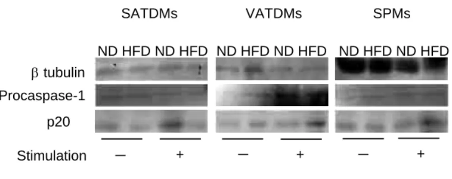
Caspase-1 activation in adipose tissue-derived
macrophagesTo investigate caspase-1 activation in SATDMs, VATDMs and SPMs from ND-fed and HFD- fed mice, Western blot analysis was performed using the culture supernatants and the lysates of these cells. The protein levels of p20 subunit of activated caspase-1 in SATDMs from HFD-fed mice decreased compared with those from ND- fed mice after stimulation. In contrast, the levels of p20 subunit in SPMs and VATDMs from HFD- fed mice were higher than those from ND-fed mice (Figure 6).
Figure 3 Histology of skin lesions of ND-fed and HFD-fed mice after inoculation with S. aureus.
(A) Skin specimens from back of non-infected mice were taken and the tissue samples were stained with hematoxylin and eosin. (B) Subcutaneous adipose tissues were immunostained with anti-F4/80 antibody, macrophages were detected as F4/80 positive cells. (C-F) Skin specimens from S. aureus-infected mice were taken and the tissues samples were stained with hematoxylin and eosin. The skin specimens were collected on day 1 (C and D) or on day 3 (E and F) after subcutaneous inoculation with 5 × 10⁵ CFU (C and E) or 1 × 10⁸ CFU (D and F) of S. aureus.
HFD ND
x100
B
C
ND HFDx40
D
Day 1
ND HFD
A
ND HFDDay 0 x40
Day 3
F
ND HFDE
x40
ND HFD
x40
x40
Sakamoto et al. Figure 3.
36 Y. Sakamoto, et al.
Discussion
It is has been known that IL-1β produced by macrophages plays a critical role in host defense against S. aureus infection
11, 12). On the other hand, IL-1β produced from visceral adipose tissue macrophages also plays a role in obesity-induced
chronic inflammatory state
2, 3). The effects of non- diabetic obesity on pathology of skin S. aureus infection, IL-1β production in SATDMs in the skin lesions, and IL-1β-dependent host defense against skin S. aureus infection are still unclear.
In this study, we investigated effects of obesity on innate immunity to skin S. aureus infection and the IL-1β production from SATDMs in HFD-
Figure 4 IL-1β production from SATDMs, VATDMs and SPMs.
SATDMs (A), VATDMs (B) and SPMs (C) were stimulated with 1 × 10⁸ CFU of FKSA for 6 h, followed by stimulation with 1 mM ATP for 12 h. IL-1β production in cell culture supernatants was determined by ELISA.
Data are represented as the medians ± interquartile range for a group of 4 to 6 mice. An asterisk represents a statistically significant difference from the ND-fed mice (P < 0.05).
Figure 5 NLRP3 mRNA expression of SATDMs, VATDMs and SPMs.
SATDMs (A), VATDMs (B) and SPMs (C) were stimulated with 1 × 10⁸ CFU of FKSA for 1 h, followed by stimulation with 1 mM ATP for 10 min. NLRP3 mRNA expression was determined by real-time quantitative PCR. Data are represented as the medians ± interquartile range for a group of 4 to 6 mice. An asterisk represents a statistically significant difference from the ND-fed mice (P < 0.05).
0 5 10 15 20 25 30 35 40
0 50 100 150 200 250 300
350 HFD
A
SATDMs ND- +
VATDMs
B
C
SPMs+ : FKSA 1 ×10⁸CFU/ml + 1 mM ATP
Stimulation - + - + - + - + - +
Sakamoto et al. Figure 4.
0.00 0.01 0.02 0.03
C
VATDMs
B A
HFD ND
+ : FKSA 1 ×10⁸CFU/ml + 1 mM ATP Stimulation
0.000 0.001 0.002 0.003 0.004
NLRP3 relative expression
0.0 0.2 0.4 0.6 0.8 1.0
- + - + - + - + - + - +
Sakamoto et al. Figure 5.
SATDMs SPMs
37 Obesity Impairs Host Defense against Skin Infection
fed non-diabetic obese mice.
Bacterial numbers in skin lesion of ND- fed mice were reduced compared with those of HFD-fed mice on day 3 after subcutaneous inoculation with 5 × 10
5CFU of S. aureus (Figure 2A). For clearance of S. aureus in skin lesion, neutrophil abscess formations associated with IL-1β are required
12). Mölne et al. reported that the neutrophil-depleted mice intradermally inoculated with S. aureus developed crusted ulcerations and exhibited increased levels of IL-6 and specific antibodies to staphylococcal cell wall components
20). In this study, inflammatory cell infiltration was impaired in subcutaneous adipose tissue of HFD- fed mice compared with that of ND-fed mice after subcutaneous infection with 5 × 10
5CFU of S.
aureus (Figure 3C and 3E) . These results suggest that impaired host defense against skin S. aureus infection in non-diabetic obese mice is associated with reduced infiltration of inflammatory cells into the infection sites. Although bacterial clearance in skin lesions was impaired in HFD-fed mice after inoculation with 5 × 10
5CFU of S. aureus, bacterial numbers in skin lesions of HFD-fed and ND-fed mice were comparable on day 3 after inoculation with 1 × 10
8CFU of S. aureus (Figure 2A). The dose of 1 × 10
8CFU of S. aureus might
be too large to clear the pathogen in innate immunity because remarkable destruction of skin tissue such as abscess formation expanded to muscle layer and ulcer formation was shown in ND-fed mice on day 3 after inoculation, although inflammatory cell infiltration which is required for clearance of S. aureus was prominent in the skin lesions of ND-fed mice on day 1 after infection (Figure 3D and 3F). On the other hand, bacterial numbers in the spleens and kidneys of ND-fed mice increased compared with those of HFD-fed mice after subcutaneous inoculation with 1 × 10
8CFU of S. aureus (Figure 2B).
After subcutaneous infection with 1 × 10
8CFU of S. aureus, inflammatory cells infiltrated into subcutaneous layer as well as muscle layer of ND-fed mice, whereas the lesions of HFD-fed mice were almost restricted in subcutaneous adipose tissue (Figure 3D and 3F). These results suggest that thick subcutaneous adipose tissue layer of HFD-fed mice could suppress the spread of bacteria after infection with high dose of S. aureus, although bacterial clearance in skin lesions was impaired in HFD-fed mice after infection with low dose of S. aureus.
IL-1β is required for host defense against S.
aureus infection to recruit neutrophils
11, 12). Several
Figure 6 Caspase-1 activation in SATDMs, VATDMs and SPMs.
SATDMs (A), VATDMs (B) and SPMs (C) were stimulated with 1 × 10⁸ CFU of FKSA for 6 h, followed by stimulation with 1 mM ATP for 1 h. Protein levels of procaspase-1 and β-tubulin in cell lysates and activated caspase-1 in cell culture supernatants were determined by Western blot analysis. p20: a subunit of activated caspase-1.
tubulin
- Procaspase-1
p20
+ SATDMs ND HFD ND HFD
- + - +
ND HFD ND HFD ND HFD ND HFD VATDMs
Stimulation
Sakamoto et al. Figure 6.
SPMs
+ : FKSA 1 ×10⁸CFU/ml + 1 mM ATP
38 Y. Sakamoto, et al.
types of inflammasome play an important role in the regulation of caspase-1 activation which is required for conversion of pro-IL-1β to mature IL-1β
21, 22). Caspase-1 activation via inflammasome was reported to be involved in the protection against the bacterial infections, such as Salmonella Typhimurium, Legionella pneumophilia and Pseudomonas aeruginosa
23-25). In this study, NLRP3 mRNA expression slightly increased in SATDMs of ND-fed mice, while the expression was significantly enhanced in VATDMs and SPMs of HFD-fed mice after stimulation with FKSA and ATP (Figure 5). It should be noted that NLRP1a and NLRC4 mRNA expression did not increase in SATDMs, VATDMs and SPMs by the stimulation (data not shown). Increased IL-1β production in SATDMs of ND-fed mice, VATDMs and SPMs of HFD-fed mice by stimulation with FKSA and ATP was coincident with higher NLRP3 mRNA expression in these macrophages by stimulation (Figures 4 and 5). Caspase-1 activation and NLRP3 mRNA expression increased in SATDMs of ND-fed mice and VATDMs and SPMs of HFD-fed mice after the stimulation (Figures 5 and 6). In contrast, SATDMs of HFD-fed mice increased neither NLRP3 mRNA expression nor caspase-1 activation after stimulation with FKSA and ATP (Figures 5 and 6). These results suggest that NLRP3 mRNA expression and caspase-1 activation of SATDMs of HFD-fed mice in skin S. aureus infection was attenuated compared with that of SATDMs of ND-fed mice.
It has been reported that activated caspase-1 production and NLRP3 inflammasome activity increased in visceral adipose tissue in diet- induced obese animal models
18, 26). Coincidence with the previous studies, VATDMs and SPMs showed strong caspase-1 activation and NLRP3 mRNA expression after stimulation with FKSA and ATP. However, SATDMs of HFD- fed obese mice did not show such activation and expression in this study. Macrophages in
adipose tissue are divided into two different profiles. M1 macrophages produce inflammatory cytokines, whereas M2 macrophages produce anti-inflammatory cytokines
27, 28). In this study, although we did not verify the difference of macrophage types in adipose tissues, different responses between SATDMs of HFD-fed and ND-fed mice in caspase-1 activation and NLRP3 mRNA expression might be due to different type of macrophages.
Our data showed that host defense of HFD- fed non-diabetic obese mice against skin S. aureus infection was impaired. IL-1β production, caspase-1 activation and NLRP3 mRNA expression of SATDMs of HFD-fed mice were attenuated compared with those of ND-fed mice after stimulation with FKSA and ATP. The present study suggests that reduction of IL-1β production in subcutaneous adipose tissues-macrophages which is associated with caspase-1 activation induced by NLRP3 inflammasome might be involved in impaired host defense against skin S. aureus infection in diet-induced non-diabetic obesity.
References
1)Hotamisligil GS, Erbay E. Nutrient sensing and inflammation in metabolic diseases. Nat Rev Immunol. 2008;8:923-34.
2)Odegaard JI, Chawla A. Mechanisms of macrophage activation in obesity-induced insulin resistance. Nat Clin Pract Endocrinol Metab. 2008;4:619-26.
3)Ouchi N, Parker JL, Lugus JJ, Walsh K. Adipokines in inflammation and metabolic disease. Nat Rev Immunol. 2011;11:85-97.
4)Falagas ME, Kompoti M. Obesity and infection.
Lancet Infect Dis. 2006;6:438-46.
5)Dodds Ashley ES, Carroll DN, Engemann JJ, Harris AD, Fowler VG Jr, Sexton DJ, Kaye KS.
Risk factors for postoperative mediastinitis due to methicillin-resistant Staphylococcus aureus. Clin Infect Dis. 2004;38:1555-60.
39 Obesity Impairs Host Defense against Skin Infection
6)Huttunen R, Laine J, Lumio J, Vuento R, Syrjänen J. Obesity and smoking are factors associated with poor prognosis in patients with bacteraemia.
BMC Infect Dis. 2007;7:13.
7)Krishnan EC, Trost L, Aarons S, Jewell WR.
Study of function and maturation of monocytes in morbidly obese individuals. J Surg Res. 1982;33:89- 97.
8)Palmblad J, Hallberg D, Engstedt L. Polymor- phonuclear (PMN) function after small intestinal shunt operation for morbid obesity. Br J Haematol. 1980;44:101-8.
9)Strandberg L, Verdrengh M, Enge M, Andersson N, Amu S, Onnheim K, Benrick A, et al. Mice chronically fed high-fat diet have increased mortality and disturbed immune response in sepsis. PLoS One. 2009;4:e7605.
10)Lowy FD. Staphylococcus aureus infections. N Engl J Med. 1998;339:520-32.
11)Miller LS, Pietras EM, Uricchio LH, Hirano K, Rao S, Lin H, O'Connell RM, et al. Inflammasome- mediated production of IL-1β is required for neutrophil recruitment against Staphylococcus aureus in vivo. J Immunol. 2007;179:6933-42.
12)Miller LS, O'Connell RM, Gutierrez MA, Pietras EM, Shahangian A, Gross CE, Thirumala A, et al.
MyD88 mediates neutrophil recruitment initiated by IL-1R but not TLR2 activation in immunity against Staphylococcus aureus. Immunity. 2006 24:79-91.
13)Ku CL, von Bernuth H, Picard C, Zhang SY, Chang HH, Yang K, Chrabieh M, et al. Selective predis- position to bacterial infections in IRAK-4-deficient children: IRAK-4-dependent TLRs are otherwise redundant in protective immunity. J Exp Med.
2007;204:2407-22.
14)Picard C, Puel A, Bonnet M, Ku CL, Bustamante J, Yang K, Soudais C, et al. Pyogenic bacterial infections in humans with IRAK-4 deficiency.
Science. 2003;299:2076-9.
15)von Bernuth H, Picard C, Jin Z, Pankla R, Xiao H, Ku CL, Chrabieh M, et al. Pyogenic bacterial infections in humans with MyD88 deficiency.
Science. 2008;321:691-6.
16)Schroder K, Tschopp J. The inflammasomes. Cell.
2010;140:821-32.
17)Muñoz-Planillo R, Franchi L, Miller LS, Núñez G. A critical role for hemolysins and bacterial lipoproteins in Staphylococcus aureus-induced activation of the Nlrp3 inflammasome. J Immunol.
2009;183:3942-8.
18)Vandanmagsar B, Youm YH, Ravussin A, Galgani JE, Stadler K, Mynatt RL, Ravussin E, et al. The NLRP3 inflammasome instigates obesity-induced inflammation and insulin resistance. Nat Med.
2011;17:179-88.
19)Nakane A, Okamoto M, Asano M, Kohanawa M, Minagawa T. Endogenous gamma interferon, tumor necrosis factor, and interleukin-6 in Staphy- lococcus aureus infection in mice. Infect Immun.
1995;63:1165-72.
20)Mölne L, Verdrengh M, Tarkowski A. Role of neutrophil leukocytes in cutaneous infection caused by Staphylococcus aureus. Infect Immun.
2000;68:6162-67.
21)Hise AG, Tomalka J, Ganesan S, Patel K, Hall BA, Brown GD, Fitzgerald KA. An essential role for the NLRP3 inflammasome in host defense against the human fungal pathogen Candida albicans. Cell Host Microbe. 2009;5:487-97.
22)Kumar H, Kumagai Y, Tsuchida T, Koenig PA, Satoh T, Guo Z, Jang MH, et al. Involvement of the NLRP3 inflammasome in innate and humoral adaptive immune responses to fungal β-glucan. J Immunol. 2009;183:8061-67.
23)Mariathasan S, Newton K, Monack DM, Vucic D, French DM, Lee WP, Roose-Girma M, et al. Differential activation of the inflammasome by caspase-1 adaptors ASC and Ipaf. Nature.
2004;430:213-18.
24)Amer A, Franchi L, Kanneganti TD, Body- Malapel M, Ozören N, Brady G, Meshinchi S, et al. Regulation of Legionella phagosome maturation and infection through flagellin and host Ipaf. J Biol Chem. 2006;281:35217-23.
25)Sutterwala FS, Mijares LA, Li L, Ogura Y, Kazmierczak BI, Flavell RA. Immune recognition of Pseudomonas aeruginosa mediated by the IPAF/
NLRC4 inflammasome. J Exp Med. 2007;204:3235-
40 Y. Sakamoto, et al.
45.
26)Stienstra R, Joosten LA, Koenen T, van Tits B, van Diepen JA, van den Berg SA, Rensen PC, et al. The inflammasome-mediated caspase-1 activation controls adipocyte differentiation and insulin sensitivity. Cell Metab. 2010;12:593-605.
27)Gordon S, Martinez FO. Alternative activation of macrophages: mechanism and functions.
Immunity. 2010;32:593-604.
28)Odegaard JI, Ricardo-Gonzalez RR, Goforth MH, Morel CR, Subramanian V, Mukundan L, Red Eagle A, et al. Macrophage-specific PPARγ controls alternative activation and improves insulin resistance. Nature. 2007;447:1116-20.