Acta Med. Nagasaki 29: 77-90
Projections to the Red Nucleus from the Telencephalon
and Diencephalon in the Rat, as Demonstrated
by the HRP and Silver-impregnation Methods
Kentaro HAYASHIDA
First Department of Anatomy, Nagasaki University School of Medicine Received for publication, June 30, 1984
Projections to the red nucleus from the telencephalon and diencephalon were examined in the rat using the horseradish peroxidase (HRP) and silver impregnation me- thods.
After injections of HRP into the red nucleus, labeled cells were found mainly in the cerebral cortex and the zona incerta (ZI). In the cerebral cortex, labeled cells were present ipsilaterally in layer V of the motor area, and, to a lesser extent, in that of the somatic sensory area. Next, large lesions were placed in the frontal cortex. Terminal degeneration was found in the rostral half of the red nucleus. In the ZI, retrogradely labeled cells were found in the caudal part bilaterally. After injections of HRP into the ZI, anterogradely labeled fibers and terminals were traced. Labeled terminals were pro- fuse in the rostral two-thirds of the red nucleus ipsilaterally and sparse contralaterally.
INTRODUCTION
Since the earlier work of von Monakow3S), the cortical projections to the red nu- cleus have been studied by numerous investigators. Anatomical and physiological studies
have elucidated the organization of the corticorubral projection in detail in the cat24)3o)33) 34)36)37) and monkey'2>13)21)22> . In the rat, however, some uncertainties still remain concern- ing the precise location of cells of origin of the corticorubral fibers. The present paper deals with the corticorubral projection, and also reports about the diencephalic projections to the red nucleus.
MATERIALS AND METHODS
A total of 40 albino rats were used in the present study. They were anesthetized intraperitoneally with Ketamine prior to the appropriate surgery.
林 田健太郎
Horseradish peroxidase procedures
In order to identify the location of the neurons projecting to the red nucleus, the retrograde tracing method employing HRP histochemistry was used. A small amount o f 50% HRP dissolved in Tris-HCI buffer (pH 8.6) was injected iontophoretically into the red nucleus in 9 rats. After about 48 hours, the animals were perfused transcardially with 0.9% saline followed by a mixture of 1.25% glutaraldehyde and 0.85% formaldehyde in 0.1 M phosphate buffer (pH 7.4). The brains were removed, stored at 4°C in the same buffer containing 30% sucrose and cut serially at 50 pm in the transverse plane with a freezing microtome. Sections were reacted with benzidine dihydrochloride by the method of MESULAM and ROSENE27), mounted on gelatinized slides and counterstained with neutral red. In another set of experiments, 10 rats were subjected to HRP injections into the ZI to trace the anterogradely labeled fibers and terminals. In 11 rats, HRP was in- jected into the cervical (6 rats) or lumbar enlargement (5 rats) of the spinal cord in order to identify the forelimb and hindlimb cortical areas. The experimental procedures were the same as described above.
Silver-impregnation procedures
In 10 rats, lesions of varying size were made in the cerebral cortex unilaterally (3 rats) or bilaterally (7 rats). After 1-2 weeks, the animals were sacrificed by perfusion with 0.9% saline followed by 10% formaldehyde. After fixation periods of 4 weeks, the brains were cut serially at 30 ,um and the frozen sections were stained using the FINK- HEIMER method').
RESULTS I. Retrograde tracing method
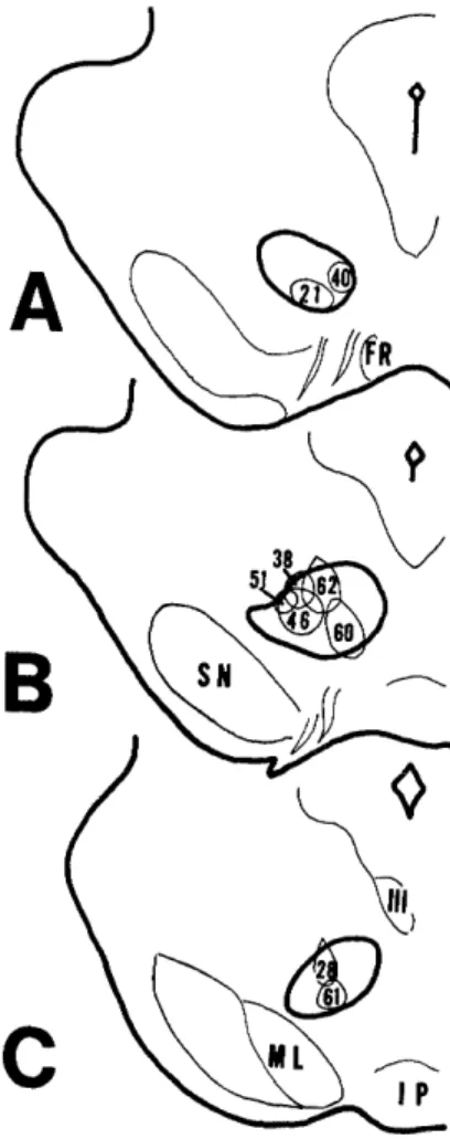
In order to localize the areas which project to the red nucleus, HRP was injected into the red nucleus. Fig. 1 shows the injection sites o f all 9 cases. In 7 cases (case 21, 38, 40, 46, 51, 60, 62), the injection sites involved the rostral two-thirds of the red nucleus, which correspond to the parvocellular portion of the red nucleus (RNp). La- beled cells were found mainly in the cerebral cortex, ZI, reticular part of the anterior pretectal nucleus and deep cerebellar nuclei. In 2 cases (case 28, 61), the injection sites involved the caudal one-third of the nucleus, which corresponds to the magnocel- lular portion (RNm). Many labeled cells were found in the deep cerebellar nuclei.
TELENCEPHALON Cerebral cortex
In case 21, 38, 40, 46 and 51, a small amount of HRP was confined to the RNp.
The number of labeled cells in the cerebral cortex was relatively small (mean 20 cells per case). Fig. 2 shows the distribution of labeled cells in case 21, in which the in- jection site was confined to the ventral part of the rostrall one-third of the red nucleus (Fig. 6). Labeled cells were found mainly in the lateral and medial agranular field (AGI and AGm) described by DONOGHUE and WISE4~, which appear to correspond to
KRIEG's area 4, 6, 10, 8 and 2420). A small number of labeled cells were also observed in the first somatic sensory cortex (SI) which appears to correspond to KRIEG's area 3 , 1 and 220). They were all present in layer V on the ipsilateral side of the injection. In the cingulate cortex, only a few labeled cells were found in this case.
Since the number of labeled cells was too small to determine the topographical organization of the corticorubral projections in these cases, a relatively large amount of HRP was injected in case 60 and 62 (Fig. 1, Fig. 3A, B). In case 60, the injection site involved the ventral part of the RNp, but there was a slight spreading of HRP into the ventrally situated medial lemniscus. In case 62, the injection site was located in the dorsolateral part of the RNp and the adjacent mesencephalic reticular formation along the track of the micropipette. In other rats, HRP was injected into the cervical or lumbar enlargement of the spinal cord to identify the forelimb and the hindlimb cortical areas. Fig. 3C shows the results. The distribution of the labeled cells in case 60 and 62 was roughly the same as in case 21. In the region which appears to be
Fig. 1. Drawings of the extent of the injection sites of HRP in the red nucleus.
the hindlimb area (arrowhead 1), the number of labeled cells was small in case 60 and 62. In the regions which appear to be the forelimb cortical area, and the second
forelimb area described by NEAFSEY and SIEVERT28), labeled cells were found in both cases (arrowhead 2, 4). In and around the second somatic sensory cortex shown by arrowhead 5, only a few labeled cells were present in each case. In the region shown
Fig. 2. Drawings of frontal sections, showing the distribution of labeled cells (dots) within the cerebral cortex and the diencephalon.
by arrowhead 3, labeled cells were seen in case 60 and 62.
The somal area of the corticorubral cells labeled with HRP ranged from 178 to 255 ftm2 (mean ± S. D. : 209±25.3 pm2), while that of the corticospinal cells ranged from 246 to 290 pmt (mean ± S. D. : 260 ± 23.1 pmt).
DIENCEP H ALON Zona incerta (ZI)
In case 21, labeled cells were found in the ZI bilaterally with ipsilateral predomi- nance. They were mainly located in the caudal part of the ZI (Fig. 2G). Further rostrally at the level of the appearance of the subthalamic nucleus (Fig. 2F), labeled cells rapidly decreased in number and were located in the medial part of the ZI, and the fields of Forel. At the level of the maximum size of this nucleus, a few cells were labeled, but none were labeled in the rostral pole of the ZI. Labeled cells were fusiform, triangular and sometimes polygonal in shape and most of them were small in size. (Fig. 8)
Fig. 3. Dorsal view of the left cerebral hemisphere, showing the distribution of labeled cells following HRP injection into the ipsilateral red nucleus (A, B, dots),
contralateral lumbar (C, open circles) and contralateral cervical enlargement
(C, dots). The dashed line represents the border between AGm and AGI, and the heavy line represents the medial border of the SI cortex.
OTHER TELENCEPHALIC AND DIENCEPHALIC STRUCTURES
A small number of labeled cells were found ipsilaterally in the subparafascicular nucleus (Fig. 2G) and entopeduncular nucleus (not illustrated), but further examinations were not made.
Fig. 4. Drawings of frontal sections (A-F) through the mesencephalon of an animal with an extensive lesion of the left cerebral hemisphere (black). Degeneration
is represented by dots.
II. Anterograde tracing methods Cerebral cortex
Since terminal degeneration was virtually absent in the red nucleus following a small lesion in the frontal cortex of the rat, a large lesion was made involving the anterior one-fourth of the cerebral cortex (Fig. 4, uppermost figure). in case 10, de- generated corticofugal fibers occupied the medial one-third of the ipsilateral cerebral pedunc le . At the rostra l pole o f the substantia nigra (Fig. 4A), a portion o f the de- generated fibers leaves the cerebral peduncle to divide into two bundles dorsally. At the mesencephalic levels, a large lateral bundle runs lateral- and dorsalwards to give
rise to the termination in the mesencephalic reticular formation and the superior col-
Fig. 5. Drawings of the extent of the injection site in the ZI (shaded areas), and the rlistrih„tion of fibers and terminals labeled anterogradely with HRP (dots).
liculus. A small medial bundle runs dorsomedially to terminate in the ventral part. of the periaqueductal gray. In the rostral one-third of the red nucleus (Fig. 4B, C), terminal degeneration was observed clearly on the ipsilateral side of the lesion. Further caudally (Fig. 4D), the degeneration became lighter. In the caudal one-third (Fig.4F), the red nucleus was free from degeneration. Contralaterally, terminal degeneration could not be found in the red nucleus.
Zona incerta
In order to confirm the incerto-rubral projection, HRP was injected into the ZI.
In case 15, the injection site was located in the caudal part of the ZI extending medio- laterally just ventral to the medial lemniscus (Fig. 5, uppermost figures). There was a spreading of HRP into the latter structure and the caudal pole of the ventrobasal complex of the thalamus along the track of the micropipette, but no spreading into the cerebral peduncle. In the mesencephalon, axon terminals labeled anterogradely with HRP were found ipsilaterally in the superior colliculus, the mesencephalic reticular formation, the periaqueductal gray, the red nucleus and the nucleus of Darkschewitsch. Labeled
fibers were seen in the mesencephalic reticular formation immediately lateral to the red nucleus and in the medial lemniscus. In the rostra l one-third o f the red nucleus (Fig.
5A, B), labeled terminals were found ipsilaterally through its entire extent, with higher density in the lateral half. They occupied the more lateral part in the middle one-third (Fig. 5C, D). In the caudal one-third of the red nucleus which is composed of a clus- ter of large neurons (Fig. 5E, F), labeled terminals were hardly found. On the con- tralateral side of the HRP injection, labeled terminals were distributed sparsely in the rostral part of the red nucleus (Fig. 5B).
DISCUSSION Cerebral cortex
Anatomically and electrophysiologically, the corticorubral projection has been studi- ed in the opossum18), rat2)8)10), cat5)9)24)30)31)33)34)36)37), monkey3)12)13)21)22), and man"). In the cat5)7)9)24)30)31)34)37) , the corticorubral fibers were reported to arise mainly from the somatic sensorimotor cortex. In addition, the projections from the gyrus proreus33) and the parietal association corteX5)27)29) were shown. In the monkey, anatomical studies') 12)21) have shown that the corticorubral fibers arise from the motor and premotor cortex, and the somatic sensory cortex. The present study shows that cells of origin of the cortico- rubral fibers in the rat are located mainly in AG1 and AGm which correspond to the motor cortex4), and to a lesser extent in the somatic sensory cortex. This is in accordance with the previous reports of the rat using the degeneration methods2)8)10)
Electrophysiological studies'"') have shown that AG1 of the rat coincides with the primary motor area and that AGm has similarities to the supplementary motor area in other mammals. Anatomical studies' )19)23) have demonstrated that AGm receives the af- ferent fibers from the mediodorsal nucleus of the thalamus in the rat. The supplementary
motor area was shown to receive afferents from the mediodorsal nucleus17), and project to the red nucleus") in the monkey.
A somatotopical organization of the corticorubral projection has been demonstrated in the cat24)3o)34)37) and monkey 12)21) . In the present study, attempts were made to investi- gate this organization in the rat using the HRP and degeneration methods. Small lesions placed in the frontal cortex did not result in degeneration in the red nucleus. Also in the HRP experiments, injections of a small amount of HRP into the red nucleus resulted in a sparse labeling of cells in the cerebral cortex. After injections of a relatively large amount of HRP into the RNp, labeled cells were moderate in the forelimb cortical area and sparse in the hindlimb cortical area. Labeled cells were also observed in the region shown by arrowhead 3 (Fig. 3A, B), which seems to correspond to the face motor area 11)28)35) KING et al.") have shown in the opossum using the silver-impregnation methods that the majority of corticorubral fibers arise from the motor-sensory forelimb cortex, while some arise from the motor-sensory hindlimb cortex, and that the definite somatoto- pical organization of the corticorubral projection cannot be established. In the present study, definite differences in the distribution of labeled cells between case 60 and case 62 could not be found. The distinct topographical organization of the corticorubral pro- jection, therefore, cannot be determined.
It has already been shown that the corticorubral fibers terminate in the RNp in the rat2)lo)18) The present results also confirm this. Although the entire red nucleus was shown to receive afferent fibers from the sensorimotor cortex in the cat24)34) and monkey 12)21), a relatively small number of cortical projections to the caudal RNm were found in the opossum by MARTIN 21) and KING et al.18) and in the monkey by CATMAN- BERRE VOET et al. 4) .
It has often been assumed in physiological studies9)36) that the corticorubral pro- jection in the cat is derived from both pyramidal tract and from non-pyramidal tract neurons. GIUFFRIDA et al') have shown in the cat that the projection to the red nucleus is much stronger from pyramidal tract neurons than from non-pyramidal tract neurons in area 4, and vice versa in area 6. HUMPHREY and RIETZ13) have provided evidence in the monkey that most of the fibers from the cortical arm area to the red nucleus are axons of non-pyramidal cells. In the present study of the rat, significant size differences between the corticorubral and the corticospinal cells labeled with HRP were noticed. The present findings are in accordance with those of the monkey by JONES and WISE 14). They have shown by the HRP method that the corticorubral neurons are smaller than the corticospina l neurons.
Zona incerta
Projection fibers from the ZI to the red nucleus have been reported in the rat32)39) and cat"). RICARDO32) has found labeling bilaterally in the RNp following injection o f tritiated amino acids into the ZI o f the rat. The present results by the HRP method confirmed his findings. On the basis of the cytoarchitecture, KAWANA and WATA- NABE16) divided the ZI into six parts : the anteropolar, dorsal, ventral, magnocellular,
caudal and posteropolar parts. The region of the ZI in which many labeled cells were observed in the present study seems to correspond to their caudal part. Some labeled cells were also present in the magnocellular part and the medial region of the dorsal and ventral part. KAELBER and SMITH") have suggested that the medial part of the ZI thand e zona incerta caudaiis, both of which project to the red nucleus, belong to the nociceptive-conducting system causing typical escape responses.
ACKNOWLEDGEMENTS
The author wishes to express his gratitude to Mr. T. OKUBO, Mr. M. KANEKO and Mrs. M. HASHIGUCHI for their skillful technical assistance.
ABBREVIATIONS
AGm: medial agranular field AGI: lateral agranular field CP: cerebral peduncle
F: fornix
FR: fasciculus retroflexus IP: interpeduncular nucleus LGN : lateral geniculate nucleus ML: medial lemniscus MT: mammillothalamic tract PC: posterior commissure RN: red nucleus
RNm: magnocellular portion of the red nucleus RNp: parvocellular portion of the red nucleus SI: first somatic sensory cortex
SM: stria medullaris SN : substantia nigra
SPf : subparafascicular nucleus ST: subthalamic nucleus ZI: zona incerta III: oculomotor nucleus
REFERENCES
1) BECKSTEAD, R. M.: Convergent thalamic and mesencephalic projections to the anterior medial cortex in the rat. J. Comp. Neurol. 166: 403-416, 1976.
2) BROWN, L. T.: Corticorubral projections in the rat. J. Comp. Neurol. 154: 149- 168, 1974.
3) CATMAN-BERREVOETS, C, E., KUYPFRS, H. G. J. M. and LEMON, R. N.:
Cells of origin of the frontal prontal projections to magnocellular and parvocellular
red nucleus and superior colliculus in cynomolgus monkey. An HRP study. Neurosci.
Lett. 12: 41-46, 1979.
4) DONOGHUE, J. P. and WISE, S. P.: The motor cortex of the rat: Cytoarchitecture
and microstimulation mapping. J. Comp. Neurol, 212: 76-88, 1982.
5) FANARDJIAN, V. V. and GORODNOV, V, L.: Electrophysiological properties of cortical synaptic inputs of rubro-spinal neurons. Neurosci. Lett. 40: 269-273,
1983.
6) FINK, R. P. and HEIMER, L.: Two methods for selective silver impregnation of degenerating axons and their synaptic endings in the central nervous system. Brain
Res. 4: 369-374, 1967.
7) FLINDT-EGEBACK, P.: An autoradiographical study of the projections from the feline sensorimotor cortex to the brain stem, J. Hirnforsch. 20: 375-390, 1979.
8) FLUMERFELT, B. A.: An ultrastructural investigation of afferent connections of the red nucleus in the rat. J. Anat. 131: 621-633, 1980.
9) GIUFFRIDA, R., LIVOLSI, G., PERCIAUALLE, V. and URBANO, A.: Pyramidal and non-pyramidal projection from cortical areas 4 and 6 to the red nucleus in the cat.
Neurosci, Lett. 40: 227-231, 1983.
10) GWYN, D. G. and FLUMERFELT, B. A.: A comparison of the distribution of cortical and cerebellar afferents in the red nucleus of the rat, Brain Res. 69: 130-
135, 1974.
11) HALL, R. D. and LINDHOLM, E, P.: Organization of motor and somatosensory
neocortex in the albino rat. Brain Res. 66: 23-38, 1974.
12) HARTMANN-VON MONAKOW, W, AKERT, K. and KONZLE, H.: Projections of precentral and premotor cortex to the red nucleus and other midbrain areas in macaca
fascicularis. Exp. Brain Res. 34: 91-105, 1979.
13) HUMPHREY, D. R. and RIETZ, R. R.: Cells of origin of corticorubral projections from the arm area of primate motor cortex and their synaptic actions in the red
nucleus. Brain Res. 110: 162-169, 1976.
14) JONES, E. G. and WISE, S. P.: Size, Laminar and columnar distribution of ef- ferent cells in the sensory-motor cortex of monkey. J. Comp. Neurol. 175: 391-438,
1977.
15) KAELBER, W. W. and SMITH, T. B,: Projections of the zona incerta in the cat, with stimulation controls. Exp. Neurol. 63: 177-200, 1979.
16) KAWANA, E. and WATANABE, K.: A cytoarchitectonic study of zona incerta in the rat. J. Hirnforsch. 22: 535-541, 1981.
17) KIEVIT, J. and KUYPERS, H. G. J. M.: Organization of the thalamocortical
connexions to the frontal lobe in the rhesus monkey, Exp. Brain Res. 29: 299-322, 1977.
18) KING, J. S., MARTIN, G, F. and CONNER, J. B.: A light and electron mi- croscopic study of corticorubral projections in the opossum, Didelphis Marsupialis
Virginiana. Brain Res. 38: 251-265, 1972.
19) KRETTEK, J. E. and PRICE, J. L.: The cortical projections of the mediodorsal nu- cleus and adjacent thalamic nuclei in the rat. J. Comp. Neurol, 171: 157-192, 1977.
20) KRIEG, W. J. S.: Connections of the cerebral cortex. 1 The albino rat. A. Topog- raphy of the cortical areas. J. Comp. Neurol, 84: 221-275, 1946.
21) KUYPERS, H. G. J. M. and LAWRENCE, D. G.: Cortical projections to the red
nucleus and the brain stem in the rhesus monkey. Brain Res. 4: 151-188, 1967.
22) LARSEN, K. D. and YUMIYA, H.: The red nucleus of the monkey: Topographic
localization of somatosensory input and motor output. Exp. Brain Res. 40: 393-404, 1980.
23) LEONARD, C. M.: The prefrontal cortex of the rat. I. Cortical projection of the mediodorsal nucleus. II. efferent connections. Brain Res. 12: 321-343, 1969.
24) MABUCHI, M. and KUSAMA, T.: The cortico-rubral projection in the cat. Brain Res. 2: 254-273, 1966.
25) MARTIN, G. F. Jr.: The pattern of neocortical projections to the mesencephalon
of the opossum, Didelphis Virginiana. Brain Res. 11: 593-610, 1968.
26) MESULAM, M. M. and ROSENE, D. L.: Differential sensitivity between blue and brown reaction procedures for HRP neurohistochemistry. Neurosci. Lett. 5: 7-14,
1977.
27) MIZUNO, N . , MOCHIZUKI, K . , AKIMOTO, C., MATSUSHIMA, R. and SASAKI, K .: Projections from the parietal cortex to the brain stem nuclei in the cat, with
special reference to the parietal cerebro-cerebellar system. J. Comp. Neurol. 147:
511- 522, 1973.
28) NEAFSEY, E. J. and SIEVERT, C.: A second forelimb motor area exists in the rat frontal cortex. Brain Res. 232: 151-156, 1982.
29) OKA. H. and JINNAI, K.: Electrophysiological study of parvocellular red nucleus neurons. Brain Res. 149: 239-246, 1978.
30) PADEL, Y., SMITH, A. M. and ARMAND, J.: Topography of projections from the motor cortex to rubrospinal units in the cat. Exp. Brain Res. 17: 315-332, 1973.
31) PIZZINI, G., TREDICI, G. and MIANI, A.: Corticorubral projection in the cat. An experimental electonmicroscopic study. J. Submicr. Cytol. 7: 231-238, 1975.
32) RICARDO, J. A.: Efferent connections of the subthalamic region in the rat. II.
The zona incerta. Brain Res. 214: 43-60, 1981.
33) RINVIK, E.: The corticorubral projection in the cat. Further observations. Exp.
Neurol. 12: 278-291, 1965.
34) RINVIK, E. and WALBERG, F.: Demonstration of a somatotopically arranged cor- tico-rubral projection in the cat. An experimental study with silver methods. J. Comp.
Neurol. 120: 393-407, 1963.
35) SANDERSON, K. J., WELKER, W. and SHAMBES, G. M.: Reevaluation of motor cortex and sensorimotor overlap in cerebral cortex of albino rats. Brain Res. 292:
251- 260, 1984.
36) TSUKAHARA, N., FULLER, D. R. G. and BROOKS, V. B.: Collateral pyramidal influences on the corticorubrospinal system. J. Neurophysiol. 32: 467-484, 1968.
37) TSUKAHARA, N. and KOSAKA, K.: The mode of cerebral excitation of red nucleus neurons. Exp. Brain Res. 5: 102-117, 1968.
38) VON MONAKOW, C.: Der rote Kern, die Haube and die Regio subthalamica bei einigen Saugetieren and beim Menschen. Arb. Hirnanat. Inst. Zurich 4: 103-226,
1910.
39) WATANABE, K. and KAWANA, E.: The cells of origin of the incertofugal projections
to the tectum, thalamus, tegmentum and spinal cord in the rat: A study using the autoradiographic and horseradish peroxidase methods. Neuroscience 7: 2389-2406,
1982.
Fig. 6. Photomicrograph of the injection site of HRP in the RNp in case 21. (bar scale: 1 mm)
Fig. 7. Photomicrograph of a HRP-labeled pyramidal cell in layer V of the cerebral cortex (arrowhead). (bar scale: 0.1 mm)
Fig. 8. Labeled cells in the ipsilateral ZI after injection of HRP into the RNp of the right side (arrowheads). (bar scale: 0.1 mm}