Acta Med. Nagasaki 45 : 15-19
Attenuation of Responsiveness to Interferon‑α Treatment by Preceded
Overactivation of Interferon-mediated Pathway in Patients Chronically Infected by Hepatitis C Virus
Keisuke NAKATA 1) , Kazuhiko NAKAO 2) , Takashi MORIKAWA 1), Kaoru INOKUCHI 1), Keisuke HAMASAKI 1),
Shotaro TSURUTA 1), Yuji KATO 1), Hiroshi YATSUHASHI 3), Yukio KUSUMOTO 4), Nobuko ISHII 2) , Katsumi EGUCHI 1)
1) First Department of Internal Medicine, Nagasaki University School of Medicine, Nagasaki, Japan.
2) Health Research Center, Nagasaki University, Nagasaki, Japan.
3) Institute for Clinical Research, Nagasaki Chuo Hospital, Nagasaki, Japan.
4) Department of Internal Medicine, Nagasaki City Hospital, Nagasaki, Japan.
The Interferon (IFN) receptor-mediated signal transduction pathways involve the two novel DNA-binding factors, inter- feron regulatory factor-1 (IRF-1) and IRF-2. Both recognize the same DNA sequence, in which the expression ratio of IRF-1 to IRF-2 is relevant to induction of IFN-inducible genes, because IRF-1 acts as a transcriptional activator and IRF-2 as a counterpart. In the present study, 54 patients with chronic hepatitis C and the age-and sex-matched 7 subjects with fatty liver as a control were subjected to analysis of the expression ratio of IRF-1 to IRF-2 mRNA in the liver tis- sue obtained at the time of livert biopsy by reverse transcription-polymerase chain reaction in combination with a restriction fragment length polymorphism assay. The expres- sion ratio of IRF-1 to IRF-2 mRNA in the liver tissue in pa-
tients chronically infected by hepatitis C virus (HCV) was significantly higher than that in control, although the val- ues did not correlate with the serum levels of HCV-RNA. Of 54 patients, 28 received IFN treatment, resulting in complete response in 8 patients. With respect to responsiveness to IFN treatment, patients who had complete response had the rela- tively lower ratios of IRF-1 to IRF-2 mRNA in the liver tis- sue, compared with those who did not. These results indicate that the IFN-mediated pathway is spontaneously activated in patients with chronic hepatitis C, and that its preceded overactivation counteracts on the efficacy of IFN treatment in these patients.
Key Words: interferon treatment, chronic hepatitis C, inter feron regulatory factor-1, interferon regulatory
factor-2, hepatitis C virus
Introduction
Interferon (IFN) treatment succeeds in viral eradica- tion in 20-30% of patients chronically infected by hepatitis C virus (HCV) 1-4). Its efficacy is closely asso- ciated with low levels of viremia or mutations in the HCV-RNA sequence, especially in the NS5A region".
However, the relationship between antiviral states against HCV and response to IFN is stil unclear. Recent ad- vances in molecular biology lead to a great deal of progress in host-defense mechanisms against viral in- fections. In the IFN system, both interferon regulatory factor-1 (IRF-1) and IRF-2 genes are induced by viral in- fections or by IFNs or, to some degrees, other cytokines
7-1o) Both gene products act as the transcriptional factors which recognize the same DNA sequence. However, since IRF-1 functions as a transcriptional activator and IRF-2 as a counterpart, the expression ratio-of IRF-1 to IRF-2 is
relevant to induction of IFN-inducible gene expression".
In the present study, the expression ratios of IRF-1 to IRF-2 mRNA in the liver tissue were analyzed in pa- tients with chronic hepatitis C.
Patients and Methods
Address Correspondence : Keisuke Nakata, M.D.First Department of Internal Medicine, Nagasaki University School of Medicine, 1-7-1 Sakamoto, Nagasaki 852-8501, Japan
TEL: +81-95-849-7260, FAX: +81-95-849-7270
Patients population
The sample population included 54 patients chroni-
cally infected by HCV who were admitted to our hospital
between 1994 and 1995; 39 were men and 15 were women, and they were 24 to 75 years of age (mean [ ± SD], 50
± 13). The 54 patients were positive for HCV-RNA by reverse transcription-polymerase chain reaction (RT- PCR) and antibody to HCV, but negative for hepatitis B surface antigen. Informed consent was obtained from all patients. Liver biopsies were performed, and the diag- nosis of chronic hepatitis was made on the basis of the histological findings in liver tissue in all patients. The serum levels of HCV-RNA were determined by a branched DNA (bDNA) assay. We also studied the age-and sex-matched 7 subjects with fatty liver who were admitted to our hospital during the same period and gave informed consent. There were 5 men and 2 women, aged 36 to 61 years (mean [ ± SD], 50 ± 10).
The 7 control subjects were negative for any viral markers tested or autoantibodies to nuclear antigens or mitochondrial antigens. Any of alcohol or drug abusers were excluded from the study.
IFN treatment
IFN treatment was performed in 28 of 54 patients with chronic hepatitis C. The 28 patients received IFN- a for 6 months in a dose of 6 million to 9 million units intra- muscularly everyday for 14 days and then three times a week for 22 wk (total dose, 480 million to 720 million units). All patients were followed, and those who per- sisted to become negative for HCV-RNA by RT-PCR together with normalization of liver function tests for more than 1 year after cessation of IFN treatment were defined as complete responders as described pre- viously6>. According to the definition, 8 of 28 patients had complete response, whereas 20 had no response be- cause they persisted or returned to be positive for HCV-RNA during the follow-up period.
Analysis of expression ratio of IRF-1 to IRF-2 mRNA in liver tissue
The liver tissue specimens obtained at the time of liver biopsy were frozen at -70°C immediately after the time of liver biopsy for later analysis of the expression ratio of IRF-1 to IRF-2 mRNA in the liver tissue by RT- PCR in combination with a restriction fragment length polymorhism assay","'. Total cellular RNA was iso- lated from the frozen samples by the guanitidium isothiocyanate method. Two p g of total RNA in a vol- ume of 10 p 1 was heated at 68°C for 3 min, chilled on
ice, then added in a reaction mixture containing 50,u M Tris-HC1 (pH8.3), 75mM KCI, 3mM MgCl2, 200p M each of dNTPs, 100pmol random hexamer, 8U of RNase in- hibitor, and 200U of M-MLV reverse transcriptase to a
volume of 20,u 1. The RT reaction mixture was incu- bated at 37C for 90 min, heated at 95C for 5min, and chilled on ice. The two pairs of oligonuleotide primers, by which IRF-1 and IRF-2 DNA sequences having a relatively higher homology'' (nucleotide 302-508 and nucleotide 162-368, respectively) were amplified in the same length (207bp) and only the IRF-2 sequence was digested by a restriction enzyme, Nsi-I (which recog- nizes ATGC A/T), resulting in 155bp and 52bp frag- ments, were used in this study as follows: for IRF-1, 5'- CCGGGGCTCATCTGGATTAA-3' and 5'-ATCTGGCAGG- GAGTTCATGG-3', and for IRF-2, 5'-CCGGGGCTCAAG- TGGCTTAA-3' and 5-ATCAGGCAAGGAATTCATGG-3'.
Briefly, the PCR was performed in a reaction volume of 50 p 1 containing 2 p 1 of the RT reaction product, 50mM KCI, 10mM Tris (pH9.0 ), 1.5mM MgCl2, 0.1%
Triton X-100, 200,u M each of dNTPs, 25pmol each of PCR primers and 2.51U of Taq DNA polymerase. The amplification was performed by 25 cycles in a pro- grammable DNA thermal cycler. Each reaction cycle involved denaturation at 92C for 30 sec, primer an- nealing at 60°C for 30 sec and primer extension at 72°C for 1 min. In the last cycle, the reaction at 72 °C con- tinued for 10 min to ensure complete DNA extension.
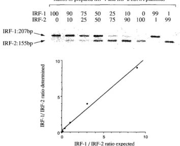
The PCR product was extracted, with phenol-chloroform and precipitated with ethanol. The precipitated prod- uct was digested by 5IU of Nsi-I at 37 °C for 2hr, and the digested product was separated by electrophoresis on a2% agalose gel. The separated bands correspond- ing to IRF-1 and IRF-2 DNA fragments were visualized with etidium bromide staining. The intensity of each band was quantified by densitometry with a image program from Macintosh 131. When IRF-1 cDNA and IRF-2 cDNA (generous gifts from professor T.Taniguchi, Tokyo University, Japan) were mixed to yield differ- ent ratios and the mixed samples were subjected to the same analysis, the ratio of IRF-1 to IRF-2 deter- mined by this method closely correlated with that in the mixed sample prepared before analysis (Figure 1).
Accordingly, each assay always contained the prepared DNA samples with the different ratios of IRF-1 to IRF- 2 to make a standard curve, and the expression ratio of IRF-1 to IRF-2 in each liver tissue sample was calcu- lated from the standard curve.
The statistical analyses were performed with Student's
t-test and the chi-square test. All P values were two-
tailed, and P values of less than 0.05 were considered
to indicate statistical significance.
Figure 1. Correlation between the ratios of IRF-1 to IRF-2 determined by our method and those expected before analy- sis. IRF-1 cDNA and IRF-2 cDNA were mixed to obtain the indicated ratios. The prepared samples were subjected to analysis as described in Methods.
Figure 3. Correlation between the expression ratio of IRF-1 to IRF-2 mRNA in the liver tissue and the serum level of HCV-RNA in patients with chronic hepatitis C. The HCV- RNA level was assayed using a branched DNA assay, and its detection limit was 0.5 megaequivalent per milliliter (Meq/ml ).
chronic hepatitis C than in control (mean [±SD], 2.48
± 1.97 vs. 0.63 ± 0.34, P<0.01), although the ratios in patients ranged widely from 0.57to 12.3 (Figure 2). The serum levels of HCV-RNA were determined by a branched DNA (bDNA) assay. Among 54 patients, the values were less than 0.5 Meq/ml (a detection limit) in 15 patients
and ranged from 0.6 to 126 Meq/ml in 39 patients. When the expression ratios of IRF-1 to IRF-2 mRNA in the liver tissue were compared with the serum levels of HCV- RNA in the 54 patients, no correlation was observed
(Figure 3).
Figure 2. Hepatic expression ratios of IRF-1 to IRF-2 mRNA in patients with chronic hepatitis C and those with fatty liver. The expression ratios were determined in 54 patients with chronic hepatitis C and 7 patients with fatty liver as a control as described in Methods. Bars are means±SD.
Results
Elevated expression ratio of IRF-1 to IRF-2 mRNA in the liver tissue in patients with chronic hepatitis C
The expression . ratio of IRF-1 to IRF-2 mRNA in the liver tissue was significantly higher in patients with
Analysis of factors involved in response to IFN in pa- tients with chronic hepatitis C
Twenty-eight of 54 patients received IFN treatment.
Of 28 patients, 8 had complete response, because these
patients persisted to become negative for HCV-RNA
together with normalization of liver function tests for
more than 1 year after cessation of IFN treatment. The
age or sex did not correlate with outcome of IFN
treatment (Table 1). However, sustained response to
IFN was observed more frequently in patients who
had relatively low levels of HCV-RNA with less than
l Meq/ml at baseline, compared with those who did not
(P<0.01). To evaluate the correlation between the ex-
pression ratio of IRF-1 to IRF-2 mRNA in the liver tis-
sue and response to IFN, the cutoff value for the ratio
was set at 2.5, which was the mean value in the 54 pa-
tients chronically infected by HCV. Seven (47%) of 15
patients who had the ratios of less than 2.5 resulted in
complete response, whereas only 1 (8%) of 13 patients
who had the ratios of 2.5 or more did. The difference was
statistically significant (P< 0.05). Moreover, when the ratio was combined with the baseline serum level of HCV-RNA for an index of response to IFN, 6 (86%) of 7 patients who had both the ratio of less than 2.5 and the aseline HCV-RNA level of less than 1Meq/ml resulted in complete response, whereas only 2 (10%) of 21 patients who did not had complete response (P< 0.001).
Table 1 Factors invoived in respones to interferon in 28 pa- tients
Variable No. of No. of x2
patients complete responders (%)
Age
<50yr. 19 5(26%)
p=0.7011
a50yr. 9 3(33%)
Sex
Male 19 6(32%)
p=0.6088
Female 9 2(22%)
Baseline HCV level
< 1 Meq/ml 13 7(54%)
p=0.0059
al Meq/ml 15 1(7%)
IRF-1/IRF-2 ratio
<2.5 15 7(47%)
p=0.0228
a2.5 13 1(8%)
Combination of HCV level and IRF-1/IRF-2 ratio
Both HCV < 1 Meq/ml 7 6(86%)
and ratio < 2.5 p=0.0001
Others 21 2(10%)
The definition of complete response was mentioned in Methods. The statistical analysis was carried out using the chi-square test.
Discussion
In this study, the expression ratio of IRF-1 to IRF-2 mRNA in the liver tissue in patients with chronic hepatitis C was significantly higher than that in con- trol. In the IFN system, IRF-1 and IRF-2 regulate both
IFN genes and IFN-inducible genes coordinately".
IFNs bind to their receptors and stimulate IRF-1 gene expression. IRF-1 activates expression of IFN genes, re-
sulting in further induction of IRF-1. However, since the 5'-upstream regulatory sequence of the IRF-2 gene contains the IRF response element, IRF-1 overexpression subsequently induces IRF-2 expression which represses IRF-1 function by competing in the same cis-acting ele- ment. In addition, the half life of IRF-1 mRNA is much shorter than that of IRF-2 mRNA". Accordingly, IRF-2 expression is always predominant over IRF-1 expres- sion in quiescent cells without stimulation, while ongo- ing stimulation leads to the increased expression ratio of IRF-1 to IRF-2. Thus, our result seems to indicate that the IFN-mediated pathway in the liver tissue is steadily activated in patients chronically infected by HCV. The expression ratios of IRF-1 to IRF-2 mRNA in
the liver tissue did not correlate with the serum levels of HCV-RNA in the 54 patients chronically infected by HCV. The lack of correlation is difficult to explain given our current understanding of the antiviral states induced by IFN. One possible explanation is that there are mechanisms underlying IFN-induced antiviral ac- tivities independent of IRF-114-161, although the activa- tion of IFN-mediated pathways is manifested by the increased expression ratio of IRF-1 to IRF-2. Consistent with this, several investigators reported that IRF-1 was necessary for the antiviral actions of IFN against some viruses, but did not affect replication of other types of viruses"". In addition, current studies have shown that
the variants of HCV-1b containing mutations in the NS5A region are susceptible to IFNS•6>. The wild-type of the NS5A product can bind to the IFN-inducible double-stranded RNA-dependent protein kinase, a po- tent inhibitor of viral replication"', and suppresses its function"'. In contrast, the mutant viral gene products
cannot, resulting in sensitizing the variant viurses to IFN19>. This may, in part, account for the lack of corre- lation between the activation of the IFN-mediated
pathway and the serum level of HCV-RNA, although the HCV-RNA sequences were not determined in this study.
By the analysis of factors involved in response to IFN in patients with chronic hepatitis C, the relatively lower level of viremia prior to IFN treatment was as- sociated with complete response to IFN in this study.
Similar results were described previously, where IFN treatment succeeded in viral eradication in 50-60% of patients with the baseline HCV-RNA levels of less than l Meq/ml, but in only approximately 10% of pa- tients with relatively higher levels of viremia1-4>. With respect to relationship between the expression ratio of IRF-1 to IRF-2 mRNA in the liver tissue and response to IFN, complete response was observed more fre- quently in patients who had the ratios of less than the cutoff value corresponding to the mean value in the 54 patients chronically infected by HCV, compared with those who did not. Moreover, 6 (87%) of 7 pa- tients who had both the low level of viremia and the expression ratio of less than the cutoff value resulted in complete response, in contrast to only 2 (10%) of 21 patients who did not. It is, therefore, possible that the measurement of the expression ratio of IRF-1 to IRF-2 mRNA in the liver tissue, particularly combined with the baseline serum level of HCV-RNA, serves as a pre- dictive marker for response to IFN in patients chroni- cally infected by HCV.
In summary, our results suggest that the activation
of the IFN-mediated pathway manifested by the in-
creased expression ratio of IRF-1 to IRF-2 mRNA in
the liver tissue occurs in a large portion of patients chronically infected by HCV, and that its preceded overactivation is related with attenuated response to IFN in these patients. The host-defense mechanisms against HCV are unlikely to result from a single-step event, but rather represent a complex process involv- ing IFN system"-22'. Given these conditions, viral gene products or altered products caused by viral gene muta- tions are considered to modify the IFN-induced antiviral activities, although the IFN-mediated activation path- ways play a crucial role in viral clearance.
Acknowledgments
We wish to thank N. Wakihama, M. Matsuo and
T.Yoshikawa for skillful technical assistance.
References
1) Hoofnagle JH. Therapy of acute and chronic viral hepatitis. Adv Intern Med 39: 241-275, 1994
2) Tsubota A, Chayama K, Ikeda K, et al. Factors predictive of response to interferon- a therapy in hepatitis C virus infection. Hepatology
19: 1088-1094, 1994
3) Yamada G, Takatani M, Kishi F, et al. Efficacy of interferon alfa therapy in chronic hepatitis C patients depends primarily on hepatitis
C virus RNA level. Hepatology 22: 1351-1354, 1995
4) Chemello L, Bonetti P, Cavaletto L, et al. Randomized trial compar- ing three different regimens of alpha-2a-interferon in chronic hepatitis
C. Hepatology 22: 700-706, 1995
5) Enomoto N, Sakuma I, Asahina Y, et al. Comparison of full-length sequences of interferon-sensitive and resistant hepatitis C virus l b:
sensitivity to interferon is conferred by amino acid substitutions in the NS5A region. J Clin Invest 96: 224-230, 1995
6) Enomoto N, Sakukma I, Asahina Y, et al. Mutations in the nonstructual
protein 5A gene and response to interferon in patients with chronic hepatitis C virus lb infection. N Engl J Med 334: 77-81, 1996 7) Harada H, Takahashi E, Itoh S, et al. Structure and regulation of
the human interferon regulatory factor 1 (IRF-1) and IRF-2 genes:
Implications for a gene network in the interferon system. Mol Cell
Biol 14: 1500-1509, 1994
8) Taniguchi T, Harada H, Lamphier M. Regulation of the interferon system and cell growth by the IRF transcription factors. J Cancer
Res Clin Oncol 121: 516-520, 1995
9) Harada H, Fujita T, Miyamoto M, et al. Structually similar but func- tionally distinct factors, IRF-1 and IRF-2, bind to the same regulatory
elements of IFN and IFN-inducible genes. Cell 58: 729-739, 1989 10) Harroch S, Revel M, Chebath J. Induction by interleukin-6 of inter-
feron regulatory factor 1 (IRF-1) gene expression through the
palindromic interferon response element pIRE and cell type-
dependent control of IRF-1 binding to DNA.
EMBO J 13: 1942-1949, 1994
11) Hamasaki K, Nakata K, Nagayama Y, et al. Changes in the preva- lence of HBeAg-negative mutant hepatitis B virus during the
course of chronic hepatitis B. Hepatology 20: 8-14, 1994 12) Stoll-Becker S, Repp R, Glebe D, et al. Transcription of hepatitis B
virus in peripheral blood mononuclear cells from persistently in- fected patients. J Viol 71: 5399-5407, 1997
13) Lennard PR. Image analysis for all. Nature 347: 103-104, 1990 14) Kimura T, Kadokawa Y, Harada H, et al. Essential and non-reductant
roles of p48 (ISGF3g) and IRF-1 in both type I and type II inter- feron responses, as revealed by gene targeting studies. Genes to
Cells 1: 115-124, 1996
15) Bovolenta C, Driggers PH, Marks MS, et al. Molecular interactions between interferon consensus sequence binding protein and mem-
bers of the interferon regulatory factor family. Proc Natl Acad Sci
USA 91: 5046-5050, 1994
16) Darnell JJE, Kerr IM, Stark GR. Jak-STAT pathways and transcriptional
activation in response to IFNs and other extracellular signaling pro- teins. Science 264: 1415-1421, 1994
17) Kimura T, Nakayama K, Penninger J, et al. Involvement of the IRF-1 transcription factor in antiviral responses to interferons.
Science 264: 1921-1924, 1994
18) Meurs E, Chong K, Galabru J, et al. Molecular cloning of charac- terization of the human double-stranded RNA-activated protein
kinase induced by interferon. Cell 62: 379-390, 1990
19) Gale M Jr, Korth MJ, Tang NM, et al. Evidence that hepatitis C virus resistance to interferon is mediated through repression of the PKR protein kinase by nonstructural 5A protein. Virology
230: 217-227, 1997
20) Hoofnagle JH, Di Bisceglie AM. The treatment of chronic viral hepati- tis. N Engl J Med 336: 347-356, 1997
21) Peters M. Actions of cytokines on the immune response and viral interactions: An overview. Hepatology 23: 909-916, 1996 22) Chisari FV. Cytotoxic T cells and viral hepatitis. J Clin Invest 99:
1472-1477, 1997