The characterization of ancestral lignin degrading enzyme
A Doctoral Thesis
Yasuyuki Semba
The characterization of ancestral lignin degrading enzyme
極限環境生物学研究室 指導教授 山岸 明彦 学位申請者 仙波 康之
1. Background
Improving enzyme stability is one of the major subjects in protein engineering. In the early stage of the protein engineering, it was done by the method so called rational engineering: improving the packing of the hydrophobic core, introducing disulfide bond and extending ion pair network. Despite of many researches, it is still a challenging task to stabilize a particular enzyme of interest. The directed evolution is the alternative method. This method is based on the evolutionary process: mutation and selection. Later, the consensus approach was proposed, which is based on the dependence of the amino acid frequency in homologous amino acid sequences. These two methods do not need physical principle and tertiary structural information of the target enzyme. Our laboratory has developed ancestral mutation method, based on the phylogenetic analysis: (1) collecting amino acid sequences from database, (2) multiple sequence alignment, (3) phylogenetic tree construction, (4) estimation of ancestral sequence, (5) introduction of ancestral amino acid residue(s) into the target enzyme, (6) Enzyme expression and characterization. Some ancestral mutants have been created starting for isocitrate dehydrogenase (ICDH), 3-isopropylmalate dehydrogenase (IPMDH), glycyl-tRNA synthetase (GlyRS) and β-amylase. The dataset of ICDH, IPMDH and GlyRS contained archaea, bacteria and eukarya. That of β-amylase was constructed with bacteria and eukarya. The ancestral mutation method based on the dataset constituted only of eukarya hasn’t been investigated.
The lignin is the polymer in the cell wall and protects plant bodies from biodegradation. In 1983, lignin degrading enzyme has been discovered from white rot fungi: lignin peroxidase (LiP), manganese peroxidase (MnP) and versatile peroxidase (VP). LiP oxidizes veratryl alcohol (VA) and the product VA+• plays a role of mediator. MnP oxidizes manganese (II) to manganese (III) and manganese (III) forms complex with dicarboxylic acid. This complex attacks to lignin. VP has both activities. These enzymes have potential to be applied to the degradation of lignin. The application of these enzymes on degrading lignin is expected to contribute to lower the energy cost and by-products. However the low stability of these enzymes has been the obstacle to the industrial application.
2. Ancestral mutants of LiP
In chapter 2 we created stable LiP by ancestral mutation method.
2.1. Method
The amino acid sequences of lignin degrading enzymes were collected from NCBI database by Protein Blast search using wild-type lignin peroxidase from Phanerochaete
chrysosporium strain UAMH3641 as a query sequence. The sequences were aligned by the
program ClustalX with its default parameters and then manually adjusted. Well-conserved regions were collected by Gblocks 0.91. The maximum likelihood tree was constructed with Treefinder and PhyML 2.4.4. The WAG substitution model was used as the substitution matrix for amino acids. Ancestral sequence was estimated by CODEML in PAML 3.14. Comparing amino acid sequences of ancestral and wild-type, eleven mutation sites were selected. The point mutations were introduced by QuikChange Lightning Site-Directed Mutagenesis Kit. Enzymes were expressed in Escherichia coli BL21 (DE3) and were accumulated as inclusion body. After washing inclusion body, the enzyme was unfolded with urea and refolded. Crude sample was purified with DEAE-sepharose and HiTrapQ columns.
The enzyme activity was measured by monitoring the oxidation of veratryl alcohol to veratryl aldehyde (veratryl alcohol + H2O2 → veratryl aldehyde + H2O). The thermal
inactivation of LiP was done by incubating enzyme at 37 oC. The resistance for H2O2 was
estimated by incubating enzyme with 0.1 mM H2O2 at 25 oC. The structural stability was
estimated by measuring circular dichroism at 222 nm.
2.2. Result
The phylogenetic tree was constructed from 198 positions of the 49 fungal peroxidase sequences. LiP from Ascomycota was used as outgroup. The sequences at the branching point of Basidiomycota and Ascomycota, two ancestors of LiP and MnP were estimated. Eleven ancestral mutants were created.
The enzyme activity of six ancestral mutants was increased. Especially the activity of H239F/T240L/I241L was 2.3-fold higher than the wild-type. The optimum temperature of H239F/T240L/I241L was increased by 10 oC than the wild-type. Five ancestral mutants showed high remaining activity than wild-type after thermal inactivation. Furthermore five ancestral mutants showed high H2O2 resistance than the wild-type. The Tm values,
2.3. Discussion
We have extended the method to select the ancestral mutation site relying on the primary amino acid sequence. We estimated the relationship between thermal stability and the
conservation of the neighboring amino acids within seven residues in the primary sequence. If a wild-type residue and the ancestral residue were identical, the likelihood value was taken as the conservation value. However, if the wild-type and ancestral residue differed then the conservation value was defined as 0. Finally, an averaged conservation value for neighboring residues on the primary amino acid sequence was calculated. This value is referred to linear ACV. The linear ACV values were plotted against the remaining activity after incubation at 37
o
C or in 0.2 mM H2O2. When the linear ACV value is greater than 0.9, mutants with improved
thermal stability were obtained at high efficiency. Three of four mutants whose linear ACV was >0.9 showed improved thermal stability. A similar trend was observed when the window size was increased to eleven residues. A similar relationship between the linear ACV value and the effect of ancestral mutation was found for the ancestral mutants of β-amylase and IPMDH reported previously. These results suggest that the mutants with higher linear ACV tend to show increased thermal stability. Thus, the linear ACV value can be used to select residues for mutation that will improve the thermal stability of the protein.
3. Ancestral lignin degrading enzyme
In chapter 3, we resurrected the ancestral lignin degrading enzyme whose amino acid sequence was entirely made of ancestral amino acids.
3.1. Method
LiP homologous 83 sequences were collected and aligned with MAFFT. The multiple sequence alignment was adjusted manually and well-conserved region was selected by Gblocks. The WAG+G+F model was selected as the amino acid substitution model by Prottest. Phylogenetic tree was constructed with PhyML. The ancestral sequence was estimated with CODEML in PAML and the gap position was estimated with GASP. Obtained ancestral sequence was named ancestral ligninase. Ancestral ligninase was expressed in E. coli BL21 (DE3). Enzyme was purified and the enzyme activities measured.
3.2. Result
The ancestral ligninase has two activities, MnP and LiP activities, although the former activity was lower than the counterpart from P. chrysosporium. The remaining LiP and MnP activities of ancestral ligninase were higher than LiP and MnP from P. chrysosporium after the 15 min heat treatment. The Tm value was defined as the half denaturation temperature. The Tm
ancestral ligninase showed higher Tm value than LiP and MnP from P. chrysosporium.
3.3. Discussion
Most residues of ancestral ligninase at the glycosylation site were the same as those of extent glycosylated enzymes. Then ancestral ligninase probably must have been glycosylated in its nascent organism. Nie et al. reported that the glycosylation is contributing to enzyme stability (Arch Biochem. Biophys. 1999. 2. 328). Because the stability of glycosylated LiP and MnP were higher than wild-type enzyme, glycosylated ancestral ligninase must also show higher stability than non-glycosylated ancestral ligninase.
In the previous studies of resurrecting ancestral enzymes, the high thermal stabilities were interpreted to represent the high environ temperature of the host organism. In the current study, the ancestral sequence represents the age around 270 million years ago (Science 2012. 336. 1715), when the whole earth temperature is not very high. However, the stability of enzyme is often much higher than the growth temperature of the host organisms. For example, the Tm
value of ribonuclease T1 from Aspergillus oryzae is 59.3 oC and the optimum growth temperature is 26 oC (J. biol. Chem. 1988. 24. 11820). The ancestral ligninase was much more stable than the growth temperature of the host.
4. Conclusion
In the ancestral mutants of LiP, we introduced ancestral mutations into wild-type LiP from P. chrysosporium to improve its thermal stability. The recombinant ancestral mutant, m10 (H239F/T240L/I241L), showed improved thermal stability comparable to that of the glycosylated wild-type enzyme. Specific activity and kcat/KM of one of the ancestral mutants,
m10, was improved by amino acid substitution. This is the first investigation to successfully improve enzyme stability by introducing ancestral residues inferred from the dataset constructed from eukaryotic sequences. The linear ACV value can be used to select ancestral residues to efficiently enhance the thermal stability of enzymes.
In the ancestral lignin degrading enzyme, we constructed dataset constructed with only Basidiomycota. By selecting position locating near peroxidases from A. ramosus and C.
cinerea as ancestral node, our ancestral ligninase showed two activities. The ancestral
Abbreviations
Anc: ancestor DTT: dithiothreitol
EDTA: ethylenediaminetetraacetic acid IPTG: isopropyl-β-D-thiogalactopyranoside
LiP: lignin peroxidase ML: maximum likelihood MnP: manganese peroxidase PCR: polymerase chain reaction PMSF: phenylmethylsulfonyl fluoride
PcLiP: lignin peroxidase from Phanerochaete chrysosporium PcMnP: manganese peroxidase from Phanerochaete chrysosporium TE: 10 mM Tris-HCl (pH8.0) and 1 mM EDTA solution
Chapter 1. General introduction
1. Stability of the enzyme
Enzyme is useful tool for civilization. For example, cellulase, lipase and protease were contained in detergent. These enzymes make detergent powerful and usage reduce. Glucose oxidase is used for glucose sensor. Enzyme has a possibility to contribute to not only industry but another field like medicine, agriculture and food. However, the designing and/or improving enzyme to be efficient for use is very difficult. In particular, the stability of the enzyme was big issue; increasing enzyme stability is the one of the great challenge of protein engineering.
1.1. The rational engineering method
Many researchers tried to improve enzyme stability.
Mathews et al. was successes in improving thermal stability of T4 lysozyme by replacing a residue by a proline (Pro, P), and replacing a glycine (Gly, G) by alanine (Ala, A)[1]. The half denaturation temperature of mutant, A82P, was increased by 0.8 oC (pH 2.0) and 2.1 oC (pH 6.5) than wild-type. These substitutions didn’t effect on enzyme activity.
Hydrogen bonds contributes to the thermodynamic stability[2]. Alber et al. used phage T4 lysozyme. Thirteen substitutions were introduced in threonine (Thr, T) 157. This residue has two hydrogen bonds between neighboring residue T157 and aspartate (Asp, D) 159. When T157 was mutated into another thirteen amino acids, the thermal stability of these variants was decreased than wild-type enzyme.
According to Sandberg and Terwilliger, interior packing and hydrophobicity was effects on the stability of protein[3]. This is shown by using gene V protein from bacteriophage F1. They introduced mutations, valine (Val, V) 35 isoleucine (Ile, I), I47V. Two single mutants and one double mutants, V35I/I47V, were decreased its stability due to change packing and hydrophobicity in hydrophobic core.
Introducing disulfide bonds was also effective[4]–[7]. Peter et al. engineered disulfide bond in T4 lysozyme[4]. Isoleucine (Ile, I) 3 was mutated to cysteine (Cys, C) to create new disulfide bond between Cys3 and Cys97. And the variant was more stable than the wild type when enzyme was treated at 67 oC. Matsumura et al. also engineered another four disulfide bonds in phage T4 lysozyme by theoretical calculations and computer modeling[7]. The protein melting temperature of two mutants was increased by 6 oC and 11 oC than wild-type. Wells and Powers engineered disulfide bonds in subtilisin, a kind of protease, from Bacillus
amyloliquefaciens[5]. Sauer et al. introduced disulfide bond at the dimer interface of the
denaturation was increased[6].
Metal ions were contributed to enzyme stability. Kuroki et al, designed and creation of a Ca2+ binding site, Gln86Asp and Ala92Asp, in human lysozyme to enhance structural stability[8]. The enzyme activity of mutant human lysozyme was 2-fold higher than that of the native lysozyme and the optimum temperature was also higher than that of wild-type. In addition, mutant human lysozyme was more stable against protease treatment. The authors conclude that the creation of the calcium binding site in human lysozyme enhances its structural stability.
Extending ion pair network is important factor for enzyme stabilities[9]–[11]. Sakuraba et
al. determined the crystal structure of L-aspartate oxidase from Sulfolobus tokodaii and
compared the structure with L-aspartate oxidase from Escherichia coli[9]. They discovered
large ion-pair network between the FAD-binding and C-terminal domains of the enzyme from
S. tokodaii, on the other hands, there are no large ion-pair network in the enzyme from E. coli.
This ion-pair network contributes to be the main factors contributing to the high thermostability.
Despite the existence of many investigations, there are no critical methods to improve enzyme stability. This caused by that mutated amino acid residue itself have unpredictable influence to another amino acid. To adapt these rational engineering methods were required too many information about the target enzyme, which means insufficient.
1.2. Direct evolutionary engineering
The direct evolutionary engineering was developed as alternative method to increase enzyme stability. This method was based on the evolution process. In this method, evolutionary library was constructed and select ideal mutants by screening or selection.
Laccase from Myceliophthora thermophila was improved by directed evolution engineering[12]. Laccase has the possibility as application for pulp bleaching and delignification for the paper industry. This laccase was expressed in Saccharomyces cerevisiae and direct evolved. Enzyme activity was enhanced 170-fold and kcat value increased 22-fold.
Not only enzyme activity but expression level was also improved by 7.7-fold.
Sutilisin S41 from psychrophilic is a kind of protease and its stability was lower than mesophilic and thermophilic homologs. Miyazaki and Arnold was tried to increase thermal stability of sutilisin S41 by direct evolution and saturation mutagenesis[13]. By directed evolution, they found the position that the possibility to increase stability. Next they applied saturation mutagenesis. As a result, thermal stable mutants were created. The half life at 60 oC of mutants and wild-type was 84 min and 8 min, respectively.
low efficiency. In random mutation library, almost variants were neutral or deleterious. So the construction of the library was devised. For example, Alcolombri et al. constructed library with ancestral mutants[16]. Flores and Ellington combined modified consensus approach[17]. However, the directed evolution method is still laborious and costly.
1.3. Ancestral mutation method
We developed ancestral mutation method as alternative method to increase enzyme stability. This method was based on the hypothesis; the universal common ancestor of life, Commonote, was thermophile and its enzyme has high thermal stability. Recently, Akanuma
et al., showed experimental evidence by reconstruction of ancestral nucleoside diphosphate
kinase (NDK) of bacteria and archaea[18]. The bacterial and archaeal NDK has high thermal stability.
The process is described below. (1) Collecting amino acid sequences of homologes from NCBI database. (2) Constructing multiple sequence alignment. (3) Phylogenetic analysis, construction of phylogenetic tree. (4) Estimating of ancestral sequence. (5) Comparing amino acid sequence between target enzyme and ancestral sequence. (6) Point or multi ancestral mutation in target enzyme (7) Expression, purification and characterization.
We succeed in increasing some enzyme stabilities by this method. Iwabata et al. used isocitrate dehydrogenase (ICDH) from Caldococcus noboribetus[19]. To estimate ancestral sequence, 18 sequences from archaea, bacteria and eukarya were used. Five ancestral mutants were created and four mutants of them were increased thermal stability than wild-type.
Watanabe et al. 3-isopropylmalate dehydrogenase (IPMDH) from Thermus
thermophilus[20]. The dataset of this study was the same as Iwabata’s dataset. 12 ancestral
mutants were created and at least six of them were increased thermal stability than wild-type. In addition, the combination of improved mutants were tested and two mutants containing multiple mutants, showed high thermal stability[21]. The additivity was observed and the mutation points were well separated in primary sequence is the key.
ICDH is related to TCA cycle and IPMDH is related to leucine biosynthetic pathway. Another type of enzymes were tried to apply ancestral mutation method. Glycyl-tRNA synthetase is related to translation. Shimizu et al. used Glycyl-tRNA synthetase from T.
thermophilus[22]. 28 sequences from archaea, bacteria and eukarya was collected and
estimate ancestral sequence. 8 mutants were constructed and 6 mutants were improved its stability than wild-type and 7 mutants were increased enzyme activity. There was the tendency that the mutants where mutation points located on the loops and β-sheets were increased stability. But this tendency was conflict with the result of IPMDH.
β-amylase is attractive enzyme to commercial usage. Yamashiro et al. introduced
identifical archaeal β-amylase sequence, no archaeal sequence was in the dataset. They constructed 18 ancestral mutants and 7 mutants were increased its stability. They didn’t estimate the commonote’s sequence, however they succeed in increasing enzyme stability.
In these investigations, ancestral amino acids were introduced single and/or double. The full length of amino acid sequence was also constructed with ancestral amino acid were constructed. Akanuma et al. resurrected full length of ancestral DNA gyrase B subunit[24]. DNA gyrase is a type II DNA topoisomerase and related to replication. B subunit has ATPase domain. The thermal stability of ancestral DNA gyrase B subunit was more similar to that of the subunit from Thermus thermophilus.
2. Lignin degrading enzymes
Recently, biofuel production from cellulosic biomass was developing. Lignin is the heterogeneous polymer in the cell wall. Lignin makes plants rigid and protects plants from biodegradation. The only fungi can brake lignin down to CO2 and H2O with secreting high
power lignin degrading enzymes[25].Lignin peroxidase, manganese peroxidase and versatile peroxidase were well known as lignin degrading enzyme. These enzymes contain heme in the center. The details of these enzymes are written in below.
2.1. Lignin peroxidase
In 1983, Tien et al., discovered lignin peroxidase[26]. Lignin peroxidase (LiP, EC 1.11.1.14) is one kind of the lignin degrading enzymes. LiP catalyzes aromatic compounds, such as veratryl alcohol (3, 4-dimethoxybenzyl alcohol, VA), 1, 4-methoxybenzene[27],
L-Dopa[28], which indicate that the substrate specificity of LiP was low. VA+ •
as mediator[29]. This mediator VA+• attacks lignin, however there is a possibility that LiP attacks lignin directly[30], [31].
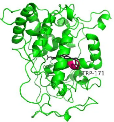
Researcher often used VA as a substrate. In this reaction, the optimum pH is <2.5. The heterologous expression techniques were developed. Doyle and Smith used Escherichia coli to express LiP from Phanerochaete chrysosporium[32]. Miki et al. also used E. coli for LiP from Trametes cervina[27]. Johnson and Li. used baculovirus and LiP from P. chrysosporium was expressed[33]. Especially, the expression system with E. coli was powerful tool, For example, for active site search. As a result, tryptophan (Trp, W) 170 was determined as active site[34]. The tertiary structure was determined[35] and shown inFigure 1-1.
2.2. Manganese peroxidase
Mn (II) + H2O2→ Mn (III) + H2O
Mn (III) form complex with dicarbonic acid like oxalic acid, malonic acid and succinic acid[36]. This complex attacks lignin. The degrading lignin occurs far from enzyme active site. Not only lignin, but also another polymer would have a possibility degraded by MnP.
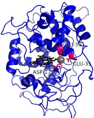
In MnP, the expression system with using E. coli was constructed[37], [38]. The active site is constructed with glutamate (Glu, E) 36, glutamate (Glu, E) 40 and aspartate (Asp, D) 180[39], [40]. The tertiary structure was determined[41] and shown inFigure 1-2.
Native MnP, secreted in fungi, is also glycosylated enzyme. The glycosylated MnP is more stable than non-glycosylated MnP[42].
2.3. Versatile peroxidase
Versatile peroxidase (VP) is hybrid enzyme. This enzyme has two activity; LiP activity and MnP activity[43]. Tertiary structure was determined[44] and it was shown inFigure 1-3.
2.4. Protein engineering of lignin degrading enzymes Researchers tried to improve MnP stability.
MnP has two or four calcium. There is a report that these calcium are very important for the enzyme stability[45]. And the peroxidase from peanut have a disulfide bond near the calcium binding site. Reading et al, focused on the region near the calcium binding site and they introduced disulfide bond to fix the structure[46]. The remaining enzyme activity of native MnP which is glycosylated MnP, recombinant MnP which expressed in E. coli and MnP which introduced disulfide bond was measured. As a result, MnP which has disulfide bond was the same remaining activity as native MnP.
Miyazaki et al., improved H2O2 stability of MnP[47], they substituted methionine (Met,
M) to leucine (Leu, L) because of methionine was sensitive for oxidizing stress. After 3 mM H2O2 treatment for 1 hour at 37 oC, the residual activity of variant and wild-type was about
30 % and 0 %, respectively.
Furthermore Miyazaki et al. used direct evolution method and select mutants improved H2O2 stability [48]. MnP was expressed in inclusion body with E. coli expression system. So
they used cell-free expression system named SIMPLEX (Single-molecule-PCR-linked in vitro expression). They selected 4 mutants improved H2O2 stability from 104 samples.
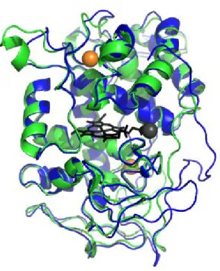
Not only improving enzyme stability, but also engineering enzyme activity was investigated. The tertiary structure was very similar to MnP and LiP. The superposition model LiP and MnP was drawn by PyMol in Figure 1-4. The RMSD (Root Mean Square Deviation) value was 0.716 Å. Timofeevski et al. were introduced LiP active site into MnP from
and VA oxidizing activity.
Conversely, Tünde and Tien introduced MnP active site into LiP[50]. Three mutations, Asn182Asp, Asp183Lys and Ala36Glu, were introduced into LiP from P. chrysosporium. This mutant was able to oxidize Mn (II) and VA. In this mutant, the optimum pH of VA and Mn (II) oxidation were also altered. The optimum pH of oxidizing VA of wild-type and mutant were 2.5 and 3.5, respectively. The optimum pH of oxidizing Mn (II) of wild-type and mutant were 4 and 5.5, respectively.
Native LiP and MnP, expressed in fungi, was glycosylated. Recombinant LiP expressed in Escherichia coli wasn’t glycosylated and its stability was lower than native LiP[42]. Then the expression system in Yeast Pichia pastoris was developed[51]. Although the MnP expressed in P. pastoris was slightly less thermo-stable than MnP from P. chrysosporium, it seems that the stability of MnP expressed in P. pastoris was higher than MnP expressed in E
coli.
Direct evolution method was applied for VP[52]. In this paper, VP from Pleurotus
eryngii was expressed in Saccharomyces cerevisiae. After 6 rounds of direct evolution, 4
Figure 1-1. The tertiary structure of the LiP.
Figure 1-2. The tertiary structure of MnP.
Figure 1-3. The tertiary structure of VP.
Figure 1-4 LiP and MnP aligned structure
PDB data was from protein data bank and aligned by PyMol.
References
[1] B. W. Matthews, H. Nicholson, and W. J. Becktel, “Enhanced protein thermostability from site-directed mutations that decrease the entropy of unfolding,” Proc. Natl. Acad.
Sci., vol. 84, no. 19, pp. 6663–6667, Oct. 1987.
[2] T. Alber, S. Dao-pin, K. Wilson, J. A. Wozniak, S. P. Cook, and B. W. Matthews, “Contributions of hydrogen bonds of Thr 157 to the thermodynamic stability of phage T4 lysozyme,” Nature, vol. 330, no. 6143, pp. 41–46, Nov. 1987.
[3] W. S. Sandberg and T. C. Terwilliger, “Influence of interior packing and
hydrophobicity on the stability of a protein,” Science, vol. 245, no. 4913, pp. 54–57, Jul. 1989.
[4] L. J. Perry and R. Wetzel, “Disulfide bond engineered into T4 lysozyme: stabilization of the protein toward thermal inactivation,” Science, vol. 226, no. 4674, pp. 555–557, Nov. 1984.
[5] J. A. Wells and D. B. Powers, “In vivo formation and stability of engineered disulfide bonds in subtilisin.,” J. Biol. Chem., vol. 261, no. 14, pp. 6564–6570, May 1986.
[6] R. T. Sauer, K. Hehir, R. S. Stearman, M. A. Weiss, A. Jeitler-Nilsson, E. G. Suchanek, and C. O. Pabo, “An engineered intersubunit disulfide enhances the stability and DNA binding of the N-terminal domain of .lambda. repressor,” Biochemistry, vol. 25, no. 20, pp. 5992–5998, Oct. 1986.
[7] M. Matsumura, W. J. Becktel, M. Levitt, and B. W. Matthews, “Stabilization of phage T4 lysozyme by engineered disulfide bonds,” Proc. Natl. Acad. Sci., vol. 86, no. 17, pp. 6562–6566, Sep. 1989.
[8] R. Kuroki, Y. Taniyama, C. Seko, H. Nakamura, M. Kikuchi, and M. Ikehara, “Design and creation of a Ca2+ binding site in human lysozyme to enhance structural stability,”
Proc. Natl. Acad. Sci., vol. 86, no. 18, pp. 6903–6907, Sep. 1989.
[9] H. Sakuraba, K. Yoneda, I. Asai, H. Tsuge, N. Katunuma, and T. Ohshima, “Structure of l-aspartate oxidase from the hyperthermophilic archaeon Sulfolobus tokodaii,”
Biochim. Biophys. Acta - Proteins Proteomics, vol. 1784, no. 3, pp. 563–571, Mar.
[10] K. S. P. Yip, T. J. Stillman, K. L. Britton, P. J. Artymiuk, P. J. Baker, S. E.
Sedelnikova, P. C. Engel, A. Pasquo, R. Chiaraluce, V. Consalvi, R. Scandurra, and D. W. Rice, “The structure of Pyrococcus furiosus glutamate dehydrogenase reveals a key role for ion-pair networks in maintaining enzyme stability at extreme temperatures,”
Structure, vol. 3, no. 11, pp. 1147–1158, Nov. 1995.
[11] M. Hennig, B. Darimont, R. Sterner, K. Kirschner, and J. N. Jansonius, “2.0 å structure of indole-3-glycerol phosphate synthase from the hyperthermophile Sulfolobus
solfataricus: possible determinants of protein stability,” Structure, vol. 3, no. 12, pp. 1295–1306, Dec. 1995.
[12] T. Bulter, M. Alcalde, V. Sieber, P. Meinhold, C. Schlachtbauer, and F. H. Arnold, “Functional Expression of a Fungal Laccase in Saccharomyces cerevisiae by Directed Evolution,” Appl. Environ. Microbiol., vol. 69, no. 2, pp. 987–995, Feb. 2003.
[13] K. Miyazaki and F. H. Arnold, “Exploring Nonnatural Evolutionary Pathways by Saturation Mutagenesis: Rapid Improvement of Protein Function,” J. Mol. Evol., vol. 49, no. 6, pp. 716–720, 1999.
[14] L. Giver, A. Gershenson, P.-O. Freskgard, and F. H. Arnold, “Directed evolution of a thermostable esterase,” Proc. Natl. Acad. Sci., vol. 95, no. 22, pp. 12809–12813, Oct. 1998.
[15] H. Zhao and F. H. Arnold, “Directed evolution converts subtilisin E into a functional equivalent of thermitase,” Protein Eng., vol. 12, no. 1, pp. 47–53, Jan. 1999.
[16] U. Alcolombri, M. Elias, and D. S. Tawfik, “Directed Evolution of Sulfotransferases and Paraoxonases by Ancestral Libraries,” J. Mol. Biol., vol. 411, no. 4, pp. 837–853, Aug. 2011.
[17] H. Flores and A. D. Ellington, “A modified consensus approach to mutagenesis inverts the cofactor specificity of Bacillus stearothermophilus lactate dehydrogenase,” Protein
Eng. Des. Sel., vol. 18, no. 8, pp. 369–377, Aug. 2005.
[19] H. Iwabata, K. Watanabe, T. Ohkuri, S. Yokobori, and A. Yamagishi, “Thermostability of ancestral mutants of Caldococcus noboribetus isocitrate dehydrogenase.,” FEMS
Microbiol. Lett., vol. 243, no. 2, pp. 393–398, Feb. 2005.
[20] K. Watanabe, T. Ohkuri, S. Yokobori, and A. Yamagishi, “Designing thermostable proteins: ancestral mutants of 3-isopropylmalate dehydrogenase designed by using a phylogenetic tree.,” J. Mol. Biol., vol. 355, no. 4, pp. 664–674, Jan. 2006.
[21] K. Watanabe and A. Yamagishi, “The effects of multiple ancestral residues on the Thermus thermophilus 3-isopropylmalate dehydrogenase.,” FEBS Lett., vol. 580, no. 16, pp. 3867–3871, Jul. 2006.
[22] H. Shimizu, S. Yokobori, T. Ohkuri, T. Yokogawa, K. Nishikawa, and A. Yamagishi, “Extremely thermophilic translation system in the common ancestor commonote: ancestral mutants of Glycyl-tRNA synthetase from the extreme thermophile Thermus thermophilus.,” J. Mol. Biol., vol. 369, no. 4, pp. 1060–1069, Jun. 2007.
[23] K. Yamashiro, S. Yokobori, S. Koikeda, and A. Yamagishi, “Improvement of Bacillus circulans beta-amylase activity attained using the ancestral mutation method.,” Protein
Eng. Des. Sel., vol. 23, no. 7, pp. 519–528, Jul. 2010.
[24] S. Akanuma, S. Iwami, T. Yokoi, N. Nakamura, H. Watanabe, S. Yokobori, and A. Yamagishi, “Phylogeny-based design of a B-subunit of DNA gyrase and its ATPase domain using a small set of homologous amino acid sequences.,” J. Mol. Biol., vol. 412, no. 2, pp. 212–225, Sep. 2011.
[25] T. K. Kirk and R. L. Farrell, “Enzymatic ‘Combustion’: The Microbial Degradation of Lignin,” Annu. Rev. Microbiol., vol. 41, no. 1, pp. 465–501, Oct. 1987.
[26] M. TIEN and T. K. KIRK, “Lignin-Degrading Enzyme from the Hymenomycete Phanerochaete chrysosporium Burds,” Sci. , vol. 221 , no. 4611 , pp. 661–663, Aug. 1983.
[28] G. Ghodake, S. Kalme, J. Jadhav, and S. Govindwar, “Purification and Partial Characterization of Lignin Peroxidase from Acinetobacter calcoaceticus NCIM 2890 and Its Application in Decolorization of Textile Dyes,” Appl. Biochem. Biotechnol., vol. 152, no. 1, pp. 6–14, 2009.
[29] P. J. Harvey, H. E. Schoemaker, and J. M. Palmer, “Veratryl alcohol as a mediator and the role of radical cations in lignin biodegradation by Phanerochaete chrysosporium,”
FEBS Lett., vol. 195, no. 1–2, pp. 242–246, Jan. 1986.
[30] T. Johjima, N. Itoh, M. Kabuto, F. Tokimura, T. Nakagawa, H. Wariishi, and H. Tanaka, “Direct interaction of lignin and lignin peroxidase from Phanerochaete chrysosporium,” Proc. Natl. Acad. Sci. , vol. 96 , no. 5 , pp. 1989–1994, Mar. 1999.
[31] M. Chen, G. Zeng, Z. Tan, M. Jiang, H. Li, L. Liu, Y. Zhu, Z. Yu, Z. Wei, Y. Liu, and G. Xie, “Understanding Lignin-Degrading Reactions of Ligninolytic Enzymes: Binding Affinity and Interactional Profile,” PLoS One, vol. 6, no. 9, p. e25647, Sep. 2011.
[32] W. A. Doyle and A. T. Smith, “Expression of lignin peroxidase H8 in Escherichia coli: folding and activation of the recombinant enzyme with Ca2+ and haem,” Biochem. J., vol. 19, pp. 15–19, 1996.
[33] T. M. Johnson and J. K.-K. Li, “Heterologous expression and characterization of an active lignin peroxidase from Phanerochaete chrysosporium using recombinant baculovirus,” Arch. Biochem. Biophys., vol. 291, no. 2, pp. 371–378, Dec. 1991.
[34] W. Blodig, A. T. Smith, K. Winterhalter, and K. Piontek, “Evidence from Spin-Trapping for a Transient Radical on Tryptophan Residue 171 of Lignin Peroxidase,” Arch. Biochem. Biophys., vol. 370, no. 1, pp. 86–92, Oct. 1999.
[35] T. Choinowski, W. Blodig, K. H. Winterhalter, and K. Piontek, “The crystal structure of lignin peroxidase at 1.70 Å resolution reveals a hydroxy group on the Cβ of
tryptophan 171: A novel radical site formed during the redox cycle,” J. Mol. Biol., vol. 286, no. 3, pp. 809–827, Feb. 1999.
[37] R. E. Whitwam, I. G. Gazarian, and M. Tien, “Expression of Fungal Mn Peroxidase in E. Coli and Refolding to Yield Active Enzyme,” Biochem. Biophys. Res. Commun., vol. 216, no. 3, pp. 1013–1017, Nov. 1995.
[38] R. Whitwam and M. Tien, “Heterologous Expression and Reconstitution of Fungal Mn Peroxidase,” Arch. Biochem. Biophys., vol. 333, no. 2, pp. 439–446, Sep. 1996.
[39] H. L. Youngs, M. D. Sollewijn Gelpke, D. Li, M. Sundaramoorthy, and M. H. Gold, “The Role of Glu39 in Mn II Binding and Oxidation by Manganese Peroxidase from Phanerochaete chrysosporium,” Biochemistry, vol. 40, no. 7, pp. 2243–2250, Jan. 2001.
[40] M. D. Sollewijn Gelpke, P. Moënne-Loccoz, and M. H. Gold, “Arginine 177 is involved in Mn(II) binding by manganese peroxidase.,” Biochemistry, vol. 38, no. 35, pp. 11482–9, Aug. 1999.
[41] M. Sundaramoorthy, K. Kishi, M. H. Gold, and T. L. Poulos, “Preliminary Crystallographic Analysis of Manganese Peroxidase from Phanerochaete chrysosporium,” J. Mol. Biol., vol. 238, no. 5, pp. 845–848, May 1994.
[42] G. Nie, N. S. Reading, and S. D. Aust, “Relative Stability of Recombinant Versus Native Peroxidases fromPhanerochaete chrysosporium,” Arch. Biochem. Biophys., vol. 365, no. 2, pp. 328–334, May 1999.
[43] S. Camarero, S. Sarkar, F. J. Ruiz-Dueñas, M. J. Martínez, and A. T. Martínez,
“Description of a versatile peroxidase involved in the natural degradation of lignin that has both manganese peroxidase and lignin peroxidase substrate interaction sites.,” J.
Biol. Chem., vol. 274, no. 15, pp. 10324–10330, Apr. 1999.
[44] M. Pérez-Boada, F. J. Ruiz-Dueñas, R. Pogni, R. Basosi, T. Choinowski, M. J. Martínez, K. Piontek, and A. T. Martínez, “Versatile Peroxidase Oxidation of High Redox Potential Aromatic Compounds: Site-directed Mutagenesis, Spectroscopic and Crystallographic Investigation of Three Long-range Electron Transfer Pathways,” J.
Mol. Biol., vol. 354, no. 2, pp. 385–402, Nov. 2005.
[46] N. S. Reading and S. D. Aust, “Engineering a Disulfide Bond in Recombinant
Manganese Peroxidase Results in Increased Thermostability,” Biotechnol. Prog., vol. 16, no. 3, pp. 326–333, 2000.
[47] C. Miyazaki and H. Takahashi, “Engineering of the H2O2-binding pocket region of a recombinant manganese peroxidase to be resistant to H2O2.,” FEBS Lett., vol. 509, no. 1, pp. 111–114, Nov. 2001.
[48] C. Miyazaki-Imamura, K. Oohira, R. Kitagawa, H. Nakano, T. Yamane, and H.
Takahashi, “Improvement of H2O2 stability of manganese peroxidase by combinatorial mutagenesis and high-throughput screening using in vitro expression with protein disulfide isomerase,” Protein Eng. Des. Sel., vol. 16, no. 6, pp. 423–428, Jun. 2003.
[49] S. L. Timofeevski, G. Nie, N. S. Reading, and S. D. Aust, “Substrate Specificity of Lignin Peroxidase and a S168W Variant of Manganese Peroxidase,” Arch. Biochem.
Biophys., vol. 373, no. 1, pp. 147–153, Jan. 2000.
[50] T. Mester and M. Tien, “Engineering of a manganese-binding site in lignin peroxidase isozyme H8 from Phanerochaete chrysosporium.,” Biochem. Biophys. Res. Commun., vol. 284, no. 3, pp. 723–728, Jun. 2001.
[51] L. Gu, C. Lajoie, and C. Kelly, “Expression of a Phanerochaete chrysosporium manganese peroxidase gene in the yeast Pichia pastoris.,” Biotechnol. Prog., vol. 19, no. 5, pp. 1403–9, 2003.
Chapter 2. Ancestral mutants of lignin peroxidase
1. Summary
Stabilizing enzymes from mesophiles of industrial interest is one of the greatest
challenges of protein engineering. The ancestral mutation method, which introduces inferred ancestral residues into a target enzyme, has previously been developed and used to improve the thermostability of thermophilic enzymes. In this report, we studied the ancestral mutation method to improve the chemical and thermal stabilities of Phanerochaete chrysosporium lignin peroxidase, a mesophilic fungal enzyme. A fungal ancestral lignin peroxidase sequence was inferred using a phylogenetic tree comprising Basidiomycota and Ascomycota fungal peroxidase sequences. Eleven mutant enzymes containing ancestral residues were designed, heterologous expressed in Escherichia coli and purified. Several of these ancestral mutants showed higher thermal stabilities and increased specific activities and/or kcat/KM than those of
2. Purpose
In this study, we applied ancestral mutation to stabilized the recombinant LiP from Basidiomycota, Phanerochaete chrysosporium strain UMHA 3641. A composite phylogenetic tree without any thermophilic organisms was constructed and used to infer the ancestral amino acids. The ancestral mutation form of LiP were then characterized and compared with wild-type recombinant LiP.
3. Material
Veratryl alcohol (VA; 2, 6-dimethoxybenzyl alcohol), oxidized glutathione and hemin chloride were purchased from Sigma-Aldrich (St Louis, MO, USA). Other chemicals were obtained from Wako Chemicals (Tokyo, Japan) and were all of analytical grade.
4. Method
4.1. Phylogenetic analysis
The amino acid sequences of ligninolytic peroxidase, including LiP (30 sequences), MnP (45 sequences) and VP (7 sequences) from Basidiomycota and LiP (3 sequences) from Ascomycota, were retrieved by a BLASTP search using wild-type lignin peroxidase of P.
chrysosporium strain UAMH 3641 as the query sequence. LiP sequence from P.
chrysosporium was provided By Professor Sonoki (Azabu University). The sequences were
aligned using the program CLUSTAL X 2.0 [1] with its default parameters and then manually adjusted. A composite LiP/MnP/VP phylogenetic tree was constructed using the
Neighbor-Joining method with the program found in the CLUSTAL X 2.0 package [1]. Well-aligned regions (198 amino acid residues) were selected using the program GBLOCKS 0.91 [2]. Forty-nine sequences of LiP (16 sequences), MnP (27 sequences) and VP (4
sequences) from Basidiomycota, and LiP (2 sequences) from Ascomycota representing main branches of the tree were selected for further analysis (Table 2-7). Composite LiP/MnP/VP phylogenetic trees were then inferred by maximum likelihood (ML) method using the
program Treefinder (October 2008) [3] and the program PhyML 2.4.4. [4]. The four-category model of discrete Gamma distribution model, which allows different substitution rates among sites, was employed. The WAG substitution model [5] was used as the substitution matrix for amino acids. Because the statistical support of some branch points were not significant, a “semi-exhaustive” search of a ML tree was then performed. We firstly assumed fixed
Because the results obtained with Treefinder and PhyML were slightly different, tree topologies with the top 50 likelihood in the Treefinder and PhyML analyses were used for further investigation. The likelihood of 100 tree topologies was assessed using CODEML program of the PAML3.14 package [8]. The best ML tree was selected and then ancestral amino acids were inferred using the same conditions.
4.2. Gene construction
The LiP cDNA from Phanerochaete chrysosporium strain UAMH 3641 cloned into pPDB-GB (pPDB-LiP) was generously provided by Prof. S. Sonoki (Department of Environmental Science, Azabu University, Japan). The LiP gene (lacking the N-terminal signal sequence) was amplified by the polymerase chain reaction (PCR). The PCR was performed using pPDB-LiP plasmid as template and the following pair of oligonucleotide primers: pLiPf (5’-GGAGATATACATATGGCCACCTGTTCCAACGGCAA-3’) and pLiPr (5’-GTGCGGCCGCAAGCTTAAGCACCCGGAGGCGGAGGG-3’). Ex Taq DNA
polymerase (Takara Bio, Ōtsu, Japan) was used for the PCR. Gene amplification involved an initial denaturation step of 94 ˚C for 3 min, followed by 25 cycles of denaturation, annealing and extension (94 ˚C for 30 sec, 55 ˚C for 1 min, and 72 ˚C for 2 min), then 72 ˚C for 7 min. The amplified wild-type LiP gene was digested with NdeI and HindIII and ligated into pET21a (+). The resulting plasmid was named pET-LiP.
The inferred ancestral amino acid residues were introduced into the expression plasmid pET-LiP by using a QuikChange Lightning Site-Directed Mutagenesis Kit (Stratagene, La Jolla, CA). The primer sequences are listed inTable 2-6. Clones harboring mutations were selected in E. coli XL10-GOLD. The sequence of each mutant was determined with an
Applied Biosystems ABI PRISM 3130x sequencer (Applied Biosystems, Foster City, CA) and the clones were used for gene expression in E. coli BL21 (DE3).
4.3. Expression and purification
Wild-type LiP protein and its ancestral mutants were produced in E. coli BL21 (DE3). Cells harboring the respective plasmid were cultured in Terrific Broth medium (12 g
polypepton, 24 g yeast extract, 4 mL glycerol, 2.31 g KH2PO4, 12.5 g K2HPO4 in 1 L)
supplemented with ampicilin (0.1 mg/mL) at 37 ˚C. When the optical density (OD 600 nm) of
the culture reached 0.6-0.8 heterologous expression was induced by addition of
isopropyl--D-thiogalactopyranoside (IPTG) to a final concentration of 0.5 mM, and the culture was then incubated for a further 4 hr at 37 ˚C. Cells were harvested and disrupted by sonication in ice cold resuspension buffer containing 10 mM Tris-HCl (pH 8.0), 1 mM ethylenediaminetetraacetic acid (EDTA), 5 mM dithiothreitol (DTT), 50 M
accumulated as inclusion bodies. Insoluble fractions were harvested and washed three times with buffer A (10 mM Tris-HCl (pH 8.0), 1 mM EDTA, 2 mM DTT and 1 % (v/v) Triton X-100). The pellets were resuspended in buffer A containing 8 M urea and incubated at room temperature for 1 hr. The denaturated-reduced protein was diluted to 2 M urea in refolding solution: 10 mM CaCl2, 0.7 mM oxidized glutathione, 30 M hemin chloride (1 mM stock
solution in 0.1 N NaOH) in 50 mM Tris-HCl (pH 8.0). The refolding mixture was incubated at room temperature for 12-14 hr with continuous stirring in the dark.
The mixture was dialyzed against 10 mM sodium acetate buffer (pH 6.0), 5 mM CaCl2
at 4 ˚C for 12-14 hr with five changes of buffer. The sample was then recovered and centrifuged at 60,000g for 20 min and the supernatant concentrated using an amicon YM-10 unit (Merck, Darmstadt, Germany).
To purify each sample, supernatant was applied onto a DEAE Sepharose Fast Flow anion-exchange column (GE Healthcare Bioscience, Piscataway, NJ) and after washing the column with the same buffer, bound proteins were eluted with 0.6 M NaCl in 10 mM sodium acetate buffer (pH 6.0) supplemented with 1 mM CaCl2. The appropriate fractions were
pooled, diluted with 10 mM sodium acetate buffer (pH 6.0) supplemented with 1 mM CaCl2,
applied onto a HiTrapQ column (GE Healthcare Bioscience) and eluted with a 0.01-0.6 M NaCl gradient in 10 mM sodium acetate buffer (pH 6.0) supplemented with 1 mM CaCl2.
Fractions with the highest activity were pooled and used for the following experiments.
4.4. Enzyme assay
Enzyme concentrations were determined by the alkaline-pyridine hemochrome method using an extinction coefficient of 34.4 mM-1 cm-1 at 557 nm for heme b [9]. Enzymatic activities were determined by monitoring the oxidation of veratryl alcohol (VA) to veratryl aldehyde. The absorbance change at 310 nm was recorded with a DU7400 spectrometer (Beckman, Fullerton, CA). The assay buffer was 50 mM tartrate (pH 2.5), 2 mM VA and 0.5 mM H2O2. Reaction was initiated by adding 0.5 mM H2O2. An extinction coefficient of 9300
M-1 cm-1 at 310 nm was used for the veratryl aldehyde [10]. One enzyme unit was defined as 1 mol veratryl aldehyde formed per minute.
The pH dependence of specific activities was measured in 50 mM tartrate buffer (pH 2.0-4.5) and in 50 mM acetate buffer (pH 4.0-5.5) at 25 ˚C. The temperature dependence of LiP activities was measured at 25, 30, 40, 50 and 55 ˚C.
The values of the Michaelis constant (KM) for the substrate VA and the catalytic
constant (kcat), were determined from steady-state kinetic analysis in the assay buffer with VA
concentrations between 50 M and 5000 M. To determine the KM value for H2O2, the
velocity data to the Michaelis-Menten equation using the Enzyme Kinetics module of SigmaPlot (Systat Software, San Jose, CA).
Thermal inactivation of LiP was studied by incubating LiP in 50 mM tartrate (pH 2.5) at 37 ˚C. Further experiments were carried out according to the previously described method where LiP was incubated in 20 mM Tris-SO4 (pH 7.9) at 51 ˚C [11].
Inactivation of LiP in the presence of H2O2 was studied by incubating LiP in 50 mM tartrate
buffer (pH 2.5) supplemented with 0.2 mM H2O2 at 25 ˚C.
4.5. Thermal melting profile
5. Result
5.1. Inference of ancestral amino acids of fungal peroxidase
Eighty-five amino acid sequences related to wild-type LiP sequences were obtained from the NCBI database and aligned (Supplementary figure 2-1). Well-aligned regions were selected by GBLOCKS. A neighbor-joining (NJ) tree was then constructed using these 85 sequences. On the basis of the NJ tree topology, 49 representative sequences, 16 LiP (including wild-type LiP), 27 MnP and 4 VP from Basidiomycota and 2 Ascomycota LiP, were selected for further phylogenetic analyses. ML trees were constructed using Treefinder and PhyML. The tree topologies were assessed by CODEML. Figure 2-1 shows the best likelihood composite phylogenetic tree of the selected fungal ligninolytic peroxidase, constructed from 198 well-aligned sites of the 49 fungal peroxidase sequences. The sources and accession numbers of the proteins are listed in (Table 2-7). In Figure 2-1, marked branches indicate the selected ancestral positions: node63 is the branching point of
Basidiomycota and Ascomycota, node60 and node62 are the two ancestors of LiP and MnP. Ancestral residues of these nodes were inferred by the ML using the tree as the effort tree. Figure 2-2 shows the sequences of inferred ancestral residues. We then compared the sequences of wild-type LiP with the ancestral sequences (Figure 2-2). Eleven selected regions where the wild-type LiP residues differed from residues of the ancestral sequences were identified. When selecting the mutation sites, the tertiary structure and spatial location of these residues were not taken into consideration. Mutants m1, m2, m3, m4, m5, m6, m7, m8, m9, m10 and m11 correspond to Ala36Glu, Val45Thr, Lys92Glu/Leu93Ala, Ala110Gln, Ala116Gly/Leu117Val, His149Asp, Gly198Ser, Gln209Met, Ile235Leu,
His239Phe/Thr240Leu/Ile241Leu, Phe254Met mutation(s), respectively. All mutation sites are shown in the tertiary structure (Figure 2-3). Each ancestral combination of residues was introduced into LiP by site-directed mutagenesis. All of the mutant enzymes were
overexpressed in E. coli and then purified for characterization.
5.2. Enzyme stability
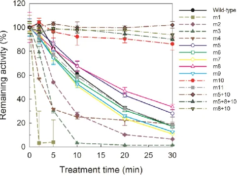
The activities of most of the ancestral mutant enzymes were similar to the wild-type enzyme. However three mutant enzymes (m1, m2 and m4) showed significantly lower activity than the wild-type enzyme at 37 °C (Figure 2-4). After incubation at 37 °C for 30 min, the wild-type enzyme retained 17 % of its initial activity, whereas the ancestral mutants, m5, m8, m10 and m11, retained 28.0, 33.5, 86.2 and 18.4 % of their initial activity, respectively (Table 2-1).
The activity of the enzymes was also measured after incubation in the presence of hydrogen peroxide. Specifically, enzymes were incubated with 0.2 mM H2O2 at 25 oC (Figure
wild-type retained 6.5 % of its initial activity, whereas four ancestral mutants, m2, m5, m10 and m11, retained 18.4, 29.6, 10.5 and 9.3 % of their initial activity, respectively (Table 2-2). Of all the LiP mutants, the ancestral mutant m4 retained the greatest proportion of enzymatic activity in the presence of H2O2, although its initial activity was very low (Table 2-2).
5.3. Steady-kinetics analysis
The specific activities of six ancestral mutant enzymes (m6, m7, m8, m9, m10 and m11) were larger than that of the wild-type LiP (Table 2-3). In particular, the ancestral mutant m10 showed significantly enhanced specific activity i.e., 2.3-fold greater than that of wild-type LiP.
The kinetic parameters of the LiPs were estimated using steady-state kinetics obtained at 25 ˚C (Table 2-3). The kcat for VA of eight ancestral mutants (m3, m5, m6, m7, m8, m9, m10
and m11) were larger than that of wild-type LiP. Ancestral mutant m10 showed remarkably improved kcat (3.4-fold higher than that of the wild-type enzyme). Eight of eleven ancestral
mutants (m3, m5, m6, m7, m8, m9, m10 and m11) showed a kcat/KM ratio (specificity
constant) for VA higher than that of the wild-type enzyme. The increased kcat/KM of ancestral
mutants was caused by an improvement of kcat. However, nine of eleven ancestral mutants
(m1, m2, m3, m4, m5, m6, m8, m9 and m11) showed improved KM values for H2O2. The kcat
for H2O2 of two mutants (m7 and m10) was higher than that of the wild-type enzyme. Eight of
the ancestral mutants (m1, m5, m6, m7, m8, m9, m10 and m11) showed high kcat/KM for H2O2.
The ancestral mutant, m10 showed improved kcat for VA and H2O2.
5.4. pH dependence
The pH dependence of the wild-type enzyme and selected ancestral mutants, m5 and m10, showed similar tendencies: the specific activities increased as the pH decreased from pH 5.5 (Figure 2-7).
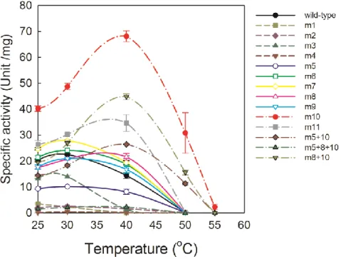
5.5. Temperature dependence
5.6. Thermal melting profile
The thermal melting profiles of the wild-type and ancestral mutant m10 enzymes were also measured (Figure 2-8). Tm values of the wild-type enzyme and m10 were 50.1 ± 0.2 oC
6. Discussion
6.1. Phylogenetic analysis
In our analysis of the phylogenetic tree, two LiPs from Ascomycota Pyrenophora
tritici-repentis were used as the outgroup for Basidiomycota peroxidases. LiPs formed a
cluster in Basidiomycota enzymes, except for BAE46585 from Trametes cervina. MnPs were clustered as two groups. The first group consists of MnPs from P.
chrysosporium and the second group consists of MnPs from Trametes versicolor. Previously,
Floudas et al., proposed an evolutionary process for the lignin degrading peroxidases of fungi [12]. The tree topology obtained from our analysis is similar to that reported by Floudas et al. According to the geologic time reported by Floudas et al., our node 63 and node 62
correspond to the Silurian period in the Paleozoic era (ca. 403 million years ago) and the Permian period in the Triassic era (ca. 270 million years ago), respectively.
6.2. Characteristics of the ancestral mutation sites
All improved mutants were located on a helix region of the protein (Table 2-4). However, a few mutations were located on loop or β sheet regions of the protein, making the correlation between thermal stability and mutation site unclear. Moreover, there was no obvious relationship between protein stability and location of the mutation site in terms of distance from the active site and calcium ions. All mutation sites were separated from the heme prosthetic group, tryptophan171 and two calcium ions. In other words, the residues locating near these factors were not selected as mutation site.
FoldX was the computer algorithm to predict the effect of mutation from tertiary structure information[13]. In Table 2-4, the total energy value of most stable mutant, m10, was the most smallest. However the correlation between total energy and stability didn’t observe.
6.3. The reasons of increasing stability
Chou and Fasman reported the secondary structural prediction of proteins from their amino acid sequence[14]. According to their report, glutamate, methionine, alanine and leucine are amino acids strong α-helix former. Phenylalanine and isoleucine are α-helix former. Proline and glycine are strong α-helix breaker. The most stable ancestral mutant was m10 and its mutation was H239F/T240L/I241L and they located on the α-helix (Table 2-4). The α-helix formation can be attributed to the increased stability induced by ancestral mutation.
located in the interior. The crystal structure analysis of ancestral LiP wasn’t done, so the 1B80 (Protein ID, [15]), the most homologous enzyme, was used to estimate the tertiary structure. The structure around I241L is shown in Figure 2-9. From the figure, the mutation I241L may have improved the packing in the hydrophobic core.
6.4. Comparing thermostability of glycosylated LiP
Figure 2-10 shows thermal inactivation time-course conditions used in a previous report
i.e., LiP was incubated in 20 mM Tris-SO4 (pH 7.9) at 51 °C [11]. Under these conditions,
wild-type LiP activity fell to less than 20 % of its original value after 10 min incubation, while ancestral mutants, m10 retained 65 % of their activity. Nie et al. reported that the thermal stability of recombinant LiP H8, which was expressed in E. coli, was lower than that of the corresponding native LiP H8 isolated from P. chrysosporium. Native glycosylated LiP H8 maintained ca. 50 % of its activity after incubation for 10 min at 51 °C, whereas recombinant LiP H8 was completely inactivated by this treatment. Wild-type LiP used in this study has 99.1 % sequence identity with LiP H8 isoform from P. chrysosporium. Recombinant LiP, m10, expressed in E. coli showed higher thermal stability than that of native LiP H8.
6.5. Comparison with the consensus approach
Watanabe et al., classified ancestral mutants of IPMDH into two groups, dominant and minor ancestral mutants [16]. The dominant ancestral mutants have the ancestral residue, which is also a consensus residue, while the minor mutants have the ancestral residue that is not a consensus residue. In this study, there were seven dominant mutants (m1, m2, m4, m5, m6, m9 and m10) and four minor mutants (m3, m7, m8 and m11) (Table 2-5). Thermostability of two mutants (m8 and m11) in four minor mutants, were improved. Two mutants (m5 and m10) among seven dominant mutants showed higher thermal stability than the wild-type enzyme. We excluded a dominant mutant m4, which showed very low activity. Accordingly, it is not possible to judge whether the consensus residue is responsible for increased thermal stability. It is important to note that the ancestral residue was introduced into the wild-type consensus residue in three mutants (m7, m8 and m11). Two (m8 and m11) of the three
mutants showed greater thermal stability over the wild-type enzyme. These results support the idea that the enhanced thermal stability is related to the ancestral residues, rather than the consensus residues.
6.6. Correlation between stability and conservation of the neighboring residues
conserved value (ACV) as a design tool. Though only four mutants have an ACV higher than 0.80, two of them showed increased thermal stability in our experiments (Figure 2-12). The ACV values of the heme, distal and proximal calcium ions were high (i.e., 0.61, 0.86 and 0.70, respectively), indicating that residues located near these factors are well conserved.
We have extended the method to select the mutation site from the primary amino acid sequence. We estimated the relationship between thermal stability and the conservation of the neighboring amino acids within seven residues in the primary sequence. If a wild-type residue and the ancestral residue were identical, the likelihood value was taken as the conservation value. However, if the wild-type and ancestral residue differed then the conservation value was 0. Finally, an averaged conservation value for neighboring residues on the primary amino acid sequence was calculated. This value is referred to as linear ACV. The linear ACV values were plotted against the remaining activity after incubation at 37 oC (Figure 2-11A) or in 0.2 mM H2O2 (Figure 2-11B). When the linear ACV value is greater than 0.9, mutants with
improved thermal stability are obtained at high efficiency. Three of four mutants whose linear ACV was >0.9 showed improved thermal stability. A similar trend was observed when the window size was increased to eleven residues (Figure 2-13).
7. Conclusion
In this study, we introduced ancestral mutations into wild-type LiP from P. chrysosporium to improve its thermal stability. The recombinant ancestral mutant, m10 (H239F/T240L/I241L), showed improved thermal stability comparable to that of the glycosylated wild-type enzyme. Specific activity and kcat/KM of one of the ancestral mutants,
Figure 2-1. Phylogenetic tree
A composite phylogenetic tree of LiP, MnP and VP. The tree includes the selected species from Basidomycota and Ascomycota. Open circles, open triangles and open squares indicates LiPs, MnPs and VPs from Basidomycota, respectively. The filled circle and triangle represent LiP and MnP from Phanerochaete
chrysosporium strain UMHA 3641, respectively. The phylogenetic tree was generated by the ML method as
Figure 2-2. Ancestral sequences
Figure 2-3. The three-dimensional structure of LiP and ancestral mutation points
Figure 2-4. Time course of thermal inactivation of wild-type LiP and ancestral mutants
Figure 2-5. Residual activity of the wild-type enzyme and ancestral mutants after incubation in the presence of H2O2.
The incubation was carried out at 25 oC in 50 mM tartrate buffer (pH 2.5) containing 2 μM enzyme supplemented with 0.2 mM H2O2. The remaining enzyme activities were measured in 50 mM tartrate buffer (pH
Figure 2-6. Temperature dependence of VA oxidation by wild-type LiP and ancestral mutants.
Reaction mixture contained 2 mM VA, 0.5 mM H2O2 and 0.2 μM of each enzyme in 50 mM tartrate buffer, pH
Figure 2-7. pH dependence of VA oxidation of wild-type and ancestral mutants of LiP.
Reaction mixture contains 2 mM VA and 0.5 mM H2O2 in 50 mM tartrate buffer, pH 2.0-4.5 (open circles) and
Figure 2-8. Thermal melting profile
Figure 2-9. Mutation site of I241L
Dark red indicates higher hydrophobic residues, sickly red indicates hydrophobic amino acid residues. Blue stick indicates I241 or L241. Black stick is heme. (A) wild-type (I241) and (B) m10 (L241).
(A)
Figure 2-10. Time course of thermal inactivation of wild-type LiP and ancestral mutants, m10.
Enzymes were incubated at 51 oC in 20 mM Tris-SO4 buffer (pH 7.9). The remaining activities were measured in 50 mM acetate buffer (pH 4.5), 2 mM VA, 0.1 mM H2O2 and 0.2 μM enzyme. Reactions were initiated by the
Figure 2-11. Correlation between thermostability and linear ACV.
The linear ACV values were plotted against remaining activity after treatment at 37 oC (Panel A). The linear ACV values were plotted against remaining activity after incubation with 0.2 mM H2O2 (Panel B). Dashed line
Figure 2-12. Correlation between thermostability and the ACV.
Figure 2-13. Correlation between thermostability and the linear ACV (window size 11).
Figure 2-14. Correlation between thermostability and the linear ACV of β-amylase.
Closed circle indicates the linear ACV value of window size 5. Open circle indicate the linear ACV value of window size 7. t1/2 values were plotted against linear ACV values. Dashed line indicates a t 1/2 of the wild-type
Figure 2-15. Correlation between thermostability and the linear ACV of IPMDH.
Closed circle indicates the linear ACV value of window size 9. t1/2 values were plotted against linear ACV.
Table 2-1. Thermal inactivation at 37 oC treatment of wild-type LiP and ancestral mutants
Specific activity (Unit/mg) Remaining activity (%) 0 min 30 min Wild-type 14.4 ± 0.0 ( 1.00 ) 2.5 ± 0.1 ( 1.00 ) 17.2 ( 1.00 ) m1 5.6 ± 0.3 ( 0.39 ) 0.0 ± 0.0 ( 0.00 ) 0.0 ( 0.00 ) m2 3.3 ± 0.1 ( 0.23 ) 0.2 ± 0.0 ( 0.08 ) 6.6 ( 0.38 ) m3 12.6 ± 0.2 ( 0.88 ) 0.2 ± 0.0 ( 0.08 ) 1.5 ( 0.09 ) m4 0.9 ± 0.0 ( 0.06 ) 0.2 ± 0.0 ( 0.08 ) 19.1 ( 1.11 ) m5 14.6 ± 0.1 ( 1.01 ) 4.1 ± 0.3 ( 1.64 ) 28.0 ( 1.63 ) m6 24.8 ± 0.7 ( 1.72 ) 4.3 ± 0.3 ( 1.72 ) 17.2 ( 1.00 ) m7 25.6 ± 0.5 ( 1.78 ) 2.8 ± 0.1 ( 1.12 ) 10.9 ( 0.63 ) m8 20.1 ± 1.5 ( 1.40 ) 6.7 ± 0.3 ( 2.68 ) 33.5 ( 1.95 ) m9 19.4 ± 0.8 ( 1.35 ) 2.5 ± 0.1 ( 1.00 ) 12.7 ( 0.74 ) m10 53.4 ± 1.6 ( 3.71 ) 46 ± 1.6 ( 18.4 ) 86.2 ( 5.01 ) m11 22.1 ± 0.7 ( 1.53 ) 4.1 ± 0.3 ( 1.64 ) 18.4 ( 1.07 ) m5+8 0.1 ± 0.0 ( 0.01 ) 0.0 ± 0.0 ( 0.00 ) 84.1 ( 4.89 ) m5+10 14.0 ± 0.3 ( 0.97 ) 14.3 ± 0.1 ( 5.72 ) 102.1 ( 5.93 ) m8+10 21.4 ± 0.2 ( 1.49 ) 20.1 ± 0.4 ( 1.40 ) 93.8 ( 5.45 ) m5+8+10 4.4 ± 0.0 ( 0.31 ) 3.9 ± 0.0 ( 1.56 ) 90.2 ( 5.24 )
Table 2-2. H2O2 resistance of wild-type LiP and ancestral mutants
Specific activity (Unit/mg) Remaining activity (%) 0 min 5 min Wild-type 12.2 ± 0.2 ( 1.00 ) 0.8 ± 0.1 ( 1.00 ) 6.5 ( 1.00 ) m1 3.9 ± 0.2 ( 0.32 ) 0.2 ± 0.0 ( 0.25 ) 4.4 ( 0.68 ) m2 2.6 ± 0.2 ( 0.21 ) 0.5 ± 0.0 ( 0.63 ) 18.4 ( 2.83 ) m3 10.0 ± 0.0 ( 0.82 ) 0.4 ± 0.0 ( 0.50 ) 4.3 ( 0.66 ) m4 0.2 ± 0.0 ( 0.02 ) 0.2 ± 0.0 ( 0.25 ) 96.9 ( 14.9 ) m5 11.3 ± 0.1 ( 0.93 ) 3.3 ± 0.1 ( 4.13 ) 29.6 ( 4.55 ) m6 12.5 ± 0.3 ( 1.02 ) 0.8 ± 0.0 ( 1.00 ) 6.2 ( 0.95 ) m7 15.3 ± 1.6 ( 1.25 ) 1.0 ± 0.2 ( 1.25 ) 6.4 ( 0.98 ) m8 9.4 ± 0.3 ( 0.77 ) 0.3 ± 0.0 ( 0.38 ) 3.3 ( 0.51 ) m9 8.8 ± 0.2 ( 0.72 ) 0.3 ± 0.0 ( 0.38 ) 3.0 ( 0.46 ) m10 32.0 ± 1.2 ( 2.62 ) 3.4 ± 0.1 ( 4.25 ) 10.5 ( 1.62 ) m11 3.3 ± 0.0 ( 0.27 ) 0.3 ± 0.0 ( 0.38 ) 9.3 ( 1.43 ) m5+8 0.0 ± 0.0 ( 0.0 ) 0.0 ± 0.0 ( 0.00 ) 54.1 ( 8.32 ) m5+10 14.3 ± 0.7 ( 1.17 ) 4.7 ± 1.2 ( 5.88 ) 32.6 ( 5.01 ) m8+10 16.7 ± 0.8 ( 1.36 ) 0.8 ± 0.2 ( 1.00 ) 5.1 ( 0.78 ) m5+8+10 5.5 ± 0.4 ( 0.45 ) 0.2 ± 0.0 ( 0.25 ) 3.5 ( 0.54 )
Table 2-3. Steady-state kinetic parameters of wild-type and ancestral mutants
Specific activity (Unit/mg)
VA H2O2
kcat (/s) KM (μM) kcat/KM (/s/μM) kcat (/s) KM (μM) kcat/KM (/s/μM)
Wild-type 18.8 ( 1.00 ) 11.6 ± 0.1 ( 1.00 ) 93.4 ± 6.2 ( 1.00 ) 0.124 ( 1.00 ) 19.4 ± 0.0 ( 1.00 ) 59.5 ± 3.5 ( 1.00 ) 0.33 ( 1.00 ) m1 8.7 ( 0.46 ) 9.6 ± 0.1 ( 0.83 ) 96.4 ± 5.3 ( 1.03 ) 0.099 ( 0.80 ) 3.9 ± 0.0 ( 0.20 ) 7.6 ± 0.4 ( 0.13 ) 0.51 ( 0.64 ) m2 1.2 ( 0.06 ) 1.7 ± 0.4 ( 0.15 ) 504.3 ± 351.3 ( 4.10 ) 0.003 ( 0.04 ) 3.3 ± 0.0 ( 0.17 ) 18.5 ± 1.9 ( 0.31 ) 0.18 ( 0.55 ) m3 12.1 ( 0.64 ) 17.4 ± 0.4 ( 1.50 ) 118.2 ± 12.1 ( 1.27 ) 0.147 ( 1.18 ) 13.4 ± 0.0 ( 0.69 ) 41.2 ± 3.9 ( 0.69 ) 0.32 ( 0.97 ) m4 0.1 ( <0.01 ) 0.2 ± 0.0 ( 0.02 ) 426.3 ± 67.2 ( 4.50 ) <0.001 ( <0.01 ) 0.1 ± 0.0 ( 0.01 ) 10.1 ± 4.9 ( 0.17 ) 0.005 ( 0.02 ) m5 14.0 ( 0.74 ) 18.9 ± 0.2 ( 1.63 ) 122.8 ± 5.6 ( 1.31 ) 0.154 ( 1.24 ) 7.2 ± 0.0 ( 0.37 ) 47.2 ± 5.5 ( 0.79 ) 0.37 ( 1.12 ) m6 26.7 ( 1.42 ) 18.7 ± 0.3 ( 1.61 ) 94.4 ± 7.4 ( 1.01 ) 0.198 ( 1.60 ) 19.2 ± 0.0 ( 0.99 ) 51.9 ± 3.5 ( 0.87 ) 0.36 ( 1.09 ) m7 27.6 ( 1.47 ) 19.6 ± 0.2 ( 1.69 ) 120.5 ± 6.2 ( 1.29 ) 0.163 ( 1.31 ) 23.5 ± 0.6 ( 1.21 ) 65.3 ± 4.3 ( 1.10 ) 0.41 ( 1.24 ) m8 21.0 ( 1.12 ) 13.8 ± 0.4 ( 1.19 ) 106.1 ± 15.6 ( 1.13 ) 0.13 ( 1.05 ) 16.5 ± 0.5 ( 0.85 ) 40.3 ± 4.2 ( 0.68 ) 0.43 ( 1.30 ) m9 19.3 ( 1.03 ) 12.3 ± 0.1 ( 1.06 ) 64.7 ± 3.3 ( 0.69 ) 0.19 ( 1.53 ) 15.7 ± 0.4 ( 0.81 ) 36.3 ± 3.0 ( 0.61 ) 0.34 ( 1.03 ) m10 43.3 ( 2.30 ) 39.7 ± 0.6 ( 3.42 ) 181.5 ± 11.6 ( 1.94 ) 0.219 ( 1.76 ) 34.2 ± 1.1 ( 1.76 ) 99.9 ± 8.6 ( 1.68 ) 0.41 ( 1.24 ) m11 25.3 ( 1.35 ) 17.6 ± 0.3 ( 1.52 ) 121.7 ± 8.6 ( 1.30 ) 0.144 ( 1.16 ) 16.4 ± 0.0 ( 0.85 ) 40.2 ± 3.1 ( 0.68 ) 0.37 ( 1.12 ) m5+8 0.0 ( 0.00 ) Not Detected 0.1 ± 0.0 ( 0.01 ) 11.0 ± 4.1 ( 0.19 ) 0.01 ( 0.04 ) m5+10 13.6 ( 0.72 ) 8.96 ± 0.3 ( 0.77 ) 139.7 ± 17.7 ( 1.50 ) 0.064 ( 1.94 ) 11.6 ± 0.4 ( 0.60 ) 74.38 ± 7.5 ( 1.25 ) 0.16 ( 0.47 ) m8+10 24.1 ( 1.28 ) 13.4 ± 0.2 ( 1.16 ) 98.5 ± 8.1 ( 1.05 ) 0.136 ( 1.10 ) 15.2 ± 0.5 ( 0.79 ) 42.24 ± 5.3 ( 0.71 ) 0.36 ( 1.09 ) m5+8+10 4.5 ( 0.24 ) 3.5 ± 0.1 ( 0.30 ) 109.7 ± 13.2 ( 1.17 ) 0.032 ( 0.26 ) 2.7 ± 0.1 ( 0.14 ) 22.7 ± 4.0 ( 0.38 ) 0.12 ( 0.36 )
Table 2-4. Characteristics of ancestral mutation sites Mutation site Accessible surface areaa (%) Secondary structureb
Distance from active site ions (Å)c Distance from Ca2+ ions (Å)c ⊿Total energy d
(kcal/mol)
Stabilitye From heme ion From w171 From distal site From proximal site
m1 A36E 14 α 13.6 21.8 21.8 13.6 0.23 - m2 V45T 1 α 10.2 15.1 8.8 18.4 0.18 - m3 K92E 63 α 19.3 28.7 16.0 26.3 1.89 - L93A 40 α 19.3 28.1 17.7 24.4 m4 A110Q 4 α 15.8 18.0 11.2 21.0 1.08 + m5 A116G 0 α 15.0 18.1 19.1 14.1 2.81 + L117V 0 α 14.3 15.1 20.1 11.7 m6 H149D 26 L 12.7 18.1 20.6 23.6 -0.51 ± m7 G198S 47 L 15.4 18.3 26.7 6.1 -0.05 - m8 Q209M 2 α 17.4 15.6 32.1 14.3 -1.78 + m9 I235L 9 β 9.7 16.8 24.1 15.6 -1.84 - m10 H239F 24 α 12.9 14.8 27.0 16.2 -2.27 + T240L 38 α 14.4 15.0 26.9 19.3 I241L 0 α 13.2 11.4 25.1 17.9 m11 F254M 4 α 17.8 9.0 29.3 12.2 0.63 +
aAccessible surface areas were calculated at the VADAR web server.
bPosition of the mutated site in the modeled LiP structure: α, β and L represent helix, sheet and loop structure, respectively. cDistances were estimated in the LiP-modeled structure.