Acta Med. Nagasaki 44 : 67-72
Case Report
Successful Resuscitation from Ventricular Fibrillation during Jogging in a Young Patient with Hypertrophic Cardiomyopathy
Noriyuki KOSE 1), Hideki FUJIWARA 1), Yuzuru HONDA 2), Yoshiyuki DOI 3), Genii TODA 3), Katsusuke YANO 3), Yoshiyuki MIYAHARA 4), Shigeru KOHNO 4)
1) Cardiology Division of Medicine, Ebisumachi Hospital 2) Division of Anesthesiology, Ebisumachi Hospital
3) Third Department of Internal Medicine, Nagasaki University School of Medicine 4) Second Department of Internal Medicine, Nagsaki University School of Medicine
A 15-year-old girl, who was previously in good health, suddenly collapsed while jogging. Immediate cardiopulmon- ary resuscitation (CPR) was initiated, and she arrived at our hospital 13 minutes later. The ventricular fibrillation (VF) on admission was reverted to sinus rhythm 18 minutes after collapse by the second cardioversion. The echocardio- gram revealed hypertrophic nonobstructed cardiomyopathy (HNOCM), although the 24hr ambulatory electrocardiogra- phic, electrophysiologic and exercise stress tests could not define the exact cause of VF. Exercise-induced ischemia
with sustained mild hypokalemia was suspected to be the cause of VF. The patient recovered consciousness three days after admission, and followed an uneventful course of treatment with oral atenolol not associating with disabling neurological deficit.
Immediate basic life support and delivery of automatic external defibrillator on the spot is needed to rescue pa- tients with out-of-hospital cardiac arrest.
Key Words: cardiopulmonary resuscitation (CPR), cardioversion, automatic defibrillator, cardiac arrest
Introduction
Hypertrophic cardiomyopathy (HCM) is a heteroge- neous disease genotypically, phenotypically, patho- physiologically and therapeutically"). Sudden death is a common complication of HCM, especially in adoles- cents and young adults. Although the annual mortal- ity rate from sudden death is 2 to 3% in adults, it is
Address Correspondence: Noriyuki Kose, M.D.
Cardiology Division of Medicine, Ebisumachi Hospital, 3-4 Ebisumachi, Nagasaki 850-0056, Japan
TEL: +81-95-824-9131 FAX: +81-95-824-9651
even higher (approximately 6%) in young patients3).
Unexpected sudden cardiac death (SCD) of young ath- letes occurs in approximately 1 of 200,000 high school
athletes per academic year'). Even if electrocardiogra- phic abnormalities were detected at annual school physical examinations, some young individuals report- edly had SCD during school physical education classes.
This was due to delayed or lack of further investiga- tion, and requires precise evaluation systems. However, the authority of permitting each young patient with hypertrophic nonobstructed cardiomyopathy
(HNOCM) to perform exercise is dependent on individ- ual physician judgement. Such judgement sometimes may differ since some patients sometimes show nei- ther severe symptoms nor significant pressure gradient of the left ventricular outflow tract.
Ventricular fibrillation (VF) is the most common mechanism of SCD, and some of the conditions facili- tating VF include bradycardia, long QT syndrome, electrocution, electrolyte imbalance, drugs, sympathetic stimulation and myocardial ischemia. If young, cardiac arrest survivors were rescued with longstanding cere- bral damages, their quality of life would be aggra- vated. We herein report a case with dramatic, success- ful resuscitation from cardiac arrest remaining no cerebral damage in a young asymptomatic female with previous unknown HCM.
Case Report
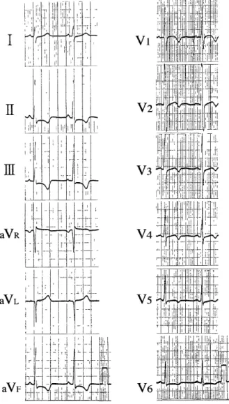
A 15-year-old, high school student, who had been well and playing volleyball during middle high school days, had an electrocardiographic abnormality noted on April 22, 1998. It showed negative T wave in leads II, III, aVf, V, through V6 and high amplitude of R wave in V, through V3 without QT interval prolonga- tion (QTc interval = 0.41 seconds) (Fig. 1). She was scheduled for admission to a university hospital on
Fig. 1, Electrocardiogram on April 22, 1998.
Negative T wave in leads II, III, Vf, V, through V6 and high amplitude of R wave in V, through V3 were shown. QTc interval was 0.41 seconds.
Fig. 2. Electrocardiogram on June 3, 1998.
Ventricular fibrillation was reverted to sinus rhythm after DC cardioversion. (a: before cardioversion ; b: after cardioversion ) DC cardioversion : direct current cardioversion
June 4, 1998 for further diagnostic investigation. She had been suffering from diarrhea for two months owing to irritable colitis. Her grandfather and uncle had left ventricular hypertrophy without definite diag- nosis. There was no family history of sudden death, congestive heart failure and lethal heart diseases.
At 10: 35 a.m. on June 3, 1998, the patient collapsed suddenly on the school grounds while jogging approxi- mately 300 meter in distance during physical educa- tion. Basic life support was started immediately by a teacher, and a first-responding fire fighter vigorously initiated CPR two minutes later. The patient was brought to our hospital by ambulance 13 minutes after collapse, with mydriasis, weak spontaneous respi- ration and E-1, V-1 and M-1 of the Glasgow Coma Scale (GCS), or 111-300-A of Japan Coma Scale (JCS).
The initial rhythm was VF, and the second direct
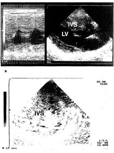
current (DC) cardioversion reverted VF to sinus rhythm 18 minutes after collapse (Fig. 2-a, b). A clini- cal time table is shown in Table 1. The arterial blood gas, complete blood cell count and serum biochemical analysis on admission to our hospital are shown in Table 2. Echocardiographycally, the wall thickness of the interventricular septum (IVS) was 22.5mm ; ante- rior left ventricular wall 19.3mm ; lateral left ventricu- lar wall 14.0mm ; posterior left ventricular wall 11.3mm (Fig. 3). Neither left ventricular outflow tract obstruction nor systolic anterior motion (SAM) of the anterior mitral valve leaflet was present. Our diagnosis was HNOCM with mild mitral regurgitation.
Since her coma scale was improved (El, V1, M4 of GCS or 111-100 of JCS) with hemodynamic stability (Table 1), the patient was transferred to the univer- sity hospital for hyperbaric oxygen therapy (HOT).
Table 1. Clinical Time Table on June 3, 1998
Time Clinical Course GCS
10:35 a. m. Collapse on the school ground E1, V1, Ml
10:40 a. m. Ambulance's arrival in the shool ground El, Vi, M1
10:48 a. m. Patient's arrival at hospital; BP: unmeasurable E1, V1, M1 10:52 a. m. Intratrcheal intubation The first delivery of 360J of DC cardioversion E1, V1, M1 10:53 a. in. The second delivery of 360J of DC cardioversion E1, V1, Ml
10:56 a.m. BP: 104/-mmHg E1, V1, M2
11:15 a.m. BP : 120/56mmHg El, V1, M4
12:50 a.m. BP : 150/88mmHg El, V1, M4
01:00 p.m. Transfer to the university hospital El, V1, M4
BP: blood pressure
DC cardioversion: direct current cardioversion GCS: Glasgow Coma Scale
However, the patient did not require HOT; due to im- proved cerebral function, and received glycerol and po- tassium L-aspartate intravenously for five days to cor- rect hypokalemia (K=2.7mEq/L in the university hospital on June 3). Three days after admission, the patient recovered to E-4, V-4 and M-6 of GCS or I-1 of JCS without neurological deficits, and the value of po-
tassium was corrected to 4.OmEq/L.
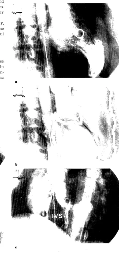
On June 17, 1998, left ventriculography and biventriculography demonstrated hypertrophic IVS
with normal coronary arteriogram and slightly ele- vated left ventricular end-diastolic pressure (LVEDP)
(Fig.4, 5, Table 3). There was no pressure gradient be- tween the left ventricular outflow tract and the aorta.
Table 2. Blood Gas, Complete Blood Cell Count and Serum Biochemical Data at 10:54 a.m. on June3, 1998
pH 6.98pH units AST 36U/f
PCO2 35.7mmHg ALT 25U/P
P02 306.4mmHg LDH 342U/i
(with 10 f /min oxygen) CK 95U/~
HC03- 8.4mmol/f Na 140mE/t
Base Excess -23.6mmol/ K 3.7mEq/e
WBC 9900/ e Cl 95mEq/4
RBC 520x 104/,U BUN 121mg/dt
Hgb 17.1g/df Creatinine 1.0mg/d4
Pit 14.7x10⁴/ £ CRP 0.4mg/de
Table 3. Hemodynamic Variables
Systole Diastole Mean EDP
PAWP mmHg * * 10
PA mmHg 25 11 16 *
RV mmHg 29 1 * 6
RA mmHg * * 3 *
LV mmHg 90 9 * 17
Ao mmHg 100 60 80 *
Ao: aorta
EDP: end-diastolic pressure PA: pulmonary arterial pressure
PAWP: pulmonary arterial wedge pressure RA: right atrium
RV: right ventricle
*: not available
Fig. 3. Echocardiogram on June 3, 1998.
Echocardiogram showed hypertrophy of the interventricular sep- tum and the anterior left ventricular wall (a : long axis view ; b:
short axis view)
A : anterior left ventricular wall IVS : interventricular septum LV : left ventricule
Ventricular tachycardia (VT), VF and supraventricular tahcycardia (SVT) were not induced by programmed ventricular stimulation, isoproterenol provacation and
by exercise stress test. The 24hr ambulatory electro- cardiogram (24hr ECG) also failed to identify any arrhythmias.
The patient was given 25mg/day of atenolol orally, and was discharged without any neurological sequelae on June 9, 1998. She has been following an uneventful course without worsening her daily quality of life.
mass ("possible HCM") (10%), aberrant coronary ar- teries (13%), other coronary anomalies (6%), ruptured
Discussion
Coronary artery disease is the most common cause of sudden death in those over the age of 35 years. In contrast, structural cardiovascular abnormalities, in- cluding HCM (36%), unexplained increased cardiac
Fig. 4. Coronary angiogram on June 17, 1998.
Coronary angiogram showed no significant stenosis of the left (a) and the right coronary arteries.
Fig. 5. Left ventriculography (a: end-diastole ; b : end-systole) and biventriculography (c) on June 17, 1998.
Marked hypertrophy of the interventricular septum was dem- onstrated.
IVS : interventricular septum
aortic aneurysm(5%), tunneled LAD(5%) and aortic valve stenosis(4%) are the major causes of sudden death in young athletes(< age 35)5'. HCM is present in about 2 of 1,000 young adults and its prevalence in men and women is 0.26 : 0.09%6'. Sudden death is often the first manifestation in an asymptomatic pa- tient with HCM, and occurs during or immediately after moderate or heavy physical activity". Although the precise mechanisms by which exercise precipitates SCD in person with HCM is unclear, potential mecha- nisms are as follows : (1) VT or SVT arise in the dis- arrayed muscle or in ischemic areas of small vessel disease ; (2) sudden hemodynamic instability occurs involving dynamic increase in the left ventricular out- flow tract obstruction ; (3) exercise-induced systemic hypotension occurs by activating ventricular baroreceptor reflex, resulting in withdrawal of sympa- thetic tone$', or abnormal vascular response to exercise occurs due to increased sensitivity of arterial baroreflexes9' ; (4) VF arising from VT is induced by ischemia due to shortening of diastole or hypotension during exercise-induced tachycardia and (5) impair- ment of coronary flow or aggravation of the left ven- tricular filling occurs during exercise, or mental excitement"). We could not identify the definite cause of cardiac arrest and VF, because neither sustained VT nor nonsustained VT were detected with electrophysio- logic study, 24hr ECG and exercise stress testing.
In a series of 115 patients immediately after resusci- tation from out-of hospital VT, resuscitated patients re- vealed that admission mean serum potassium value was 3.70__F0.72(SD) mEq/L, compared with 4.09±0.6 6 mEq/L in acute myocardial infarction, and 4.17±0.
35 mEq/L in patients with coronary heart disease"'.
Exercise-induced ischemia with sustained mild hypokalemia owing to irritable colitis may be a cause of V F in our case.
The mainstay treatment of HCM is drug therapy using beta-blockers, calcium antagonists and other drugs including disopyramide, diuretics and antivitamin K agents"). The benifits of beta-blockers are significant reduction in nonfatal cardiac arrest in the short term trials and sudden cardiac death in long term trials, which are likely due to relief ischemia, re- duction of heart rate and maintenance of favorable autonomic nervous system balance"). Amiodarone is reported to improve symptoms and to prevent sudden death in patients with HCM. In the study of amiodarone treatment refractory to conventional drug therapy(beta-blockers and calcium antagonist), eight out of 50 patients treated with amiodarone died dur- ing mean follow-up period of 2.2± 1.8 years, and the survival rate of patients with VT was significantly
worse than that of patients without VT 13'. Decrease in peak left ventricular filling rate within 10 days of amiodarone therapy was associated with subsequent sudden death13'. While recent studies have indicated that nonsustained ventricular tachycarida in asymptomatic patients without additional risk factors, such as a positive family history of sudden death or syncope should not be treated prophylactically with amiodarone, and symptomatic patients with sustained ventircular tachycardias and/or syncope related to ventricular arrhythmias should undergo implantable cardioverter-defibrillator (ICD) implantation"'. Elliott et al. showed that ICD was probably superior to low dose amiodarone in patients with HCM who survived a cardiac arrest owing to an episode of VT/VF"'.
Kron et al. also emphasized that the ICD represented an effective treatment approach for young patients less than 20 years old with life-threatening ventricular tachyarrhythmias16'. The patient of our case will need to implant ICD when a VT/VF episode occurs in the future.
Weaver et al. reported that both the period from col- lapse until initiation of basic life support and the dura- tion of basic life support before delivery of the first defibrillatory shock were shorter in patients who sur- vived compared with those died (3.6±2.5 versus 6.1
± 3.3minutes and 4.3±3.3 versus 7.3±4.2 minutes) "'.
Mortality increased by 3 % each minute until CPR was begun and by 4% a minute until the first shock was diliveredt8'. Survival from VF can be improved by shortening the delay of CPR initiation and to defibril- lation. Therefore, it is necessary to increase the num- ber of qualified persons who can use an automatic ex- ternal defibrillator and to authorize them to attempt it without the permission of a physician in some urgent cases. Furthermore, many doctors should have interest in CPR, and actively educate and enlighten the general citizen on the basic life support maneuvers required to decrease morbidity and mortality from serious dis- eases.
- A part of this manuscript was presented at the 6th annual Nagasaki Emergency Medical Association on Sep. 12, 1998.-
References
1) Wigle ED, Rakowski H, Kimball BP, Williams WG : Hypertrophic cardiomyopathy. Clinical spectrum and
treatment. Circulation 92: 1680-1692.1995.
2) Spirito P, Chiarella F, Carratino L, Berisso MZ, Bellotti P, Vecchio C: Clinical course and prognosis of hypertrophic
cardiomyopathy in an outpatient population. N Engl J Med 320: 749-755, 1989.
3) Mckenna WJ, Franklin RC, Nihoyannopoulos P, Robinson KC, Deanfield JE: Arrhythmia and prognosis in infants,
children and adolescents with hypertrophic cardiomyo-
pathy. J Am Coll Cardiol 11: 147-153, 1988.
4) Maron BJ: Cardiovascular risks to young persons on the athletic field. Ann Inern Med 129: 379-386, 1998.
5) Maron BJ, Shirani J, Poliac LC, Mathenge R, Roberts WC, Mueller FO : Sudden death in young competitive athletes.
JAMA 276: 199-204, 1996.
6) Maron BJ, Gardin JM, Flack JM, Gidding SS, Kurosaki TT, Bild DE : Prevalence of hypertrophic cardiomyopathy
in a general population of young adults. Echocardiogra-
phic analysis of 4111 subjects in the CARDIA Study.
Coronary Artery Risk Development in ( Young ) Adults.
Circulation 92 : 785-789, 1995.
7) Maron BJ, Roberts WC, EPstein SE. Sudden death in hypertrophic cardiomyopathy : a profile of 78 patients.
Circulation 65 : 1388-1394, 1982.
8) Frenneaux MP, Counihan PJ, Caforio ALP, Chikamori T, McKenna WJ. Abnormal blood pressure response during
exercise in hypertrophic cardiomyopathy. Circulation 82
: 1995-2002, 1990.
9) Counihan PJ, Frenneaux MP, Webb DJ, McKenna WJ.
Abnormal vascular responses to supine exercise in
hypertrophic cardiomyopathy. Circulation 84 : 686-696,
1991.
10) Sugishita Y, lida K, Matsuda M, Ajisaka R, Ito I, Koshinaga J, Ueno M. Sudden death in hypertrophic
cardiomyopathy, a guideline to prevention in daily life.
Acta Cardiol 43 : 677-688, 1988.
11) Thompson RG, Cobb LA. Hypokalemia after resuscitation
from out-of-hospital ventricular fibrillation. JAMA 248 2860-2863, 1982.
12) Delahaye JP, Azzano O. Hypertrophic obstructive
cardiomyopathy: current treatment, indications and re- sults. Press Med 23 : 925-927, 1994.
13) Fananapazir L, Leon MB,Bonow RO, Tracy CM, CannonRO 3d, Epstein SE. Sudden death during empiric
amiodarone therapy in symptomatic hypertrophic
cardiomyopathy. Am J Cardiol 67 : 169-174, 1991.
14) Kuck KH. Arryhthmias in hypertrophic cardiomyopathy.
Pacing Clin Electro-physiol 20: 2706-2713, 1997.
15) Elliott PM, Sharma S, Varnava A, Polonieckie J, Rowland E, McKenna WJ. Survival after cardiac arrest or sus-
tained ventricular tachycardia in patients with
hypertrophic cardiomyopahty. JACC 33 : 1596-1601, 1999.
16) Kron JO, Oliver RP, Norsted S, Silka MJ. The automatic
implantable cardioverter-defibrillator in young patients. J Am Coll Cardiol 16 : 896-902, 1990.
17) Weaver WD, Cobb LA, Hallstrom AP, Fahrenbruch C, Copass MK, Ray R. Factors influencing survival after
out-of-hospital cardiac arrest. J Am Coll Cardiol 7 : 752-
757, 1986.
18) Weaver WD, Cobb LA, Hallstrom AP, Copass MK, Ray R, Emery M, Fahrenbruch C. Consideration for improving
survival from out-of-hospital cardiac arrest. Ann Emerg
Med 15 : 1181-1186, 1986.