• 検索結果がありません。

The effect of the gastrointestinal hormones on colonic mucosal blood flow

N/A
N/A
Protected

Academic year: 2021

シェア "The effect of the gastrointestinal hormones on colonic mucosal blood flow"

Copied!
6
0
0

読み込み中.... (全文を見る)

全文

(1)

Acta Med. Nagasaki 39: 125-130

The effect of the gastrointestinal hormones on colonic mucosal blood flow

Tatsuhide HOSHIKO, Kazuya MAKIYAMA and Noboru YOSHIDA

Second Department of Internal Medicine, Nagasaki University School of Medicine

The effect of intravenous administration of various gas- trointestinal hormones or peptides on the colonic mucosal blood flow was investigated by a reflex spectrum apparatus (TS-200, Sumitomo Denko Co). Various gastrointestinal hormones (pentagastrin, secretin, substance P, vasoactive intestinal polypeptide) were administered via the femoral vein at different doses. The hormones were administered over 30 minutes using a chronofuser at a rate of 0.1 ml/min. Serial measurements of cecal mucosal blood flow were performed.

Saline was administered to the control group.

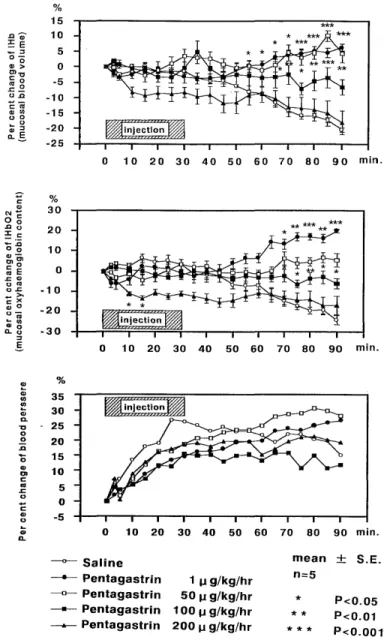
1) Pentagastrin was administered at doses of lµg/kg/hr, 50µg/kg/hr, 100µg/kg/hr, and 200µg/kg/hr. Cecal mucosal blood flow decreased when the dose of pentagastrin was increased. The intravascular supply of oxygen also decreased in a dose dependent manner. Each dose of the gastrointesti- nal hormone caused a reduction in blood pressure.

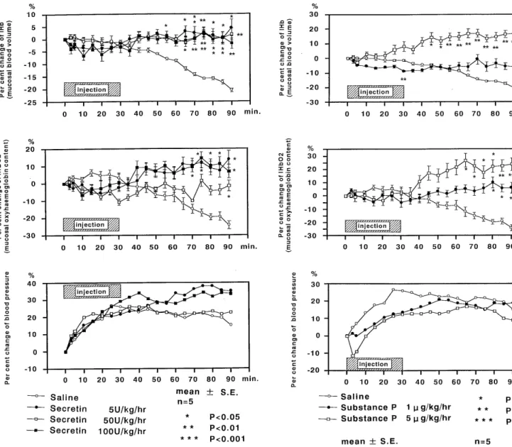
2) Secretin was administered at doses of 5µg/kg/hr, 50µg/

kg/hr, and 100µg/kg/hr. Each tested dose of this gastrointes- tinal hormone acted to maintain cecal mucosal blood flow.

The blood pressure remained unchanged throughout the experiment.

3) Substance P was administered at doses of 1µg/kg/hr and 5µg/kg/hr. Cecal mucosal blood flow and the intravascular oxygen supply increased after administration of this gastro- intestinal hormone. The blood pressure decreased transiently at the start of administration, but later gradually returned to the baseline values.

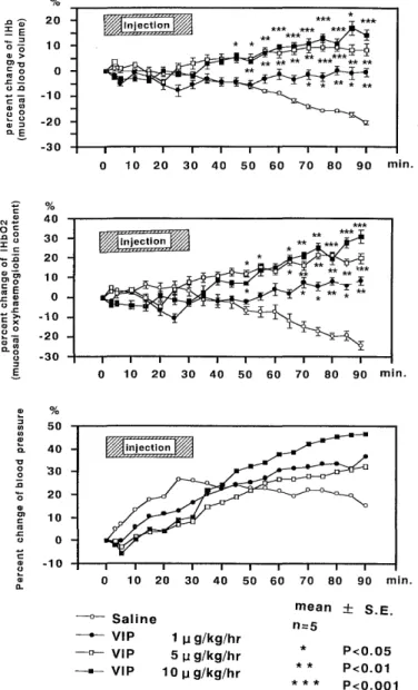
4) Vasoactive intestinal polypeptide (VIP) was adminis- tered at doses of lµg/kg/hr, 5,ug/kg/hr, and 10µg/kg/hr. VIP caused the cecal mucosal blood flow to increase in a dose- dependent manner. The intravascular oxygen, supply also increased signific antly after administration of this gastroin- testinal hormone. The blood pressure initially de creased after administration of each test dose of VIP, after which it gradually started to increase.

5) To identify the factors responsible for the increase in cecal mucosal blood flow at the start of VIP administration at a dose of 5µg/kg/h of VIP, the blood concentrations of VIP, cyclic AMP, phospholipase, prostaglandin E2, prosta-

glandin:E,  and  6 keto‑prostaglandin:F1α  were  examined  at

regular intervals. However, no significant changes with regard to these substances were noted.

Key words : gastrointestinal hormone pentagastrin, secre- tin, substance P, VIP, colonic mucosal blood

flow

I . Introduction

The effect of the gastrointestinal hormones on the mucosal blood flow in the upper digestive tract have been studied by many investigators. However, the effect of the gastrointestinal hormones on the mucosal blood flow in the lower digestive tract, especially that of the colonic mucosal blood flow, remains to be clarified. Gastro- intestinal hormones are considered to be humoral factors

that regulate complex digestive processes. Thus, it is speculated that these hormones also influence colonic function. In this study, we examined the effects of the various gastrointestinal hormones and peptides on colonic mucosal blood flow using a spectrum analyzer.

1 1. Materials and Methods

1) Experimental animals

Male Wistar rats weighing about 300 g were used.

2) Experimental methods

Rats were anesthetized by intraperitoneal administra- tion of pentobarbital (30 mg/kg). They were then sub- jected to laparotomy while their blood pressure was monitored at the left common carotid artery. The cecum was resected at a location contralateral to its attachment to the mesentery. Feces were carefully removed from the resected cecal specimens so as not to damage the mucosa.

The resected cecal specimens were then washed with physiological saline and fixed to the table where measure- ments were to be made. After placing an optical fiber over the cecal mucosa, the cecal blood flow was measured using a TS-200 tissue spectrum analyzer (Sumitomo Denko Co.).

The following experiments were performed after the mucosal blood flow had stabilized.

a) The effect of the gastrointestinal hormones on colonic mucosal flow

Various doses of the gastrointestinal hormones (penta-

(2)

gastrin, secretin, substance P [SP] , vasoactive intestinal polypeptide [VIP]) were continuously infused via the femoral vein for 30 minutes. Mucosal blood flow was serially measured to 90 minutes after the start of the administration of the gastrointestinal hormones or 60 minutes after the completion of infusion. The various test substances were dissolved in physiological saline and were infused at a rate of 0.1 ml/min using a chronofuser. 02 saturation (SaO2) was used as an indicator of the oxygen

content in blood, and IHb was used an indicator of blood flow. The product of SaO2 and IHb was designated as IHbO2, which served as an indicator of the oxygen supply in blood. The measured values were expressed as the rate of change after administration of the test substances.

b) Changes in the various factors during increases in blood flow The cause for the significant increase in blood flow after continuous infusion of VIP (5,ug/kg/h) was by serial measurements of the concentrations of VIP, cyclic AMP, phospholipase, prostaglandin E2, prostaglandin E, and 6 keto-prostaglandin FL a. The measured values were expressed as rates of change after the administration of VIP. The same measurements were made after infusion of physiological saline in the control group.

3) Statistical analysis

Data were expressed as means ± SE. The t-test was used for the comparison of data. Differences were considered significant when the P value was less than 0.05.

111. Results

The findings with regard to blood flow which were measured and recorded by a spectrum analyzer showed occasional markedly disruptive waves. Despite the appear- ance of these waves which were thought to be due to peristalsis, the results were generally satisfactory. The blood flow was represented by the means of the measured values.

A transient increase in the colonic mucosal blood flow was noted initially in the control group during continuous infusion of physiological saline. However, the colonic mucosal blood flow soon started to decrease gradually.

The blood pressure showed gradual increases during this interval.

a) Administration of the gastrointestinal hormones The colonic mucosal blood flow was lower in the group which was administered pentagastrin (ICI pharma) at a dose of 1,ug/kg over 30 minutes compared to that in the group that was administered physiological saline (Fig. 1).

However, the colonic mucosal blood flow started to increase 60 minutes after administration of pentagastrin.

A 6 % increase in colonic mucosal blood flow was noted 90 minutes after administration of pentagastrin. Administ-

Figure 1. The effect of pentagastrin on colonic mucosal blood volume, mucosal oxyhaemoglobin content and blood pres- sure. Pentagastrin was continuously injected for 30 min. at a concentration of 1 ,ug/kg/hr (closed circles), 10 ,ug/kg/hr

(open squares), 100,ug/kg/hr (closed squares) and 200 gg/kg/

hr (closed deltas). Open circles were saline injection to control rats. Solutions were administered using the same flow rate in femoral vein of anesthetized rats. Index of colonic mucosal haemoglobin concetration (upper panel)

and colonic mucosal oxyhaemoglobin content (middle panel) were measured by a tissure spectrum analyzer during monitoring of blood pressure (lower panel) during 90 min.

ration of pentagastrin at a dose of 50 ,ug/kg/h resulted in a 4 % increase in the colonic mucosal blood flow at the end of infusion, after which it started to decrease gradually.

The colonic mucosal blood flow was at its mininmum 60

minutes after administration of pentagastrin at a rate and

dose of 50 ,ug/kg/hr. However, after 60 minutes, the

(3)

Figure 2. The effect of exogenous secretin on colonic mucosal blood volume, mucosal oxyhaemoglobin content and blood pressure. The concentration of secretin was 5 U/kg/hr (closed circles), 50 U/kg/hr (open squares) and 100 U/kg/hr (closed squares). The conditions were similar to those in Fig. 1.

Figure 3. The effect of exogenous substance P on colonic mucosal blood volume, mucosal oxyhaemoglobin content and blood pressure. The concentration of substance P was 1 ug/kg/hr (closed circles) and 5 ,ug/kg/hr (open squares).

The conditions were similar to those in Fig. 1.

colonic mucosal blood flow was at its minimum 60 min- utes after administration of pentagastrin at a rate and dose of 50,ug/kg/h. However, after 60 minutes, the colonic mucosal blood flow began to increase again. A 4 % was noted 90 minutes after the administration of pentagastrin at a rate and dose of 50 ,ug/kg/hr. Slight reductions in the blood pressure and blood flow were noted 15 minutes after the administration of the hormone at a rate and dose of 100,ug/kg/hr. The blood flow continued to decrease 60 and 90 minutes after administration. The blood pressure and blood flow were markedly reduced soon after the start of administration of pentagastrin at a dose and rate of 200,ug/kg/h. These reductions were significant compared

to those seen after administration of physiological saline.

However, the levels of the blood pressure and blood flow were similar in the group administered pentagastrin at a dose and rate of 200 ,ug/kg/hr and in the control group, 60 minutes after administration of the gastrointestinal hormone.

The effect of secretin (Sigma) at doses of 5 ,ug/kg/hr,

50 ,ug/kg/hr, and 100,ug/kg/hr on blood pressure was less

compared to that of physiological saline (Fig. 2). The

mucosal blood flow was unchanged before and after

administration of secretin. The effect of secretin on cecal

mucosal blood flow was greater than that of physiological

saline.

(4)

The administration of substance P (SP) (Sigma) at doses of 1,ug/kg/hr, and 5 ,ug/kg/hr produced a transient reduction in blood pressure at first. Subsequently, the blood pressure started to increase again, albeit gradually.

At a dose of 1/ug/kg/h of SP, the mucosal blood flow was reduced 15 ~ and 30 minutes after the start of its admini- stration. The mucosal blood flow still showed a 5 % reduction 45 minutes after infusion. After this, the muco- sal blood flow showed a slightly higher increase than that in the control group. The mucosal blood flow started to increase soon after administration of SP at a dose of 5/ug/kg/h. A state of increased mucosal blood flow was maintained until 90 minutes after the administration of SP. This increase in blood flow was statistically signifi- cant compared to that in the control group. The rats died soon after the administration of SP at a dose of 10 ,u g/

kg/h, and this portion of the experiment could not be completed.

The blood pressure was slightly lower during admini- stration of VIP (Sigma) than during the administration of physiological saline (Fig. 4). The blood flow slightly decreased during the administration of 1,ug/kg/h of VIP.

However, the blood flow gradually returned to its pread- ministration level after infusion of the gastrointestinal hormone. The blood flow was reduced at first when either 5 or 10 ,ug/kg/h of VIP was administered. However, the blood flow showed small increases 20 minutes after the administration of VIP at these doses. Later, the blood flow was observed to be markedly increased in this group.

b) Measurements of various factors

Factors which might have contributed to the increase in blood flow during the administration of VIP were investi- gated. For this purpose, the blood levels of VIP, cyclic AMP, phospholipase, prostaglandin E2, prostaglandin E, and 6 keto-prostaglandin F] a were measured. The results of these measurements showed no significant changes with regard to these factors after the experiments.

N. Discussion

Gastrointestinal hormones are thought to be humoral transmitters which regulate complex digestive functions.

There have been many reports with regard to the effect of these hormones on mucosal blood flow in the upper diges- tive tract, especially the stomach'). However, the effect of these gastrointestinal hormones on mucosal blood flow in the lower digestive tract, especially that in the colon, has not been fully investigated. In this study, we evaluated the effect of the gastrointestinal hormones (or gastrointesti- nal peptides) on mucosal blood flow in the colon.

Mucosal blood flow at different colonic sites have been studied for many years. Steinberg et al. measured the mucosal blood flow at different sites in the rat large intestine using the' Rb clearance technique. They reported

Figure 4. The effect of exogenous VIP on colonic mucosal blood volume, mucosal oxyhaemoglobin content and blood pressure. The concentration of VIP was 1 ,ag/kg/hr (closed circles), 5 ,ug/kg/hr (open squares) and 10 ,ug/kg/hr (closed squares). The conditions were similar to those in Fig. 1.

that the mucosal blood flow in the cecum, colon, and rectum (and sigmoid colon) was 0.5 ± 0.19 ml/min/g, 0.6 ± 0.17 ml/min/g, and 0.2 ± 0.20 ml/min/g, respecti- vely"). Coxon et al. showed that the mucosal blood flow at

different colonic sites in pigs all similar, except at the

sigmoid colon, using the... Xe clearance technique"). Wang

et al") found no difference in the regional blood flow at

different colonic sites in healthy adults, except at the

rectum where the blood flow was significantly less than at

other sites. Given that the mucosal blood flow is similar

at all colonic sites, we chose to measure the regional blood

flow at the cecum, where measurements could be most

easily made.

(5)

Methods of measuring the regional blood flow in the colonic membrane consist of the isotope clearance tech- nique and the hydrogen gas clearance technique. Absolute values of the regional blood flow may be obtained using these techniques. However, there are drawbacks with regard to these techniques which included the long time required for making measurements and the difficulty in serial measurements. In contrast, the TS-200 tissue spec- trum analyzer is easy to operate and can be used for serial noninvasive measurements of mucosal blood flow. Each measurement was made over a 5 second interval and the mean value was taken to be the blood flow. . Even if differences in pulse waves had been present, the blood flow could be measured by taking the average.

Low dose administration of pentagastrin also resulted in an increase in mucosal blood flow. However, the blood pressure was unchanged at this low dose. The blood flow peaked 30 minutes after the administration of pentagas- trin at a dose of 50,ug/kg/hr. Discontinuation of infusion was followed by reduction in blood flow in a dose- dependent manner. However, administration of 200 ,ug/kg/

h of pentagastrin resulted in reductions of both the mucosal blood flow and the blood pressure. The admini- stration of a low dose of pentagastrin produced an in- crease in mucosal blood flow. Higher doses resulted in reductions of blood flow as well as blood pressure. After the completion of pentagastrin infusion, its blood level

decreased in a dose-dependent manner.

Many studies have confirmed that pentagastrin in- creases the mucosal blood flow of the upper digestive tract"). It is believed that this gastrointestinal hormone mainly acts to increase the gastric blood flow and secre- tion. We showed that these substances modified the arterial flow in different ways according to the affected organ system. Pentagastrin had the effect of increasing the colonic mucosal blood flow up to a certain concentra- tion, just as it did the gastric blood flow. However, concentrations above this critical level reduced the colonic mucosal blood flow instead. We speculated 3 reasons which might explain this phenomenon. First, the 200 ,u g/kg/hr dose resulted in a much higher blood concentration than is the case when a conventional dose of 5 or 6 ,ug/kg/hr was used. The abnormally high blood level may have caused the reduction of the colonic mucosal blood flow. Second, the secondary metabolic products of pentagastrin may have played a role in reducing the colonic mucosal blood flow.

Third, the increase in gastric blood flow due to pentagastrin could have produced an imbalance in the blood distribution in the abdominal organs, which in turn may have resulted in the reduction of the colonic mucosal blood flow.

Secretin was discovered in the porcine duodenal and jejunal mucosa by Baylis and Starling in 1902". Its struc-

ture was determined by Mutt and Jorpes in 19662). Secretin is a normal chain peptide which is composed of 27 amino

acids. The 27 amino acids comprise 11 kinds of amino acids and has a molecular weight of 3055. This gastrointestinal hormone acts to inhibit gastrin secretion, but enhances duodenal secretion. Secretin also increases the blood flow in the celiac artery') and superior mesenteric artery') 16). The arterial blood in the right side of the colon and cecum is supplied by the superior mesenteric artery. Naturally, we expected an increase in the blood flow in these areas as a result of administration of secretin. In our experiments secretin caused an increase in blood flow, regardless of its

concentration. This increase in the blood flow was en- hanced by treatment with atropine. From this, one may infer that the observed increase in blood flow was a manifestation of the secondary metabolic effect of secretin on the intestinal glands. The increase in blood flow may also be mediated by the release of intermediate metabolites having vasoactive properties"). In this study, we did not investigate the secondary factors that might have been associated with the effect of secretin. Although secondary factors may play a role in causing an increase in mucosal blood flow, their effects were not clearly seen in our experiments.

Substance P (SP) was first extracted from intestinal and brain tissues by Euler and Gaddum in 193118). Today, it is known as a potent vasoactive substance. In 1975 Pernow and Rosell intraarterially administered SP to dogs which resulted in an increase in the mucosal blood flow").

Hallberg and Pernow reported in 1975 that the intravenous administration of SP increased the blood flow in the carotid artery, hepatic artery, mesenteric artery, and portal vein without compromising the renal circulation"".

Gronstad et al.') showed that small intestinal blood flow increased after SP administration in cats by endoluminal perfusion. This experimental observation indicates that SP acts as a vasodilator, regardless of its route of admini- stration. SP triggers relaxation of the cerebral vasculature in the presence of vascular endothelial cells, just as acetylcholine does'). Furchgott and Zawadzki f oud that acetylcholine did not show a vasodilating effect in ex- tracted blood vessel specimens'. They ascribed this phe- nomenon to endothelial cell injury that might have been caused by the investigator during the experiment. They hypothesized that acetylcholine manifests its vasodilating action by releasing an unknown vasodilating substance from the vascular endothelial cells. Given that the vasodilating action of SP is dependent upon the integrity of the vascular endothelial cells, it is speculated that the vasodilating effect of SP is caused by an endothelium- derived relaxing factor (EDRF) or prostaglandin (PG) I2.

PGI2 seems to mediate the vasodilating action of SP. PGI2 is not only produced in the vascular endothelial cells, but is also synthesized in the subendothelial tissues. Thus, removal of the vascular endothelial cells is not sufficient for complete inhibition of the vasodilating action of SP.

Because of this, it is likely that EDRF is associated with

(6)

the vasodilating effect of SP. It has been shown that as least a portion of the EDRF is constituted by nitric acid.

Howevr, much still remains unknown with regard to the nature of EDRF, and need to be further investigated.

VIP, extracted and purified from porcine intestine in 1970 by Said and Mutt, is a peptide which acts as a vasodilator and enhances blood flow in porcine intestin").

The cardiovascular, gastrointestinal, endocrine, meta- bolic, and hematological effects of VIP have been identi- fied so far. Duckles and Said et al. 171 and Lee et al.') found that acetylcholine did not dilate cerebral vessels of mon- keys when their endothelial cells were experimentally removed. However, dilatation of these vessels by VIP and neural stimulation still occurred even after the endothelial cells were removed. This observation suggests that noncholinergic vasodilation of cereblral vessels may be mediated by VIP. VIP activates adenylate cyclase and increases the level of intracellular cyclic AMP in vascular smooth muscle. An increase in the level of intracellular cyclic AMP results in vasodilation'). There have been many reports that VIP increases hepatic and portal venous blood flow').

VIP causes only a slight increase in the blood flow in the superior mesenteric artery") " ). Initially, we expected mark- ed increases in colonic mucosal blood flow after the administration of VIP. However, our findings showed the vasodilating effects of VIP to be weaker than the other gastrointestinal hormones that we tested in this study.

Cerebrovascular vasodilation by VIP may be prevented if the production of PG is inhibited by indomethacin'). This suggests that cerebrovascular vasodilation may be related to the production of PGI2 in the vascular walls. However, no significant changes regarding the blood concentration of 6 keto-PGF, a were noted in our experiments (6 keto- PGF a is produced during the synthesis of PGI2). Thus, our findings failed to support the hypothesis that PGI2 is somehow related to cerebrovascular vasodilation. If microdialysis had been performed in an experimental system confined to the intestinal circulation, our findings may have been very well different from what we have obtained in this study.

We are indebted to professor Kohei Hara for his support in conducting this study and to the members of our department and the Experimental Animal Center for their cooperation.

References

1) Bayliss, W. M. and Starling E. H.: The mechanism of pancreatic secretion. J. Physio., 28: 325-328, 1902.

2) Mutt V. and Jorpes J. E.: Secretin isolation and determination of structure (abstr.) Proceedings of fourth internstional symposium on

the chemistry of natural products. Stockholm, Sweden, 1966.

3) Beijer H. J. M., Hulstaert P. E., Brouwer F. A. S., and Charbon G.

A. : The effect of secretin on peripheral arterial blood flow. Arch. Int.

Phermacodyn. 240, 269-277, 1979

4) Said S. and Mutt V.: Polypeptide with broad biological activity:

Isolation from small intestine. Scince, 169: 1217-1218, 1970

5) Wei E. P,, Kontos H. A. and Said S. I.: Mechanism of action of vasoactive intestinal polypeptide on cerebral arterioles. Am. J. Physiol.

239: 765-768, 1980.

6) Lee T. J. F. and Saito A.: Vasoactive intestinal polypeptide-like

substance: The potential transmitter for cerebral vasodilation. Science 224 : 898-901, 1984.

7) .LF urchgotto R. F. and Zawadzki J. V.: The obligatory role of endothelial cells in the relaxation of arterial smooth muscle by

acethylcholine. Nature, 288: 373-379, 1980.

8) Edvinsson L., Fredholm BB., Hamel E., Jansen I. and Verrecchia C.:

Perivascular peptides relax cerebral arteries concomitant with stimula-

tion of cyclic adenosine monophosphatase accumulation or release of

an endothelium-derived relaxing factor in cat. Neurosci Lett, 58:

213-217, 1985

9) Sumi S., Inoue K., Doi R., I M., Higashiide S., Minode S. and Tobe T.: Effect of exogenous VIP on hepatic blood flow in conscious dogs,

Vessels. 13; 115-118, 1990

10) Sumi S., Inoue K., Kogire M., Doi R., Takaori K., Morita K., Miyosi 'I' ., Suzuki T. and Tobe T.: Experimental study on splanchinic

circuration by ultrasonic transit time volume flow meter and doppler

flow meter ; Jpn. J. gastroenterol. 84; 2579-2586, 1987

11) Inoue K, Sumi S, Doi R, Kogire M., Takaori K. and Tobe T.: Effect of synpathetic chicken and poricrine vasoactive intestinal peptide on pancreatic blood flow indogs, Biomed. Res., 9, suppl. 3 ; 125-129, 1988 12) Wang K., Matsunaga T., Sakai., Utino H. and Miyake T.: Local

hemodynamics in the human large intestine, Gastroenterol. Endosc.,

28 ; 542-549, 1986

13) Steiner S. H. and Mueller G. C. E.: Distribution of blood flow in the digestive tract of rat, Circ. Res., 9 : 99-102, 1961

14) Coxson J. E., Dicksin C. and Taylor I.: Changes in intestinal blood flow during the development of chronic large bowel obstruction, Br. J.

Surg., 71 : 795-798, 1984.

15) Moriga. M.: Secretin, Medicina, 11: 2209-2211, 1979.

16) Fasth, S., Filipsson S., Hulten L., Martinson J., : The effect of gastrointestinal hormones on small intestinal motility and blood

flow ; Experientia, 29: 982-984, 1973

17) Duckles S. P. and Said S. I., Vasoactive intestinal peptide as a neurotransmitter in the cerebral circulation.: European J. Phamacol.,

78: 371-374, 1982

18) Euler von U. S. and Gaddum J. H.: An unidenstified depressor substance in certain tissur extract : J. Phamacol. 192 : 74-87, 1931 19) Pernow B. and Rosell. S.: Effect of Substance P on blood flow in

canine adipose tissue and skeletal muscle. ; Acta Physiol Scand, 93:

139-141, 1975

20) Gronstad K. 0., Dahlstrom A. Jaffe B. M., Zinner M. j., Ahlman H.

Studies on the mucosal hyperemia of the ferine small intestine observed

at endoluminal perfusion with substance P, Acta physiol Scand., 128:

97-108,1986

21) Hallberg D. & Pernow B., Effect of substance P on various vascular bed in the dog. : Acta physiol stand. 93, 277-285, 1975

22) Moriga M. ; Effect of gastrointestinal hormones on gastric secretion.

Seminor of gastroenterology, 37: 191-203. 1989

Figure  1.  The  effect  of  pentagastrin  on  colonic  mucosal  blood  volume,  mucosal  oxyhaemoglobin  content  and  blood   pres-sure
Figure  2.  The  effect  of  exogenous  secretin  on  colonic  mucosal  blood  volume,  mucosal  oxyhaemoglobin  content  and  blood  pressure
Figure  4.  The  effect  of  exogenous  VIP  on  colonic  mucosal  blood  volume,  mucosal  oxyhaemoglobin  content  and  blood  pressure

参照

関連したドキュメント

The inclusion of the cell shedding mechanism leads to modification of the boundary conditions employed in the model of Ward and King (199910) and it will be

Pour tout type de poly` edre euclidien pair pos- sible, nous construisons (section 5.4) un complexe poly´ edral pair CAT( − 1), dont les cellules maximales sont de ce type, et dont

Later, in [1], the research proceeded with the asymptotic behavior of solutions of the incompressible 2D Euler equations on a bounded domain with a finite num- ber of holes,

“Breuil-M´ezard conjecture and modularity lifting for potentially semistable deformations after

Then it follows immediately from a suitable version of “Hensel’s Lemma” [cf., e.g., the argument of [4], Lemma 2.1] that S may be obtained, as the notation suggests, as the m A

We study the classical invariant theory of the B´ ezoutiant R(A, B) of a pair of binary forms A, B.. We also describe a ‘generic reduc- tion formula’ which recovers B from R(A, B)

The normalized velocity profiles of H-B and Casson fluids for different values of the power law index z c and yield stress n flow i through circular tube and ii between parallel

Q discrep : Predefined empirical constant corresponding to the minimum value of the module of total discrepancy between estimated gas supply volumes, which is of practical