Acta med. Nagsaki. 12: 99-111
The Influence of Mersalyl and some Sulfhydryl Group Reagents on the Pacemaker Action Potential and the Contraction in Isolated Guinea Pig Atria.
Michio TANI
Department of Pharmacology, Nagasaki University School of Medicine, Nagasaki, Japan
Received for publication, March 4, 1968
In order to analyze the cause of a cardiotoxic action of mersalyl, its direct effects of transmembrane action potential in cardiac pacemaker cells and on contractility of the atrial muscle fibers were investigated in isolated guinea pig hearts. The effect was compared with those of sod. arsenite, iodoacetamide, N-ethylmaleimide and iodosobenzene acetate. All of these SH reagents caused a shortening of the duration and a reduction of the propa- gated action potential. The former change is considered as a cause of the rhythm accerelating effect of these reagents, and the latter as a cause of arrhythmia. The reagents act on the contractility different ways, while iodoacetamide and sod. arsenite having a marked depolarizing charactor cause the contracture.
It has long observed that the mercurial preparation has the strong cardiotoxic effects i. e. , its high concentration causes cardiac arrhy- thmia. That results acute death in animals and also in human beings following the intravenous infusion of mercurial preparations (1, 2, 3).
And the the toxic action has been attributed to a blocking action of sulfhydryl radicals in the tissue (4). Recently, E. MUSCHOLL and others reported the effect of mercurial preparations on transmembrane action potential in diaphragm muscle fibers and cardiac proper fibers (5, 6).
However, nothing is known about the direct action of the agents and other types of sulfhydryl reagents upon cardiac pacemaker cells.
In the present study attempts were made to observe influences of mercurial preparations on the membrane activity of the pacemaker cell in isolated guinea pig atria with an aid of intracellular microelectrode technique and on the contractility of atrial muscle fibers, because it is thought as the most basic problem to consider the mode of the cardio- toxic action. And the effects of the mercurial preparation is compared with those of other types of sulfhydryl reagents subjected to analyze parmacologic ally.
*谷 美智士
METHODE and MATER.I'ALS ;
The hearts taken from exsanguinated :-;guinea pigs of either sex
weighing 180g to 350g were used throughout,. _; the experiment. The atrium was quickly separted from the ventricle and placed in an oxygen satu- rated nutrient fluid bath at about 30°C. ,Isometric record of contractions
of the naturally beating atria was carried out by using a strain gage transducer (Shinkoh UL-10-120) and y,; an automatic balancing strainre - corder (Shinkoh AS-2) with an initial tension c 600mg.
Transmembrane action potentials"of the pacemaker cell in the right atrium were led from its pacemaker region via intracellular microelec- trode of suspended type (with 80,u copper vsiire) to a low imput high impedance amplifier (Nihonkohden MZ-3B) and displayed in two cathod ray oscilloscopes, utilizing one for record, the other for monitor. The location of the pacemaker area and' the procedure of its exposure will be described in the next chapter. In the cu 'rent study, stable records of a pacemaker action potential having a. distinct prepotential were at least 10mV conveniently.
The solution composed of NaC1. 154mM;, KC1 5.6mM, CaC12 2.2mM, Glucose 5mM, NaHCO3 5.95mM per liter-was used as a nutrient med- ium. The chemicals used in this experiment were mersalyl sodium (Sigma), iodoacetamide (Hani Co~');"iodosobenzene acetate (Wako Co.), sodium arsenite (Hani Co.) and N-ethylmaleimide (Sigma).
RESULTS
A. Location of pacemaker cells-}in the 'guinea pig.
A. P. de Carvalho and et al- have studied extensively about the location of specialized fibers in 'the rabbit atrium and have drawn its distribution map (7). However -it„ was confirmed in the current investi- gation that the map could not .be adapted for the guinea pig atrium.
Although a report concerning activity of. pacemaker cells of the guinea pig atrium was already published- from -our laboratry, yet precise loca-
tion of the area has not been. -Presented'._ So the approximate site and procedure of the exposure are for the first time displayed in this paper.
A right atrium was isolated from ' the heart together with interat- rial septum and the tricuspid 'valve. In the atrium, an incision from
atrioventricular orfice to superior vena cava was taken, then the cut
was extended along with free,-, anterior edge of the atrium from atrio-
ventricular orfice to the trianglar, cusp. of the right atrium. The disse-
cted atrium was fixed with stainless steel needles on a cork block in a
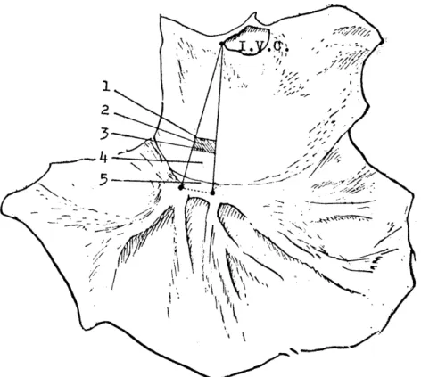
horizontal bath. The appearance is likely to Fig. 1. In this drawing,
a shaded trapezoidal portion was found as the most probable site which
gives a typical pattern of the pacemaker action potential having a dia-
stolic slow depolarization. In the practice, impalement of a microelec-
Fig. 1 . Schematic drawing the site of the pacemaker area in endocardial side of the right guinea pig atrium. A triangle is postulated between one point at the
ostium of inferior vena cava and two points on the crista terminalis inter-
secting with large endocardial foldings. And the trapezoidal portion was
found as the most probable site of the pacemaker area in guinea pig atria.
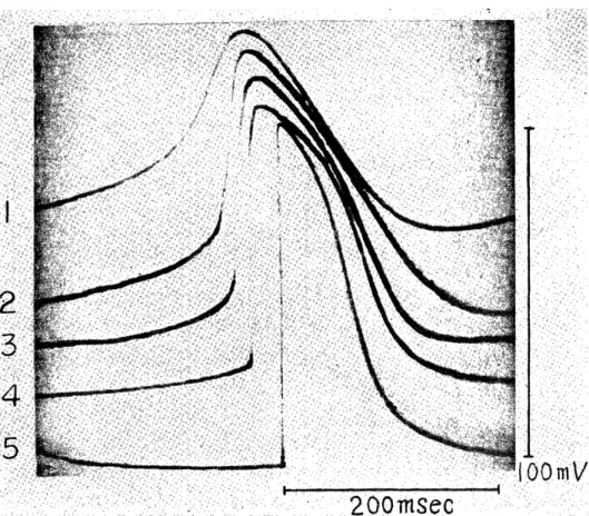
trode was made at arbitrary point in this portion and some places had to be changed to have the sufficient prepotentials. Actual records of the transmembrane action potentials taken from different points in this portion in one experiment were shown in Fig. 2.
B. Effects of sulfhydryl group reagents on the isolated guinea pig atrium.
1. Mersalyl (mersalyl sodium).
A gradual decrease in the contractile tension and a slight transient increase in the atrial beat followed by a decrease were produced by application of mersalyl to the bath in a final concentration of 0.8mM.
Five to fifteen minutes after the application, the contraction has abru- ptly stopped in most instances. The contractions could be obtained by a repetitive electrical stimulation even in this state of atrium, but these contractions decreased again in process of time. And a slight elevation of resting tension was observed after the contractile response had been inhibited markedly (Fig. W.
The pacemaker propagated action potential reduced gradually its
Fig. 2. Transmembrane action potentials recorded from the pacemaker area and its vicinity in an experiment. Tracing 1 to 5 correspondly represents a record
from the point of the same number in Fig. 1.
Fig. 3. A. Effect of mersJyl 0.8mM (0.4mg/ml) on the contractile tension of guinea pig atria. Upper numbers indicated time in minutes after ap-
plication, and the lower indicated atrial beats every 20 sec..
STIM.: electrical driven stimuli (120 c/min).
R: restraining the stimulation for 10 sec..
B. Effect of mersalyl 0.8mM on the transmembrane action potential of the pacemaker area in the guinea pig atrium. Each photograph after the
application were superimposed upon the original record. The last pho-
tograph was superimposed upon original recrod and upon that 10
minutes later.
M. TANI
Fig. 4. A. Effect of sodium arsenite 15.5mM (2mg/ml) on the contractile tension of guinea pig atria. Upper numbers indicated time in minutes after application, and the lower indicated atrial beats every 20. sec. .
STIM.: electrical driven stimuli.
B. Effect of sodium arsenite 15.5mM on the transmembrane action potential of the pacemaker area in the guinea pig atrium. Each photograph after
the application were superimposed upon the original record. The last
photograph was superimposed upon original 'record and upon that 10
minutes later.
height and duration after application of mersalvl, but the prepotential remained in no change for first few minutes. Consequently rate of slow depolarization of the prepotential became so slow, that a marked redu-
ction of pacemaker rhythm was resulted. Height of the prepotential was not influenced so much until the pacemaker`rhythm being so slow, but it has also diminished finally (Figs, .3B).
2. Sodium arsenite.
Marked increase of contractile tension and irregurality of the beat were observed transiently within few minutes after appling sodium arsenite(15.5mM), and then increase o f the resting tension became so marked and the beat was stopped completely about 5 minutes later.
But electric stimulation could activate the atrium to contract even tho- ugh the resting tension increased considerably (Fig. 4A). Change in the pacemaker action potential appeared first as a shortening of the propagated action potential with slight accerelation of the rhythm, which was followed by marked elevation of the, resting potential. Con- sequently height of the action potential reduced markedly, and it was no longer to activate the contractile atrial fiber`s (Fig. 4B).
3. lodoacetamide.
Almostly similar change in contractile tension by appling of sodium arsenite was also produced by iodoacetamide (6..5mM), that is, marked and transient increase of the contractile tension and elevation of the resting tension was observed (Fig. 5A).
Height of the prepotential increased slightly during first few minu- tes was followed by a marked reduction. Speed rate of slow deporali- zation of the prepotential was slightly accerelated, and consequently frequency of the pacemaker rhythm was accerelated moderately. Howe- ver, only a slight shortening of the duration in the propagated action potential was observed. And reduction both in'the resting potential and in the action potential was observed finally (Fig. 5B).
4. N-ethylmaleimide(NEM).
Contractile tension of the guinea pig atrium was increased moder-
ately with a slight accerelation of the beat by,appling of NEM in a final
concentration of 1.2.10-1mM . The resting tendon was elevated gradually
and the effect lasted over a period of ten to twenty minutes. More
marked increase in contractile tension was produced by heigher conce-
M. TANI
Fig. 5. A. Effect of iodoacetamide 6.5mM (1.2mg/ml) on the contractile tension of guinea pig atria. Upper numbers indicated time in minutes after appl-
ication, and the lower indicated atrial beats every 20 sec..
STIM.: electrical driven stimuli.
B. Effect of iodoacetamide 6.5mM on the transmembrane action potential of the pacemaker area in the guinea pig atrium. Each photograph after
the application were superimposed upon the original record. The last
photograph was superimposed upon original record and upon that 10
minutes later.
Fig. 6. A. Effect of NEM on the contractile tension of guinea pig atria.
1. NEM 1.2 • 10-1mM (1.5. 10-2mg/ml)
2. NEM 6 . 10-1mM (7.5 • 10-2mg/ml)
3. NEM 8 • 10-1mM (O.lmg/ml)
Upper numbers indicated time in minutes after application, and the lower indicated atrial beats every 20 sec..
STIM.: electrical driven stimuli
B. Effect of NEM 1.2 • 10-1mM on transmembrane action potentials of the pacemaker area in the guinea pig atrium. Each photograph after the
applicatiop were superimposed upon the original record,
ntration such as 8.10-1 mM, and the effect lasted shorter, causing with irregularity of the beat and elevation of the resting tension (Fig. 6A).
The pacemaker action potential, its height of the prepotential was lowered temporarily after NEM application, then elevated moderately, and duration of the propagated action potential was shortened slightly, but speed rate of the slow depolalization was not influenced. Finally
the resting potential reduced moderately (Fig. 6B). It is considered as a concomitant change with elevation of the resting tension in the atr- ium.
Iodosobenzene acetate (4mM) showed the similar effect with those of NEM (1.2.10-1mM) on the contractile tension and on the configura- tion of action potential.
DISCUSSION
The study on transmembrane action potential of cardiac pacemaker cells of the guinea pig heart has already been published from our labo- ratory (8). But an exact location of the pacemaker area of a guinea pig atrium has never been pointed out, because sizes of both the tip of a microelectrode and the area in the guinea pig atrium are very small.
So the first attempt was made to determine the exact location of the pacemaker site in the guinea pig atrium. Though impalement of the microelectrode into the pacemaker area is suggested by A. P. de Carvalho (7) and T. C. West (9) in a rabbit atrium, it failed to get any evidence of the pacemaker action potential. The present mapping of the pacemaker in the guinea pig atrium cleary indicated that there were a considerable species difference between the rabbit and the guinea pig in site of the pacemaker.
All SH group reagents used in the current experiment give changes in configuration of the pacemaker action potential. The changes found in effects of the reagents throughout are a shortening on duration of the propagated action potential and a reduction on height of the action potential. The transient rhythm accerelating effect of the reagents seen in soon after its administration could adequately be explainable by the former change. The latter change, the reduction of the action poten- tial, may participate in producing of arrhythmia, since it is very pro-- bable that the inhibited and propagated action potential in the pacemaker cell could not activate cardiac proper fibers to contraction, so an ectopic excitation replaces the normal rhythm. On the height of the prepoten- tial and speed rate in slow depolarization, these SH reagents do not show any uniform effect, so reducing in height, and shortening in duration of the propagated action potential and diminution of the resting potential are of the general effects attributable to the SH group blocking action.
The reagents showing marked reduction of the resting potential
such as iodoacetamide and sod. arsenite produce also a marked eleva- tion of the mechanical resting tension of the atrium. This suggests that the elevation of the resting tension is due to depolarizing charactor of these agents.
A positive inotropic action found in these reagents with the excep- tion of mersalyl calls an attention through its mechanism of the phar- macological action. Although the reason is not explainable, it seems to result from any inhibitor action of them. Because it has been already known that some durgs having inhibitory qualities such as quinidine (10), fluoride (11-15), cardiotonic glycoside (16) etc. also exert a po- sitive inotropic action. And another possibility has appeared recently
(17), that some of these agents could interact directly with adrenergic receptor, because NEM was suggested as an antiadrenergics through its blocking action of the receptor in a guinea pig heart.
Differences in the mode of action of each SH blocking agent on the atrium could be attributable to degree of penetration into the tissue
(18, 19), mode of binding to tissue SH (20, 21), specificity of the SH blocking action and other charactors of each reagent.
ACKNOWLEDGEMENT The author is gratefull to professor Dr. Y. NAKA- ZAWA and assistant professor Dr. A. UENO for their helpfull ccmments and
suggestions.
REFERENCES
1) DEGRAFF, A.C.: Diuretics. JA. H. A. 136(16), 1025(1948).
2) FARAH, A. and MARESH, G.: The influence of sulfhydryl compounds on diuresis and renal and cardiac circulatory changes caused by mersalyl. J. Pharmacol. 92,
73 (1948).
3) LEHMAN, R.A.: Further studies on the acute toxicity of mercurial diuretics. Proc.
Soc. Exp. Biol. Med. 64,'428(1947).
4) PITTS, R.F. and SARTORIUS, O.W.: Mechanism of action and therapeutic use of diuretics. Pharmacol. Rev. 2, 161(1950).
5) STEIN, E., MAGIN, J. and KLEINFELD, M.: Effect of mersalyl on isolated per- fused guinea pig atria. Am. J. Physiol. 199(3), 460 (1960).
6) MUSCHOLL, E.: Die Wirkung thiolopriver Substanzen auf Membran-und Aktionspo- tential des Rattenzwerchfells. Naunyn-Schmiedeberg's Arch. exp. Path. u. Pharmak.
235, 23 (1958).
7) DE C.°.RVALHO, A.P., DE MELLO, W.C. and HOFFMAN, B.F.: Electrophysio-
logical evidence for specialized fiber types in rabbit atrium. Am. J. Physiol.
196(3), 483 (1959).
8) KIDO, M.: Changes in the transmembrane action potential's pattern of the speci- alized fiber in guinea pig atria during anaphylaxis in vitro. Acta Medica Naga-
sakiensia. 9, 29 (1964).
9) WEST, T.C.: Ultramicroelectrode recording from the cardiac pacemaker. J. Pha- rmacol. Exp. Ther. 115, 283 (1952).
10) KRUTA, V.: Importance of the interval-strength relationship for the evaluation of cardiac inotropic effects of drugs. Second International Pharmacological Meeting.
5 (Pharmacology of Cardiac Function), 45 (1964).
11) BENNETT, D.R. and CHENOWETH, M.B.: Summary of completed studies on the mechanism of the positive inotropic action of fluoride and oubabain. ,I. Pharmacol,
Exp. Ther. 106, 373 (1952).
12) LOEWI, 0.: On the mechanism of the positive inotropic action of fluoride, oleate and calcium on the frog's heart. J. Pharmacol. Exp. Ther. 114, 90 (1955).
13) KATZUNG, B., ROSIN, H. and SCHEIDER, F.: Frequency-force relationship in the rabbit auricle and its modification by some metabolic inhibitors. J. Pharmacol.
Exp. Ther. 120, 324 (1957).
14) RICE, L.I. and BERMAN, D.A.: Malonate and fluoride effects on metabolism and contraction of electrically stimulated heart strips. Am. J. Physiol. 200 (4), -727
(1961).
15) CoviN, J.M. and BERMAN, D.A.: Metabolic aspects of the positive inotropic action of fluoride on rat ventricle. J. Pharmacol. Exp. Ther. 125, 137 (1959).
16) HAJDU, S. and LEONARD, E.: The cellular basis of cardiac glycoside action.
Pharmacol. Rev. 11, 173 (1959).
17) UENO, A. and NAKAZAWA, Y.: Adrenaline blocking action of N-ethylrnaleimide
and its reversal with BAL in isolated uinea pig. Folia Pharmacol. Jap. 63(2), 60 § (1967).
18) SMYTH, D.G., NAGAMATSU, A. and FRUTON, J.S.: Some react ons of N-ethylma- leimide. J. Am. Chem. Soc. 82, 4600 (1960).
19) JACOB, H.S. and J:.NFL, J.H.: Effects of sulfhydryl inhibition on red blood cells.: 1. Mechanism of hemolysis. J. Clin. Invest. 41, 779 (1962).
20) NEILANDS, J.B. and STUMPF, P.K.: Outlines of enzyme chemistry (second edi- tion), (1962).
21) BARRON, E.S.G. and SINGER, T.P.: Studies on biological oxidation.; XIX. Sul- fhvdrvl enzymes in carbohydrate metabolism. J. Biol. Chem. 157. 221 (1945).