A New Approach for Analysing the Patterns of Thermoregulatory Responses in Heat Acclimated Rabbits
Mariko FUJIWARA, Nobu OHWATARI and Mitsuo KOSAKA
Department of Environmental Physiology, Institute for Tropical Medicine, Nagasaki University, Nagasaki 852, Japan
Ye-WIN*
D epartment of Physiology, Institute of Medicine 2, Rangoon,Burma
Abstract: Rabbits were reared in a hot environment (30℃, 60% R. H.) for more than 6 months as a model of the heat acclimated animals. And, to clarify the mechanisms of physiological modifications which appeared during and/or after heat acclimation, various thermoregulatory responses to change in ambient temperature have been investigated in those rabbits. After 6 months of continuous heat exposure, change in rectal temperature in response to general heating (40℃) and cooling (10℃) of heat acclimated rabbits was smaller than that of control rabbits (control rabbits were reared in thermoneutral environ- ment; 25℃, 60%). Furthermore, remarkable differences were observed in threshold temperature for starting vasodilation or vasoconstriction of the ear skin. These patterns were analysed to define the thermoregulatory responses to heat acclimation.
Key words: Heat acclimated rabbits, Rectal temperature. Cutaneous vasodilation and vaso‑
constriciton, Thermoregulat℃ry pattern analysis, Habituation
INTRODUCTION
Homeothermic animals are able to regulate their body temperature within a certain range under changing external and internal conditions, and they develop adaptive modi- fications of already exisiting regulatory processes (Ashby, 1966). For many years attempts were made to determine these physiological mechanisms which appeared during and/or after thermal acclimation (Hensel, 1973, 1981). These modifications consist of not olny
R eceived for Publication, July 15, 1985.
Contribution No. 1653 from the Institute for Tropical Medicine, Nagasaki University.
*Participant in JICA-sponsored Research Training for Tropical Medicine, 1984- 1985
the functional changes in regulatory system or capacities of effectors, but also the morphological changes such as changes in body size, shape or fatty layer. To analyse this problem, various thermoregulatory responses such as cutaneous vasodilation or vaso- constriction, changes in respiratory frequency and rectal temperature to thermal stimulation have been investigated in heat acclimated rabbits (Fujiwara et al., 1985). So far, these responses were sequentially followed up in the time course of thermal stimulation. How- ever, relationships between the thermal stimuli and the thermoregulatory effects are some- what difficult to detect directly. Therefore, in the present experiments, a new approach was induced for analysing the patterns of thermoregulatory responses to change in ambient temperature of heat acclimated rabbits, and these underlying mechanisms were discussed.
MATERIALS AND METHODS
Two groups of male albino rabbits weighing from 2.5 kg to 2.8kg were used. For first two weeks, both groups of animals were reared in a thermoneutral environment
(25°C, 60%), being checked their general conditions. They were fed by 150g of commercial rabbits chow/day, and taken water ad libitum. Body weight, food and water intake, respiratory frequency and rectal temperature were measured at 10 o'clock in every morning. After this period, one group of rabbits was started to expose in a hot environment (30°C, 60%) as heat acctimated rabbits, and the other remained under the thermoneutral conditions as control.
The present experiments were carried out in an environmental control chamber without anesthesia. Both heat acclimated and control rabbits were lightly restrained just around the cervical region, and submitted to the following experimental program of general thermal stimulation. Namely, change rate of ambient temperature (Ta) was set to be 0.5°C/min, and was automatically programmed from 10°C to 40°C as follows; for
the first half an hour, the environmental control chamber was kept at 25°C. Then, the ambient temperature was raised to 40°C within 30 minutes. The temperature of 40°C was maintained for another half an hour. Next, the temperature was lowered to 10°C in the same rate of temperature change. It took about one hour to reach 10°C. The temperature was again raised to 25°C and kept in the same condition for 30 minutes. In each
experimental procedure, 240 minutes were required to thermal stimulation.
Temperatures of the rectum (Tre), ear skin (Tea), and ambient (Ta) were con-
tinuously measured with the thermistor probes. Blood pressure (BP) was measured from
the femoral artery, and heart rate (HR) was] calculated from its pulse. Respiratory
frequency (RF) was detected from the electrical resistance changes of a strain gauge
attached around the abdomen (For details, see Ohwatari et al. , 1983).
RESULTS
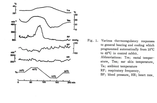
Figure 1 represents the simultanous recordings of the various thermoregulatory re- sponses to general heating and cooling in a control rabbit. At the bottom of this figure, experimental program of thermal stimulation was shown. Change in Tre induced by the general heating and cooling was about 1.6°C. The ear skin temperature changed markedly, and was responsible to the change in Ta. Increase of respiratory frequency (RF) started at 40°C of Ta, and diminished toward the initial level according to the general cooling (10°C). However, the change pattern of RF to the displacement of Ta was not always consistent in each experiment. Furthermore, blood pressure (BP) and heart rate (HR) were fairly constant by general thermal stimulation. Therefore, in the present experi- ment, comparison was made between the changes of Tre and Tea to general heating and cooling.
In case of heat acclimated rabbits, these change patterns of thermoregulatory re- sponses to general thermal stimulation were similar to those of a control rabbit shown in Figure1.
In Figure 2, changes in rectal and ear skin temperatures of the same control rabbit shown in Figure 1 are continuously plotted against the time course to Ta displacement.
The ambient temperature in the horizontal axis was shifted gradually from 25°C to 40°C, than lowered to 10°C, and backed to 25°C again in the same program of thermal stimulation shown at the bottom of Figure 1. Solid circles (à") and open circles (Q) represent the starting and ending points of each trial, respectively. At the start of thermal stimulation
(solid circles of which Ta shown at 25°C), Tea in Figure 2-A and Tre in Figure 2-B were 35.9°C and 38.6°C, respectively. 35.9°C of Tea indicated cutaneous vasodilation of
°c 40r
sain-1 4SOr
§0^
à"*T 350r 250 1-
Tre
Tea
RF
*w mmHg
BP ^
**
'100
Ta ast. 40<*T 25°C
Mot;
60 120 180 240 mlR
Various thermoregulatory responses to general heating and cooling which programmed automatically from 10°C to 40°C in control rabbit.
Abbreviations: Tre; rectal temper- ature, Tea; ear skin temperature, Ta; ambient temperature
RF; respiratory frequency,
BP; blood pressure, HR; heart rate
Tea 401
pc
30
20
Tre 40
°C
39
38
B
°c Tea 40
20
10 Tre 40
°C
39
38
7U Changes in rectal (A) and ear skin
(B) temperatures of the same control rabbit shown in Fig. 1 to the time course of Ta displacement.
Abbreviations are same as in
Fig. 1.
A
B