A Comparative Study of Blood Flow in the Cerebellum and Brainstem between Machado ‑Joseph Disease and
Olivopontocerebellar Atrophy
Nobuyoshi F UKUMITSU,Masahiko S UZUKI,Yasuhiko I TO, Yasuyuki I GUCHI,and Yutaka MORI
Department of Radiology,The Jikei University School of Medicine Division of Neurology,Department of Internal Medicine,
The Jikei University School of Medicine
ABSTRACT
In recent years,the neurogeni c and pathological differences between Machado‑Joseph disease (MJD)and sporadic olivopontocerebellar atrophy(OPCA)have been clarified. We performed N‑ isopropyl‑p‑[I‑123]iodoamphetamine(IMP)SPECT on 9 patients with MJD and 12 patients with OPCA. The blood flow of the cerebellum in the MJD group was significantly decreased than that of the control group(p<0.0001). That of OPCA gr oup was significantly decreased than those of the control and MJD groups(p<0.0001,respectively) . The blood flow of the brainstem in the MJD group was significantly decreased than that of the control group(p<0.001). That of OPCA group was significantly decreased than those of the cont rol and MJD groups(p<0.0001,respectively). The blood flow of cerebellum and brainstem in the OPCA group were much decreased than those of MJD group. IMP distribution pattern in MJD pat ients obviously differed from that of OPCA patients. (Jikeikai Med J 2002;49:157‑62)
Key words:Machado‑Joseph disease,Olivopontocerebellar atrophy, I‑IMP SPECT,cerebellum, brainstem
I NTRODUCTION
In 1972,Nakano et al.reported the hereditary spinocerebellar degenerati on in Portuguese or descen- dants of Portuguese from the Portuguese Azores Islands as Machado diseas e. Rosenberg et al.also reported the hereditary spi nocerebellar degeneration in emigrants from the Azor es Islands as Joseph dis-
ease. The above mentioned diseases were consid- ered to be the same disease and have been called Machado‑Joseph disease( MJD). There have been many reports of this diseas e,MJD,since 1980ʼs . MJD is an autosomal dominant disorder in which the symptoms that occur range f rom gait disturbance,
opthalmoplegia,bulging eyes,faciolingual fascicula- tion and myokimia.
When Dejerine and Thomas described two patients with ataxia during l ife and atrophy of the olives,pons,and cerebell um at autopsy,they com-
mented that their new disease was“neither heredi- tary,nor familial,nor congenital,it comes on at an advantaced age.Its etiology i s obscure” . One of their patients had an immobi le face,decreased arm movement,and late incont inence. Although many families with dominantly inherited atrophy of olives, pons,and cerebellum were later described,there have also been a large number of patients with no family history (often interpreted as evi dence for recessive
Received for publication,September 13,2002
福光 延吉,鈴木 正彦,伊藤 保彦,井口 保之,森 豊
Mailing address:Nobuyoshi FUKUMITSU,Department of Radiology,The Jikei University School of Medicine,3‑25‑8,Nishi‑Shimba- shi,Minato‑ku,Tokyo,105‑8461,Japan.
E‑mail address:GZL13162@nifty.ne.jp
157
inheritance), usually with some combination of levodopa‑unresponsive par kinsonism and autonomic failure as well as ataxia,l umped under the rubric
“olivopontocerebellar atrophy”(OPCA) .
In recent years,gene analysis has progressed at a surprising rapid pace. Mol ecular genetics studies became active in 1990ʼs,and the gene locus for MJD was reported to be locat ed in chromosome 14q in 1993 . CAG repeat in 14q32. 1 was reported,in 1994,
to expand 2‑3 times normality .
Both MJD and OPCA have neurodegeneration in cerebellum and brainstem,and t hey are reported to have decreased blood flow i n cerebellum and brain- stem . However,the reports before gene analysis was progressed had several questions in patients selec-
tion. In our previous study,the blood flow of cerebel- lum and brainstem were reported to be decreased in MJD patients who were di agnosed by gene analysis . There were no comparative studies of cerebral blood flow of MJD and OPCA pat ients who were diagnosed by gene analysis. In this study,we investigated the distribution pattern of cer ebral blood flow of MJD and OPCA patients, us ing N‑isopr o p y l‑p‑[ I]
iodoamphetamine(IMP)SPECT.
MATERIALS AND METHOD
Materials
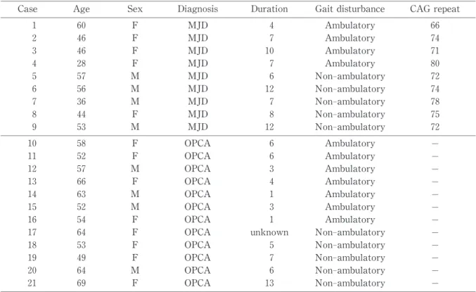
Ni ne patients with MJD and 12 patients with OPCA were studied and compar ed to 7 normal con- trols. Table 1 shows the patients of MJD and OPCA in detail.
The subjects with MJD were those who were diagnosed through family hi story and gene analysis. The MJD group was as follows:age range 28‑60 years;mean age,47.3±10. 5 years,4 male patients and 5 female patients. As far as gait disturbance is concerned,4 ambulatory pat ients and 5 non‑am- bulatory patients. The morbid term of the MJD group was 8.1±2.7 years. Pat ients who could walk with or without help wer e classified as ambulatory patients,and those who coul d not walk even with help were classified as non‑ambul atory patients. MJD patients showed CAG expans ion of the repeat‑number
(from 66‑80).
We based the diagnosis of OPCA on a history of
Table 1. The patients of MJD and OPCA.
Case Age Sex Diagnosis Duration Gait disturbance CAG repeat 1 60 F MJD 4 Ambulatory 66 2 46 F MJD 7 Ambulatory 74 3 46 F MJD 10 Ambulatory 71 4 28 F MJD 7 Ambulatory 80 5 57 M MJD 6 Non‑ambulatory 72 6 56 M MJD 12 Non‑ambulatory 74 7 36 M MJD 7 Non‑ambulatory 78 8 44 F MJD 8 Non‑ambulatory 75 9 53 M MJD 12 Non‑ambulatory 72 10 58 F OPCA 6 Ambulatory − 11 52 F OPCA 6 Ambulatory − 12 57 M OPCA 3 Ambulatory − 13 66 F OPCA 4 Ambulatory − 14 63 M OPCA 1 Ambulatory − 15 52 M OPCA 3 Ambulatory − 16 54 F OPCA 1 Ambulatory − 17 64 F OPCA unknown Non‑ambulatory − 18 53 F OPCA 5 Non‑ambulatory − 19 49 F OPCA 7 Non‑ambulatory − 20 64 M OPCA 6 Non‑ambulatory − 21 69 F OPCA 13 Non‑ambulatory −
sporadically occurring progressive deterioration of cerebellar function manifes ted by at least two of the following features:limb at axia,gait ataxia,ocular dysmetria,and ataxic dys arthria. The diagnosis required the exclusion of at axia,medications,toxins, cerebellar degeneration,multiple sclerosis,or other diseases that can cause progr essive cerebellar ataxia.
We took a detailed family history to ensure that the disorder was sporadic. The di agnosis was assisted by finding cerebellar and br ainstem atrophy in MRIs. Those OPCA subjects who were suspected of having spinocerebellar degenerati on due to neurological find- ings by a neurologist were excluded from having existing hereditary spinoce r e b e l l a r d e g e n e r a t i o n through gene analysis. The OPCA gr oup was as follows:age range 49‑69 years;mean age,58.4±6.6 years,4 male patients,8 f emale patients. As far as gait disturbance is concerned,7 ambulatory patients,5 non‑ambulatory patients. The mor bid term of the OPCA group was 5.0±3.3 years.
Normal controls had no neurologic finding and normal MRIs. All patient s were performed after obtaining the patientsʼcons ensus. The normal con-
trol group was as follows:age range 39‑71 years;
mean age,56.0±14.1 years,4 male patients,3 female patients.
Methods
Int ravenous infusion of 222 MBq IMP for 1 min- ute was done. SPECT scan was performed,with mid‑scan time set up to 40 minutes after infusion of IMP.
The SPECT scanner used was a RC‑2600I(HITA- CHI Corp.,Tokyo,Japan),equipped with a two‑head rotating gamma camera. A l ow‑e n e r g y, high‑ resolutign collimator was used which provided a measured system spatial r esolu t i o n o f 7.8 m m FWHM.
Data was obtained from 64 projections(60 sec/ projection in a 64×64 matrix). Images were col- lected using a 20% energy window,centered on 159 keV photopeak of I. Pr efiltered raw data(Butter- worth filter)was used to construct transaxial sections using a filtered back‑proj ection algorithm (Ramp
filter). Attenuation correction was performed using Changʼs method (μ=0.067 cm ),scatter correction was performed using an el liptical approximation method.
Image slices were set up parallel to the or- bitomeatal(OM)line and obtained at approximately 4‑mm intervals through the whole brain. Regions of interest(ROIs)were establ ished in the brainstem, bilateral cerebellar and occipital cortices. The ROIs of bilateral cerebellar corti ces were established on the selected slice cut surfaces ,3‑4 slices above the OM line,in which the mid‑level of the cerebellum and pons were passed through. The ROI s established in the cerebellar cortices were the ones which accumulation was highest in the cerebell ar cortices. The ROI that was established in the brai nstem was the one matched for pons. When the ROI of pons was established,the same slice cut surface whi ch cerebellar cortices were established was selected,and t he most appropriate slice cut surface was decided from three consecutive slices,which were the slice cut surface first selected and ones 1 slice above and below. The size of ROIs was 2 pixel×2 pixel(Fig.1) . The blood flow of the cerebellum and occipital lobes were defined as the mean of bilateral blood flow. The blood flow of the cerebellum divided by the blood flow of the occipital
Fig.1. ROIs of cerebellar hemispheres,brainstem and occipital lobe.