ORIGINAL RESEARCH
Detection of changes in the periaqueductal gray matter of patients with episodic migraine using quantitative diffusion kurtosis imaging: Preliminary findings
Kenji Ito1, Masako Kudo2, Makoto Sasaki1, Ayumi Saito2, Fumio Yamashita1, Taisuke Harada3, Suguru Yokosawa4, Ikuko Uwano1, Hiroyuki Kameda1, Yasuo Terayama2
1Division of Ultra-high Field MRI, Institute for Biomedical Sciences, Iwate Medical University, 2-1-1 Nishitokuta, Yahaba, Iwate, Japan
2Department of Neurology and Gerontology, Iwate Medical University, 19-1 Uchimaru, Morioka, Iwate, Japan
3Department of Diagnostic and Interventional Radiology, Hokkaido University Hospital, N14 W5 Kita-ku, Sapporo, Hokkaido, Japan
4Research & Development Group, Hitachi, Ltd., 1-280 Higashi-Koigakubo, Kokubunji, Tokyo, Japan
Correspondence to:
Kenji Ito, PhD
Division of Ultra-high Field MRI, Institute for Biomedical Sciences, Iwate Medical University, 2-1-1 Nishitokuta, Yahaba, Iwate 028-3694, Japan
E-mail: [email protected], Phone: +81-19-651-5111, Fax: +81-19-908-8021
Abstract
Introduction: The periaqueductal gray mater (PAG) is considered to play an important role in generating migraine, but findings from imaging studies remain unclear.
Therefore, we investigated whether diffusion kurtosis imaging (DKI) can detect changes in the PAG of migraine patients.
Methods: We obtained source images for DKI from 20 patients with episodic migraine and 20 healthy controls using a 3T magnetic resonance imaging scanner. Mean kurtosis (MK), fractional anisotropy (FA), and mean diffusivity (MD) maps were generated, and the values of the PAG and other deep gray and white matter structures were
automatically measured using an atlas-based region-of-interest analysis. The metrics of these structures were compared between the patients and controls.
Results: The MK and MD values of the PAG were significantly increased in the migraine patients compared with the controls (p < 0.05). The FA values were not significantly different. There were no significant differences in the metrics of the other structures between the patients and controls. The MK values of the PAG were
significantly positively correlated with both age and the untreated period in the patient group under univariate analysis (r = 0.53 and 0.56, respectively; p < 0.05) but not multivariate analysis.
Conclusions: DKI detected significant increases in the MK and MD values of the PAG in patients with migraine, which suggests that structural changes in the PAG are
associated with the pathophysiological mechanisms of migraine.
Keywords: migraine; headache; periaqueductal gray matter; diffusion kurtosis imaging
Introduction
Migraine is a common disabling headache disorder characterized by paroxysmal attacks of unilateral throbbing headache, autonomic dysfunction, and sensory hypersensitivities, and is occasionally accompanied by auras. Although many pathophysiological
mechanisms of migraine have been proposed [1], the dysfunction of the descending pain inhibitory system in the brainstem has been considered to play a critical role. The
periaqueductal gray matter (PAG) is the primary center for the descending pain inhibitory system [2], and has been proposed as a possible generator or modulator of migraine attacks via the modulation of the trigeminovascular nociceptive system [3-5].
This assumption is supported by evidence that electrical stimulation of the PAG in humans results in analgesia and, in some cases, migraine-like symptoms[6,7].
Changes in the PAG of migraineurs have been investigated using several imaging modalities. A PET study revealed activation of the brainstem, including the PAG, during migraine attacks [3]. A structural MR imaging study using voxel-based morphometry (VBM) showed an apparent increase in gray matter density of the
brainstem area, including the PAG, but only in patients with aura [5]. Susceptibility MR imaging studies suggested that iron accumulates in the deep nuclei, including the PAG, of episodic/chronic migraine patients [4,8]. Diffusion tensor imaging (DTI) studies revealed that decreased fractional anisotropy (FA) of the trigeminal somatosensory pathway and PAG occurred in patients with episodic/chronic migraine [9], while major white matter structures showed no substantial changes in the DTI metrics of migraine patients [10].
Although these aforementioned studies indicate that structural and/or functional abnormalities can occur in the PAG of migraineurs, the techniques used in these studies
are limited in their ability to readily detect subtle alterations of the PAG. For example, PET has low spatial resolution. VBM introduces unavoidable errors during
segmentation of the brainstem structures and susceptibility imaging introduces non- specific physiological signal alteration. In addition, DTI reveals minimal anisotropic characteristics of gray matter structures. Recently, diffusion kurtosis imaging (DKI), which is an extension of DTI, has been introduced to detect minute histological changes of complex brain structures in various neurological disorders by quantifying the degree of non-Gaussian water diffusion [11]. Thus, we aimed to investigate whether DKI can detect subtle structural abnormalities in the PAG of patients with episodic migraine to help differentiate patients with migraine from healthy individuals.
Material and Methods Subjects
From February to October 2013, MR examination including DKI was performed in 20 patients with episodic migraine (age range, 23–55 years [median = 42 years]; 2 men and 18 women). These patients were recruited from the headache clinic of the neurological division of our hospital. The clinical diagnosis of the patients was confirmed by board-certified neurologists specializing in headache based on the consensus criteria of the International Classification of Headache Disorders 2nd Edition (ICHD-II) [12]. We excluded those who had stroke events, other neurological/psychiatric disorders, or contraindications for an MR examination. The disease duration and the untreated period of the disease ranged from 1.5–35 years (median, 10 years) and 0.25–35 years (median, 7 years), respectively. The frequency of migraine attacks during the one month before the MR examination ranged from 0–29 times (median, 1.6 times). Seven patients
received prophylactic medication (4 Ca-antagonists, 1 beta-blocker, 2 anti-epileptics) and 18 patients received abortive medication (16 triptan, 2 nonsteroidal anti-
inflammatory drugs). In addition, 20 age-matched healthy subjects without any
neuropsychiatric disorders (age range, 23–53 years [median = 40 years]; 3 men and 17 women) were recruited. We performed all examinations after obtaining an approval from the institutional ethics committee (No. H24–157) and written informed consent from each subject.
Imaging protocol
All subjects underwent MR examination in a 3-Tesla scanner (Discovery MR750, GE Healthcare, Milwaukee, WI, USA) with an 8-channel head coil. DKI source images were obtained using a single-shot spin-echo echo-planar imaging (EPI) technique with the following scanning parameters, which were optimized by a previous study [13]:
repetition time/echo time: 4000/110 ms; motion probing gradients: 20 directions with a duration of 31.0 ms and a separation of 39.8 ms; b values: 0, 1000, and 2500 s/mm2; field of view: 24 cm; matrix size: 128 × 128; reconstructed matrix size: 256 × 256; slice thickness: 3.0 mm without interslice gaps; number of slices: 28; number of excitations:
4; reduction factor of parallel imaging: 2; and acquisition time: 10 min 12 s. Structural images, such as three-dimensional T1-weighted, T2-weighted, and fluid-attenuated inversion recovery images, also were obtained.
Image analyses
One of the authors (K.I.), who was unaware of the subject’s status (i.e., migraine patient or healthy control), performed DKI processing in a blinded fashion. Diffusion metric maps, such as mean kurtosis (MK), FA, and mean diffusivity (MD), were calculated using an in-house software program that was developed by one of the authors (S.Y.) and
was used in previous studies [13,14]. These maps were then non-linearly registered to the Johns Hopkins University Eve (JHU-Eve) atlas template of DiffeoMap Ver. 1.8 (MRI Studio, https://www.mristudio.org) using the Advanced Normalization Tools Ver.
1.9 (Penn Image Computing & Science Lab, University of Pennsylvania;
http://picsl.upenn.edu/software/ants). The region of interest (ROI) associated with PAG, which was manually segmented on the averaged images of the healthy controls by one of the authors (F.Y.), was also applied to the JHU-Eve atlas. The mean MK, FA, and MD values were then automatically calculated using the ROIs on the atlas regarding deep gray matter structures such as the PAG, caudate nucleus (CN), putamen (PU), globus pallidus (GP), thalamus (TH), substantia nigra (SN), and red nucleus (RN) as well as white matter structures such as the anterior and posterior limb of the internal capsule (aIC, pIC) and corpus callosum (CC), which were selected based on the results in the previous studies [3-5,8,9,15-17] (Fig. 1).
Statistical analyses
Statistical analyses were performed using JMP 11.2.0 (SAS Institute, Cary, NC, USA).
Either the Mann-Whitney U test or Fisher’s exact test was used to compare MK, FA, and MD values as well as demographics between the migraine and control groups. In addition, the ability of the DKI metrics for differencing migraine patients from healthy subjects was examined by the multiple logistic regression analysis. The relationships between the DKI metrics and clinical characteristics of the patients were evaluated with univariate and multivariate regression analyses. Variables with p < 0.2 in the univariate analysis were selected for multivariate analysis. To determine the sensitivities and specificities of the DKI metrics for discriminating migraine patients from healthy subjects, receiver operating characteristic (ROC) analyses were performed, in which
cut-off values were determined using Youden’s index. Differences in the area under the ROC curves (AUCs) were examined using the DeLong test [18]. For all statistical analyses, significance was set at p < 0.05.
Results
Although all the subjects underwent the MR examination, 6 subjects were excluded for the following reasons: 1 patient due to that aura was reported by him, 2 healthy controls due to head motion of more than 2 degrees of rotation or 2 mm of translation in any direction during the DKI acquisition [19] and 1 patient and 2 healthy controls due to failed registration to the atlas template. No pathological findings, including asymptomatic infarction, were observed on the structural images of any patients. However, mild white matter hyperintensity (Fazekas grade 1–2) was observed in 1 patient and 4 controls. The demographics of the remaining subjects who were eligible for further quantitative analyses are summarized in Table 1. There were no significant differences in age and sex among the patients and the controls (p = 0.85, Mann-Whitney U-test; p = 0.59, Fisher’s exact test; respectively).
The quantitative analyses revealed no significant differences in the MK, FA, and MD values of the CN, PU, GP, TH, SN, RN, aIC, pIC, and CC between the migraine and control groups (p = 0.08–0.99 [MK], 0.09–0.94 [FA], and 0.49–0.96 [MD]; Mann-Whitney U test) (Fig.1,2). In contrast, the MK and MD values of the PAG were significantly increased in the migraineurs compared with the controls (p = 0.031 and 0.012, respectively; Mann-Whitney U test), although there were no significant differences in FA values (p = 0.406; Mann-Whitney U test) (Fig.1, 2). In addition, the multiple logistic regression analysis showed that only the elevated MK and MD values
of the PAG significantly contributed to discriminating the migraineurs from the controls (odds ratios, 1.44 and 1.57; 95% confidence intervals, 1.07–2.09 and 1.13–2.47; p = 0.027 and 0.021; respectively). In the patient group, the age and duration of the
untreated period were significantly positively correlated with the MK values of the PAG in the univariate analysis (r = 0.53 and 0.56, respectively and p = 0.024 and 0.013, respectively). There were no significant relationships observed in the multivariate analysis (Table 1).
The AUCs of the MK and MD values of the PAG to discriminate the migraine patients form the controls were 0.72 and 0.75, respectively, which were not significantly different from each other (p = 0.75, DeLong test). The sensitivities and specificities of the MK in the PAG were 72.2% and 62.5%, respectively (cutoff value, 0.694) and those of the MD in the PAG were 72.2% and 81.3%, respectively (cutoff value, 0.992 × 10⁻³ mm2/s).
Discussion
In this study, we detected changes in the DKI metrics of the PAG in patients with episodic migraine. The MK and MD values, but not the FA values, of the PAG were significantly increased in the migraineurs compared with healthy controls. In contrast, there were no significant differences in the DKI metrics of other deep gray matter structures and white matter structures between the two groups. Therefore, our study revealed alterations in the diffusion kurtosis and diffusivity selectively of the PAG in patients with episodic migraine.
Many imaging studies have been conducted in patients with migraines.
Asymptomatic white matter abnormalities and infarct-like lesions as well as volume
loss of various gray/white matter structures are more common in migraineurs,
particularly those with aura, than in healthy controls [20,21]. However, these structural changes are non-specific and are considered to be a consequence of repetitive migraine attacks. Increased iron accumulation was reported not only in the PAG but also in other deep brain nuclei, i.e., PU, GP, and RN, of migraineurs and was related to the patient age and duration of the disease [4,8]. Therefore, it remains unclear whether non-specific iron deposition plays a causative role in the development of migraine or is a
consequence of repetitive attacks. DTI studies have reported both positive and negative results in terms of alterations of white matter structures in migraineurs [10,15-17,22], which suggests that DTI changes are unlikely to characterize migraine. Therefore, our results of selective alterations in DKI metrics of the PAG represent a specific and pathognomonic imaging finding to evaluate patients with migraine.
In this study, the PAG of the migraineurs exhibited substantial increases in the MK and MD values with unchanged FA values. In general, MK is augmented in
conditions with increased cell density or tissue complexity, such as reactive gliosis [23], and MD increases are associated with reduced cell density, demyelination, and
increased extracellular water content [24,25]. Hence, simultaneous increases in MK and MD values are rarely observed in common neurological disorders. We hypothesize that increased MK and MD in the PAG can occur due to concomitant gliosis and
demyelination induced by repeated overactivation and/or free radical damage of the PAG during repetitive migraine attacks over a long period of time [3,4]. Although no studies have investigated pathological changes in the PAG of migraineurs, our
hypothesis is partially supported by an apparent increase in gray matter density of the PAG in a previous VBM study [5], because gliosis and/or demyelination can appear as
gray matter signal on VBM source images.
Regarding the diagnostic performance of DKI, the ROC analyses revealed that the MD values of the PAG were able to differentiate the migraineurs from the healthy individuals with a relatively high sensitivity and specificity despite the absence of MD correlations with clinical characteristics. In contrast, the MK values of the PAG had suboptimal specificity but were positively associated with the duration of the untreated period of the patients, which suggests that long-term uncontrolled repeated migraine attacks can induce MK changes of the PAG. Thus, MK and MD may reflect different pathological processes of the PAG. A combined use of these metrics may be helpful in the clinical assessment of migraine in terms of differentiation and disease progression.
Our finding that FA values were not significantly changed in migraineurs was inconsistent with that of a previous study in which migraine patients had significantly lower FA values of the PAG [9]. This discrepancy can be attributed to differences in sample size, patient characteristics, and/or the analytical approach between the two studies. In addition, a low FA value of the PAG (approximately 0.24) indicates nearly isotropic diffusion in healthy subjects, which suggests an inherent inaccuracy to detect its structural changes. Therefore, MK and MD are considered more reliable markers to assess PAG lesions in patients with migraine than FA.
This study has several limitations. First, out cohort included no patients with chronic migraine and few patients with aura. In addition, the patients in this study had no structural abnormalities such as white matter hyperintensity and infarct-like lesions except one patient. Therefore, we were unable to investigate differences in the DKI metrics of the PAG between migraine subtypes or clarify the potential clinical roles of the DKI measurement in the management of migraine. Second, this study included
patients with a wide range of disease duration, which could have a diluting effect on the alterations in the imaging metrics of the patients. Third, this study used a cross-sectional design. Therefore, the longitudinal changes in the DKI metrics of the PAG and their clinical implications remain unclear. Fourth, we did not compare DKI metrics with other imaging markers such as iron concentration on susceptibility imaging, structural
changes on VBM, functional/metabolic changes on functional MR imaging, MR spectroscopy, or PET. Therefore, the advantages of DKI over other techniques remain unknown. Finally, the spatial resolution of DKI appears to be insufficient for the accurate measurement of the PAG. The single-shot EPI technique we used as source images for DKI is technically forced to restrict its in-plane resolution to diminish unfavorable image distortions and susceptibility artifacts. In addition, relatively thick sections, i.e., 3 mm, should be used to maintain sufficient signal-to-noise ratios even on images with high b values of 2500 s/mm2. Therefore, the DKI metrics of the PAG in this study may include errors due to partial volume effects, which may deteriorate the
accuracy of our results. In addition, errors during image registration for the ROI
measurement may affect the results. We registered the patient images to the atlas for the ROI measurement because the images of the healthy subjects had been already
registered for generating the PAG-ROI. Although being unconventional, the approach registering the brain images to the atlas has been sometimes used in previous studies [26,27] and appears no substantial disadvantages regarding accuracies of diffusion metrics when compared with the method registering the atlas to the brain images [28,29] . To establish the clinical significance of the DKI technique in the management of patients with migraine, such as differentiation of subtypes, selection of medication strategies, and prediction of patient outcome, further longitudinal prospective studies
with a larger sample size of patients with episodic/chronic migraine with and without aura using sophisticated high-resolution multimodal imaging techniques are needed.
Conclusion
DKI detected significant increases in MK and MD values of the PAG in patients with episodic migraine, which suggests that minute structural changes of the PAG are associated with the pathophysiological mechanisms of migraine.
Conflict of Interest
S.Y. is an employee of Hitachi, Ltd. M.S. has received an honorarium and research grant from Hitachi Medical Corp.
Acknowledgments
This work was partially supported by a Grant-in-Aid for Strategic Medical Science Research (S1491001) from the Ministry of Education, Culture, Sports, Science, and Technology of Japan.
Ethical standards and patient consent
We declare that all human studies have been approved by the institutional ethics committee and have therefore been performed in accordance with the ethical standards laid down in the 1964 Declaration of Helsinki and its later amendments. We declare that all patients gave informed consent prior to inclusion in this study.
References
1. Tfelt-Hansen PC, Koehler PJ (2011) One hundred years of migraine research: major clinical and scientific observations from 1910 to 2010. Headache 51 (5):752-778.
doi:10.1111/j.1526-4610.2011.01892.x
2. Hoskin KL, Bulmer DC, Lasalandra M, Jonkman A, Goadsby PJ (2001) Fos expression in the midbrain periaqueductal grey after trigeminovascular stimulation. J Anat 198 (Pt 1):29-35
3. Weiller C, May A, Limmroth V, Juptner M, Kaube H, Schayck RV, Coenen HH, Diener HC (1995) Brain stem activation in spontaneous human migraine attacks. Nat Med 1 (7):658-660
4. Welch KM, Nagesh V, Aurora SK, Gelman N (2001) Periaqueductal gray matter dysfunction in migraine: cause or the burden of illness? Headache 41 (7):629-637 5. Rocca MA, Ceccarelli A, Falini A, Colombo B, Tortorella P, Bernasconi L, Comi G, Scotti G, Filippi M (2006) Brain gray matter changes in migraine patients with T2- visible lesions: a 3-T MRI study. Stroke 37 (7):1765-1770.
doi:10.1161/01.STR.0000226589.00599.4d
6. Hosobuchi Y, Adams JE, Linchitz R (1977) Pain relief by electrical stimulation of the central gray matter in humans and its reversal by naloxone. Science 197 (4299):183-186 7. Raskin NH, Hosobuchi Y, Lamb S (1987) Headache may arise from perturbation of brain. Headache 27 (8):416-420
8. Kruit MC, Launer LJ, Overbosch J, van Buchem MA, Ferrari MD (2009) Iron accumulation in deep brain nuclei in migraine: a population-based magnetic resonance imaging study. Cephalalgia 29 (3):351-359. doi:10.1111/j.1468-2982.2008.01723.x 9. DaSilva AF, Granziera C, Tuch DS, Snyder J, Vincent M, Hadjikhani N (2007)
Interictal alterations of the trigeminal somatosensory pathway and periaqueductal gray matter in migraine. Neuroreport 18 (4):301-305. doi:10.1097/WNR.0b013e32801776bb 10. Neeb L, Bastian K, Villringer K, Gits HC, Israel H, Reuter U, Fiebach JB (2015) No microstructural white matter alterations in chronic and episodic migraineurs: a case- control diffusion tensor magnetic resonance imaging study. Headache 55 (2):241-251.
doi:10.1111/head.12496
11. Jensen JH, Helpern JA (2010) MRI quantification of non-Gaussian water diffusion by kurtosis analysis. NMR Biomed 23 (7):698-710. doi:10.1002/nbm.1518
12. Headache Classification Subcommittee of the International Headache S (2004) The International Classification of Headache Disorders: 2nd edition. Cephalalgia 24 Suppl 1:9-160
13. Yokosawa S, Sasaki M, Bito Y, Ito K, Yamashita F, Goodwin J, Higuchi S, Kudo K (2015) Optimization of scan parameters for reducing acquisition time of diffusion kurtosis imaging at 1.5 T. Magnetic Resonance in Medical Sciences
14. Ito K, Sasaki M, Ohtsuka C, Yokosawa S, Harada T, Uwano I, Yamashita F, Higuchi S, Terayama Y (2015) Differentiation among parkinsonisms using quantitative diffusion kurtosis imaging. Neuroreport 26 (5):267-272. doi:Doi
10.1097/Wnr.0000000000000341
15. Yuan K, Qin W, Liu P, Zhao L, Yu D, Zhao L, Dong M, Liu J, Yang X, von Deneen KM, Liang F, Tian J (2012) Reduced fractional anisotropy of corpus callosum
modulates inter-hemispheric resting state functional connectivity in migraine patients without aura. PLoS One 7 (9):e45476. doi:10.1371/journal.pone.0045476
16. Li XL, Fang YN, Gao QC, Lin EJ, Hu SH, Ren L, Ding MH, Luo BN (2011) A diffusion tensor magnetic resonance imaging study of corpus callosum from adult
patients with migraine complicated with depressive/anxious disorder. Headache 51 (2):237-245. doi:10.1111/j.1526-4610.2010.01774.x
17. Yu D, Yuan K, Qin W, Zhao L, Dong M, Liu P, Yang X, Liu J, Sun J, Zhou G, von Deneen KM, Tian J (2013) Axonal loss of white matter in migraine without aura: a tract-based spatial statistics study. Cephalalgia 33 (1):34-42.
doi:10.1177/0333102412466964
18. DeLong ER, DeLong DM, Clarke-Pearson DL (1988) Comparing the areas under two or more correlated receiver operating characteristic curves: a nonparametric approach. Biometrics 44 (3):837-845
19. Yendiki A, Koldewyn K, Kakunoori S, Kanwisher N, Fischl B (2013) Spurious group differences due to head motion in a diffusion MRI study. Neuroimage 88C:79-90.
doi:10.1016/j.neuroimage.2013.11.027
20. Bashir A, Lipton RB, Ashina S, Ashina M (2013) Migraine and structural changes in the brain: a systematic review and meta-analysis. Neurology 81 (14):1260-1268.
doi:10.1212/WNL.0b013e3182a6cb32
21. Hougaard A, Amin FM, Ashina M (2014) Migraine and structural abnormalities in the brain. Curr Opin Neurol 27 (3):309-314. doi:10.1097/WCO.0000000000000086 22. Szabo N, Kincses ZT, Pardutz A, Tajti J, Szok D, Tuka B, Kiraly A, Babos M, Voros E, Bomboi G, Orzi F, Vecsei L (2012) White matter microstructural alterations in migraine: a diffusion-weighted MRI study. Pain 153 (3):651-656.
doi:10.1016/j.pain.2011.11.029
23. Zhuo J, Xu S, Proctor JL, Mullins RJ, Simon JZ, Fiskum G, Gullapalli RP (2012) Diffusion kurtosis as an in vivo imaging marker for reactive astrogliosis in traumatic brain injury. Neuroimage 59 (1):467-477. doi:10.1016/j.neuroimage.2011.07.050
24. Valonen PK, Lehtimaki KK, Vaisanen TH, Kettunen MI, Grohn OH, Yla-Herttuala S, Kauppinen RA (2004) Water diffusion in a rat glioma during ganciclovir-thymidine kinase gene therapy-induced programmed cell death in vivo: correlation with cell density. J Magn Reson Imaging 19 (4):389-396. doi:10.1002/jmri.20026
25. Hemanth Kumar BS, Mishra SK, Trivedi R, Singh S, Rana P, Khushu S (2014) Demyelinating evidences in CMS rat model of depression: a DTI study at 7 T.
Neuroscience 275:12-21. doi:10.1016/j.neuroscience.2014.05.037
26. Nowrangi MA, Okonkwo O, Lyketsos C, Oishi K, Mori S, Albert M, Mielke MM (2015) Atlas-based diffusion tensor imaging correlates of executive function. J Alzheimers Dis 44 (2):585-598. doi:10.3233/JAD-141937
27. Adluru N, Destiche DJ, Lu SY, Doran ST, Birdsill AC, Melah KE, Okonkwo OC, Alexander AL, Dowling NM, Johnson SC, Sager MA, Bendlin BB (2014) White matter microstructure in late middle-age: Effects of apolipoprotein E4 and parental family history of Alzheimer's disease. Neuroimage Clin 4:730-742.
doi:10.1016/j.nicl.2014.04.008
28. Stebbins GT, Murphy CM (2009) Diffusion tensor imaging in Alzheimer's disease and mild cognitive impairment. Behav Neurol 21 (1):39-49. doi:10.3233/BEN-2009- 0234
29. Oishi K, Faria A, Jiang H, Li X, Akhter K, Zhang J, Hsu JT, Miller MI, van Zijl PC, Albert M, Lyketsos CG, Woods R, Toga AW, Pike GB, Rosa-Neto P, Evans A,
Mazziotta J, Mori S (2009) Atlas-based whole brain white matter analysis using large deformation diffeomorphic metric mapping: application to normal elderly and
Alzheimer's disease participants. Neuroimage 46 (2):486-499
Figure legends
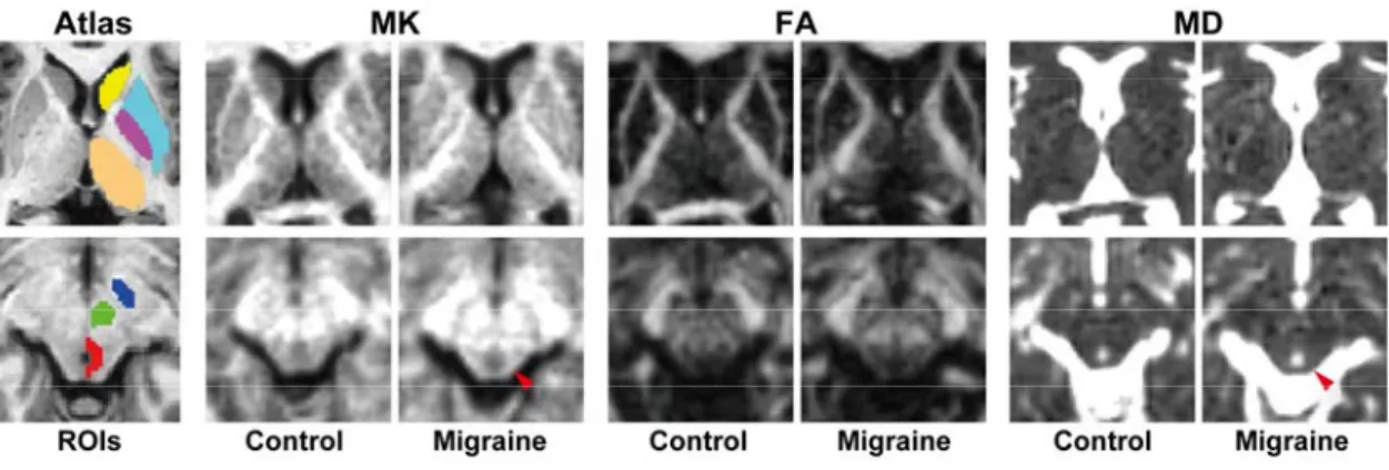
Fig. 1 Diffusion kurtosis maps of representative migraine patients and healthy control subjects and regions-of-interest of the deep gray matter structures.
The control subject was a 42 year-old woman. The patient with episodic migraines without aura was a 47 year-old woman. Her disease duration was 26 years with an untreated period of 26 years, and she experienced approximately 12 attacks per month. The atlas-based regions-of-interest (ROIs) of the left caudate nucleus (yellow), putamen (light blue), globus pallidus (purple), thalamus (beige), substantia nigra (blue), red nucleus (green), and left half of the periaqueductal gray matter (PAG) (red) are shown on the Johns Hopkins University Eve T1-weighted images. The signal intensity of the PAG ROI in the migraineur appears slightly increased on the MK and MD maps (arrowheads).
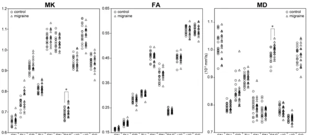
Fig. 2 Diffusion kurtosis metrics of the deep gray matter structures in patients with episodic migraines.
The MK and MD values of the periaqueductal gray matter (PAG) in the migraine group (triangles) are significantly increased compared with the control group (circles), while those of the other deep nuclei such as the caudate nucleus (CN),
putamen (PU), globus pallidus (GP), thalamus (TH), substantia nigra (SN), red nucleus (RN), anterior and posterior limbs of the internal capsule (aIC, pIC), and corpus
callosum (CC) exhibit no significant differences between the groups. *p < 0.05 (Mann- Whitney U-test)
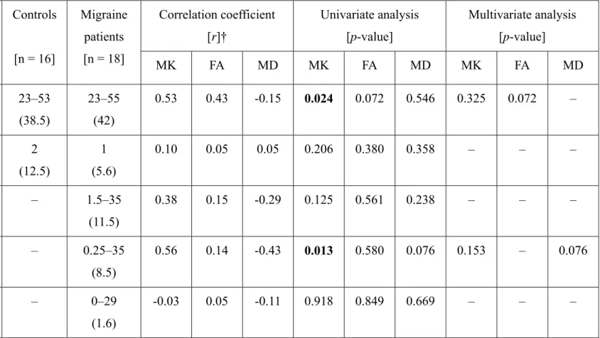
Table 1. Demographics of the migraine patients and healthy controls and their associated diffusion kurtosis metrics of the periaqueductal
gray matter.
Controls
[n = 16]
Migraine patients [n = 18]
Correlation coefficient [r]†
Univariate analysis [p-value]
Multivariate analysis [p-value]
MK FA MD MK FA MD MK FA MD
Age [years] 23–53 (38.5)
23–55 (42)
0.53 0.43 -0.15 0.024 0.072 0.546 0.325 0.072 –
Sex [men] 2
(12.5)
1 (5.6)
0.10 0.05 0.05 0.206 0.380 0.358 – – –
Disease duration [years]
– 1.5–35
(11.5)
0.38 0.15 -0.29 0.125 0.561 0.238 – – –
Untreated period [years]
– 0.25–35
(8.5)
0.56 0.14 -0.43 0.013 0.580 0.076 0.153 – 0.076
Attack frequency [times/month]
– 0–29
(1.6)
-0.03 0.05 -0.11 0.918 0.849 0.669 – – –
The data are presented as the range (median) or number (%); FA, fractional anisotropy; MD, mean diffusivity; MK, mean kurtosis; †per 10 years or per 10 times.