Evaluation of the efficacy of Ajuga decumbens extract supplement in a
rabbit osteoarthritis model and in individuals with knee discomfort
( )
Yoko Sawada
2017Laboratory of Veterinary Surgery, Department of Veterinary Clinical Medicine,
Contents Page No. Publications 1 Abstract 2 General introduction 5 References 8 Chapter I.
Ajuga decumbens stimulates differentiation of mesenchymal stem cells and
regenerates cartilage deficient injury in a rabbit osteoarthritis model
Abstract 12
1. Introduction 13
2. Materials and Methods 15
3. Results 20
4. Discussion 25
References 28
Figures 33
Chapter II.
Evaluation of the efficacy of Ajuga decumbens extract supplement in individuals with knee discomfort associated with physical activity: a
randomized, double-blind, placebo-controlled study
1. Introduction 47
2. Materials and Methods 48
3. Results 54
4. Discussion 60
References 64
Figures and Tables 70
Conclusions 85
Publications
Title Ajuga decumbens stimulates differentiation of mesenchymal stem cells and regenerates cartilage deficient injury in a rabbit osteoarthritis model
Author Yoko Sawada, Atsushi Sugimoto, Tomohiro Osaki, Yoshiharu Okamoto Journal: Experimental and Therapeutic Medicine, April, 2017 accepted
Title Evaluation of the efficacy of Ajuga decumbens extract supplement in individuals with knee discomfort associated with physical activity: a randomized, double-blind, placebo-controlled study
Author Yoko Sawada, Atsushi Sugimoto, Takehito Hananouchi, Norimasa Sato, Isao Nagaoka
Abstract
The dietary supplement glucosamine hydrochloride (GlcN) has been advocated as a safe and effective option for the management of symptoms of osteoarthritis (OA). Furthermore, the combination of GlcN with other supplements is expected to be more effective in relieving the OA symptoms. Therefore, to achieve a more effective therapy for OA, we investigated the substances having a beneficial effect on OA. Ajuga decumbens (AD), a natural herb, has been used long in Japan for pain relief. Recently, AD was reported to have potential for preventing osteoporosis (Ono et al., 2008); however, AD has not been investigated for its effects on damaged cartilage tissue.
In our previous report, we examined the effects of orally administered AD extract (ADE) alone or the combination with GlcN using rabbits with
experimentally -ecdysone (BED), a component of ADE. We revealed that ADE enhanced the regeneration of subchondral bone in this model. Moreover, the combination of ADE with GlcN synergistically increased the regeneration of cartilaginous matrix and subchondral bone. Interestingly, BED exhibited the similar effects on the regeneration of damaged subchondral bone, suggesting that BED is an active component of ADE. Together these observations indicate that the combination of ADE with GlcN synergistically exhibits a therapeutic effect on OA due to the regeneration of the cartilaginous matrix and subchondral bone (Sawada et al., 2014). In the present study, we concentrated the effective fraction of the extract to give extra ADE (EADE), which included 20-hydroxyecdysone, an active component of ADE. Firstly, we evaluated the effect of EADE on the acceleration of healing in
experimental cartilage injury. A model of cartilage injury was surgically created by introduction of three holes, one in the articular cartilage of the medial trochlea and two in the trochlear sulcus of the distal femur. Rabbits used in the experiment were divided into four groups (n = 3), namely the control group, the ADE group, the low dosage of EADE (Low EADE) group, and the high dosage of EADE (High EADE) group. ADE and EADE were dissolved in tap water and each dosage was orally administered every day for 3 weeks. After the experimental period, the cartilage matrix was strongly regenerated in the Low and High EADE groups. The average number of osteoclasts per 100 counts of osteoblast in subchondral bone decreased in the High EADE group to a greater extent compared to the control group. EADE also stimulated chondrogenic differentiation of mesenchymal stem cells and induced
in vitro proteoglycan production. Moreover, EADE significantly attenuated the
stimulation of prostaglandin E2 production by
interleukin-Therefore, EADE might represent a remarkable improvement on the currently-used ADE.
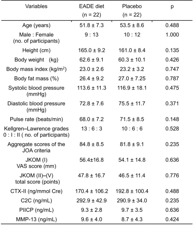
Secondary, we evaluated the efficacy and safety of the oral administration of EADE supplement to individuals with knee discomfort associated with physical activity. A randomized, double-blind, placebo-controlled study was conducted using 48 subjects. The subjects were randomly allocated to an EADE diet group (oral administration of EADE-containing diet, n = 24) and a placebo group (n = 24), and the intervention was continued for 12 weeks. Finally, 22 subjects in the placebo group and 22 subjects in the EADE diet group assessed to be eligible for assessment of the efficacy of supplement. The knee function was assessed by changes in the scores of the Japanese Knee Osteoarthritis Measure (JKOM) questionnaire and the
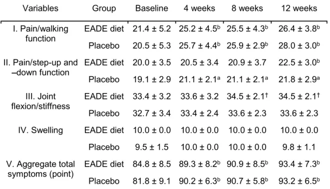
scores of the Japan Orthopedic Association (JOA) criteria as well as by analyzing the levels of type II collagen synthesis and degradation biomarkers (Procollagen II C-terminal propeptide, Degradation products of C-terminal telopeptide of type II collagen, C-terminus of the piece of type II collagen cleavage product and Matrix metalloproteinase -13). Outcomes were measured at the baseline and 4, 8, and 12 weeks from the start of administration. The subscale II (Joint flexion/stiffness) of the JOA criteria was substantially improved in the EADE diet group compared with the placebo group at 8 and 12 weeks during the intervention. Moreover, in the subgroup analyses using subjects with less knee discomfort, the subscale II (Pain/stiffness) and IV (General activities) scores of JKOM were significantly improved and Total score was substantially improved in the EADE diet group compared with the placebo group. Finally, no adverse effects were identified for the administration of EADE. These observations suggest that the administration of EADE-containing diet is safe and improves the joint function (flexion and stiffness) and general activities in the subjects with mild knee discomfort.
In conclusion, EADE could be a promising candidate as a functional food, which is useful for the joint health. The chondroprotective and anti-inflammatory actions EADE contribute to improve the knee join function, and general activities and QOL in the subjects with mild knee discomfort.
General Introduction
Osteoarthritis (OA) is a major cause of pain and disability in the aging population; however, the disease pathogenesis remains incompletely understood (Sunita et al., 2012). Because OA is a multifunctional process, characterized by changes in joint structure and function in which mechanical factors play a central role (Martin et al., 2001), treatment of patients with OA is difficult.
The dietary supplement glucosamine hydrochloride (GlcN) has been advocated as a safe and effective option for the management of OA symptoms (Reginster et al., 2001). GlcN possesses potential cartilaginous matrix regeneration properties but not bone matrix regeneration properties (Tamai et al., 2002). In addition, GlcN alone or in combination with other supplements does not reduce pain effectively in patients with OA of the knee (Clegg et al., 2006). Exploratory analyses suggested that the combination of GlcN and chondroitin sulfate is effective in a subgroup of patients with moderate-to-severe knee pain (Clegg et al., 2006). To achieve a more effective therapy for OA, we investigated ingredients that may have a beneficial effect on OA pain and bone regeneration. Ajuga decumbens (AD), a natural herb, which has long been used as a medicine for pain relief in Japan, was selected as a candidate. Recently, AD was reported to have potential for preventing osteoporosis (Ono et al., 2008); however, AD has not been investigated for its effects on damaged cartilage tissue.
Active components such as 8-acetylharpagide (Konoshima et al., 2000), cyasterone, and ecdysterone have been identified in AD (Imai et al., 1969); however, the main active component of AD remains unidentified. By referring to previous reports, we focused on phytoecdysones, which are plant-derived ecdysteroids found
in invertebrates and plants (Imai et al., 1969). In mammals, ecdysteroids have been reported to exert a protein anabolic effect (Todorov, et al., 2000). Moreover, the ecdyst -ecdysone (BED) has been reported to have beneficial effects on joints, epiphyseal cartilage, and trabecular bone in ovariectomized rats (Kapur et al., 2010). However, these beneficial effects have not been studied in damaged cartilaginous tissues. In the previous study, we investigated the effect of orally administered AD extract (ADE) and possible synergetic effects of orally administered ADE and GlcN on damaged cartilaginous tissues. Moreover, we investigated the active components in ADE. Additionally, we orally administered BED to an experimental rabbit model with artificially-induced cartilaginous injury and compared the results to a group of rabbits orally administered with GlcN and ADE. After the experimental period, the cartilage matrix was strongly regenerated in the Low and High EADE groups. The average number of osteoclasts per 100 counts of osteoblast in subchondral bone was decreased in the High EADE group to a greater extent than in the control group. Moreover, EADE stimulated chondrogenic differentiation of mesenchymal stem cells and induced in vitro proteoglycan production. Finally, EADE significantly attenuated the stimulation of prostaglandin E2 production by
interleukin-(Sawada et al., 2014).
Chapter I, we concentrated the effective fraction of the extract to give extra ADE (EADE), which included 20-hydroxyecdysone, an active component of ADE, to make ADE stronger effect and evaluated the effect of EADE on the acceleration of healing in experimental cartilage injury. Moreover, we evaluated whether the EADE stimulates differentiation of mesenchymal stem cells and have anti-inflammation effect. Because EADE has powerful effect for healing in experimental cartilage injury
and activates osteogenesis in subchondral bone, thus, we hypothesized that EADE changed the chondrogenic differentiation and some anti-inflammation effects sere associated with cartilage matrix regeneration (Sawada et al., 2017).
Chapter II, we evaluated the efficacy and safety of the oral administration of EADE supplement to individuals with knee discomfort associated with physical activity. Because ADE has been shown to have anti-inflammatory action on a rat arthritis model (Ono et al., 2008). Besides, ADE promotes the osteoblastic differentiation of cultured osteoblasts (Ono et al., 2008) and the repair of articular cartilage and subchondral bone in a cartilage damaged model (Sawada et al., 2014, 2017). Moreover, EADE has beneficial effect of osteogenesis and anti-inflammatory effect (Sawada et al., 2017). However, these beneficial effects on the joint have not been studied in humans. In this study, we investigated the effect of orally administered EADE on the subjects, who experienced the knee discomfort associated with physical activity but clinically did not need the treatment. Dietary supplement containing EADE was administered to the subjects, and the effect on the knee joint was evaluated using OA scores (the Japanese Knee Osteoarthritis Measure and the Japan Orthopedic Association criteria). In addition, type II collagen degradation (Degradation products of terminal telopeptide of type II collagen, C-terminus of the piece of type II collagen cleavage product), synthesis (Procollagen II C-terminal propeptide) markers and matrix metalloproteinase-13 were analyzed to evaluate the effects of EADE on cartilage metabolism. Moreover, to make the effect of EADE more clear, subgroup analyses of the subjects with less knee discomfort (Kellgren Lawrence grades of 0 I and >75 points of JOA score) were performed. The subscale II (Joint flexion/stiffness) of the JOA criteria was substantially improved
in the EADE diet group compared with the placebo group at 8 and 12 weeks during the intervention. Moreover, in the subgroup analyses using subjects with less knee discomfort, the subscale II (Pain/stiffness) and IV (General activities) scores of JKOM were significantly improved and total score was substantially improved in the EADE diet group compared with the placebo group. No adverse effects were identified for the administration of EADE. In conclusion, these observations suggested that the administration of EADE-containing diet was safe and improved the joint function (flexion and stiffness) and general activities in the subjects with mild knee discomfort. (Sawada et al., 2016).
References
Sunita, S., David, A. (2012). Walsh Osteochondral alterations in osteoarthritis. Bone, 51,204 211
Martin, JA., Buchwalter, JA. (2001). Roles of articular cartilage aging and chondrocyte senescence in the pathogenesis of osteoarthritis. Iowa Orthop J, 21, 1-7
Reginster, JY., Deroisy, R., Rovati, LC., Lee, R.L., Lejeune, E., Bruyere, O., Giacovelli, G., Henrotin, Y., Dacre, JE., Gossett, C. (2001). Long-term effects of glucosamine sulfate on osteoarthritis progression, a randomized, placebo-controlled clinical trial. Lancet, 357,251-256
Tamai, S., Miyatake, K., Okamoto, Y., Takamori, Y., Sakamoto, K., Minami, S. (2002). Enhanced healing of cartilaginous injuries by glucosamine hydrochloride. Carbohydr Polym, 48, 369-378
Clegg, D.O., Reda, D.J., Harris, C.L., Klein, M.A., , J.R., Hooper, M.M., Bradley, J.D., Bingham, III.C.O., Weisman, M.H., Jackson, C.G., Lane, N.E., Cush, J.J., Moreland, L.W., Schumacher, Jr.H.R., Oddis, C.V., Wolfe, F., Molitor, J.A., Yocum, D.E., Schnitzer, T.J., Furst, D.E., Sawitzke, A.D., Shi, H., Brandt, K.D., Moskowitz, R.W., Williams, H.J. (2006). Glucosamine, chondroitin sulfate, and the two in combination for painful knee osteoarthritis. N Engl J Med, 354, 795-808
Ono, Y., Fukaya, Y., Imai, S., Yamakuni, T. (2008). Beneficial Effects of Ajuga decumbens on Osteoporosis and Arthritis. Biol Pharm Bull, 31, 1199-1204
Konoshima, T., Takasaki, M., Tokuda, H., Nishino, H. (2000). Cancer chemopreventive activity of an iridoid glycoside, 8-acetylharpagide, from Ajuga
decumbens. Cancer Lett, 157, 87-92
Imai, S., Toyosato, T., Sakai, M., Sato, Y., Fujioka, S., Murata, E., Goto, M. (1969). Isolation of Cyasterone and Ecdysterone from Plant Materials. Chemical Pharm Bull, 17, 340-342
Todorov, I.N., Mitrokhin, Y.I., Efremova, O.I., Sidorenko, L.I. (2000). The influence of ecdysterone on the biosynthesis of proteins and nucleic acids in mouse organs. Khimiko Farmatsevticheskii Zhurnal, 34, 3-5
Kapur, P., Wuttke, W., Jarry, H., Seidlova-Wuttke, D. (2010). Beneficial effects of b-Ecdysone on the joint., epiphyseal cartilage tissue and trabecular bone in ovariectomized rats. Phytomedicine, 17, 350 355
Sawada, Y., Sugimoto, A., Fukuda, K., Osaki, T., Minami, S. (2014). Oral administration of Ajuga decumbens extract has a synergetic effect with glucosamine on cartilaginous injury in a rabbit osteoarthritis model. J Chitin Chitosan Sci, 2, 191-196
Sawada, Y., Sugimoto, A., Osaki, T., Okamoto Y., (2017). Ajuga decumbens stimulate differentiation of mesenchymal stem cell and regenerate cartilage deficient injury in Rabbit osteoarthritis model. Exp Ther Med, Accepted.
Chapter I.
Ajuga decumbens stimulates differentiation of mesenchymal stem cells and
regenerates cartilage deficient injury in a rabbit osteoarthritis model
Abstract
In our previous study, Ajuga decumbens extract (ADE) showed potential to decrease the number of osteoclasts in subchondral bone damage and had a synergistic effect with glucosamine on cartilaginous injury in a rabbit osteoarthritis model. In the present study, we concentrated the effective fraction of the extract to give extra ADE (EADE), which included 20-hydroxyecdysone, an active component of ADE, and evaluated the effect of EADE on the acceleration of healing in experimental cartilage injury. A model of cartilage injury was surgically created by introduction of three holes, one in the articular cartilage of the medial trochlea and two in the trochlear sulcus of the distal femur. Rabbits used in the experiment were divided into four groups (n = 3), namely the control group, the ADE group, the low dosage of EADE (Low EADE) group, and the high dosage of EADE (High EADE) group. ADE and EADE were dissolved in tap water and each dosage was orally administered every day for 3 weeks. After the experimental period, the cartilage matrix was strongly regenerated in the Low and High EADE groups. The average number of osteoclasts per 100 counts of osteoblast in subchondral bone was decreased in the High EADE group to a greater extent than in the control group. Moreover, EADE stimulated chondrogenic differentiation of mesenchymal stem cells and induced in vitro proteoglycan production. Finally, EADE significantly attenuated the stimulation of prostaglandin E2 production by
interleukin-Therefore, EADE may represent a remarkable improvement on the currently-used ADE.
1. Introduction
Osteoarthritis (OA) is a chronic degenerative joint disorder causing pain, stiffness, and limitations in the range of motion. OA is a major public health problem in the elderly, particularly OA of the knee, which is one of the most common forms of OA and estimated to affect up to 25 million people in Japan (Yoshimura et al., 2009). OA is caused by articular cartilage degeneration, include fibrillation of the articular surface, and a decrease in the size and aggregation of proteoglycan (PG) (Buckwalter et al., 2010, Buckwalter et al., 1994, Martin et al., 2001). These alterations are principally the result of changes in chondrocyte function due to aging that decrease the ability of the cells to maintain the tissue, including a decrease in synthetic activity (Martin et al., 2001). The dietary supplement D-glucosamine hydrochloride (GlcN) has been used as a safe and effective treatment for the management of OA symptoms (Reginster et al., 2001). GlcN decreases chemical mediators in chondrocytes (Nakamura et al., 2004), brings about cartilaginous matrix regeneration in an OA model, and restores the articular surface at the site of injury through its effects on PG and collagen (Tamai et al., 2002, Naito et al., 2010). Conversely, GlcN alone, or in combination with other supplements, does not effectively reduce pain in patients with OA of the knee (Clegg et al., 2006). However, tentative analyses have suggested that the combination of GlcN and chondroitin sulfate is effective in a subgroup of patients with moderate-to-severe knee pain (Clegg et al., 2006). To develop a more effective supplement for the treatment of OA,
we investigated a compound that may have a beneficial effect on this disease.
Ajuga decumbens (AD), a naturally-occurring herb, which has long been used
as a medicine for pain relief in Japan, was selected as a candidate that could be an effective supplement in the treatment of OA. In a previous study, AD extract (ADE) showed potential in the prevention of osteoporosis (Ono et al., 2008), decreasing the number of osteoclasts following subchondral bone damage and having a synergistic effect with GlcN on cartilaginous injury in a rabbit OA model (Sawada et al., 2014). 20-hydroxyecdysone, an active component of ADE, has also been shown to decrease the number of osteoclasts following subchondral bone damage in cartilaginous injury (Sawada et al., 2014). As 20-hydroxyecdysone has beneficial effects on epiphyseal cartilage tissue and trabecular bone in ovariectomized rats (Kapur et al., 2010), ADE was effective when used for subchondral bone regeneration. However, the mechanism of regeneration within the cartilage matrix induced by ADE is unclear, and its effect on such regeneration was somewhat insufficient in the previous study. The role of chondrocytes seems to be the most important factor with respect to repair in cartilage degradation (Mardones et al., 2015). The chondrocytes are present in hyaline cartilage and develop from the highly regulated differentiation of mesenchymal stem cells (MSCs), mesodermal-derived stem cells present in several fet al and adult tissues (Mardones et al., 2015). MSCs have recently been applied to the treatment of OA in clinical trials because of their regeneration potential and anti-inflammatory effects (Mardones et al., 2015). Several studies have demonstrated the therapeutic effects on OA resulting from transplantation of chondrogenic-differentiated MSCs (Mardones et al., 2015). Although transplanted native MSCs can be problematic because of their multipotent
differentiation activity, the use of pre-differentiated MSCs enhances the speed of defect healing (Ham et al., 2015). Furthermore, some compounds affect the differentiation of MSCs to chondrocytes and also have therapeutic effects on joint injury (Johnson et al., 2012).
In the present study, to increase the efficacy of ADE in the treatment of OA, we concentrated the effective fraction of the extract which includes 20-hydroxyecdysone to form extra ADE (EADE), and evaluated its effect on cartilage in the cartilage injury rabbit model. In addition, to more thoroughly investigate the mechanisms of the regeneration process in cartilage injury, the ability of EADE to induce differentiation of MSCs and its anti-inflammatory effect in chondrocytes was assessed.
2. Materials and Methods
2.1. Preparation of ADE and EADE
The whole dried AD plant was refluxed with aqueous ethanol and AD was extracted to give ADE. EADE was obtained from the extract using semi-polarity resins with the concentrated fraction having >1 wt% concentration of 20-hydroxyecdysone. The ADE and EADE were purchased from Matsuura Yakugyo Co., Ltd. (Aichi, Japan).
2.2. Animal model
The animal model was established using our previously published method (Osaki et al., 2012, Sawada et al., 2014). Twelve-week-old clinically healthy Japanese Albino female rabbits were purchased from Shimizu Laboratory Supplies
Co., Ltd. (Kyoto, Japan) with a body weight of approximately 2.0 kg, and they were acclimated for 1 week in the laboratory environment. The use of the animals and the procedures followed were approved by the Animal Research Committee of Tottori University.
2.3. Experimental procedures
An analgesic (xylazine hydrochloride, 10 mg/kg) was administered as premedication. After sedation, induction of anesthesia was performed in an anesthetizing box with a mixture of 5% isoflurane in oxygen. Anesthesia was maintained by inhalation of a mixture of 3% isoflurane in oxygen using a mask. The fur at the left knee joint was clipped and the area disinfected with chlorhexidine solution (Hibiscrub, Sumitomo Dainippon Pharma Co., Ltd., Osaka, Japan) and 70% alcohol. Approaching from the lateral portion of the knee joint, an incision was made vertically on the skin from the central part of the femur toward the tibial tuberosity. The articular capsule was incised, and the patella of the stifle joint was exposed completely by artificially dislocating the patella toward the medial side. Three holes measuring 2 mm in diameter and 4 mm in depth were made using a hand drill (Micro-engine BL-F, Osada Medical Co., Ltd., Tokyo, Japan) at the articular cartilage of the medial trochlea (one hole) and the trochlear sulcus (two holes) of the distal femur. The wound was rinsed afterwards with saline solution, and the articular capsule was sutured and closed with a synthetic absorbent thread (3-0 PDSII, Johnson & Johnson, Tokyo, Japan). The subcutaneous tissues and skin were sutured with nylon (USP 3-0 suture, Suprylon, Vomel, Germany). During the 1-week period after the operation, the wound surface was disinfected by povidone iodine (Isodine, Meiji confectionery,
Tokyo, Japan) was subcutaneously administered twice a day to prevent infection.
2.4. ADE and EADE administration
The experimental subjects were divided into four groups as follows: the control group, the ADE group, the low dosage of EADE (Low EADE) group, and the high dosage of EADE (High EADE) group. ADE contained 0.04% 20-hydroxyecdysone and EADE contained 1.38% hydroxyecdysone. 500 mg ADE/kg/day (0.2 mg 20-hydroxyecdysone/kg/day) was administered to the ADE group; 500 mg EADE/kg/day (0.69 mg 20-hydroxyecdysone/kg/day) was administered to the Low EADE group; and 500 mg EADE/kg/day (6.9mg 20-hydroxyecdysone/kg/day) was administered to the High EADE group. ADE and EADE were dissolved in tap water and each dosage was orally administered from the bottle every day for 3 weeks. The control group had free access to tap water. At 3 weeks post-operation, the rabbits were euthanized by
Ltd., Osaka, Japan) through intravenous injection. The stifle joints were opened and were macro- or microscopically observed at the site of operation for assessment of the injured cartilage.
2.5. Assessment of macroscopic changes
For the macroscopic analysis, the extent of restoration within the defective holes was scored as previously reported (Gao et al., 2008). The restoration score was as follows: 0 points: less than 50%; 1 point: 50 60%; 2 points: 60 80%; 3 points: 80 100%. The degree of restoration was scored separately for the trochlear sulcus
and the medial trochlear ridge for each subject to give a group mean, and the average score of these two areas combined was also calculated.
2.6. Assessment of histological changes
Histological assessment was performed using the femurs of the rabbits within each group. The recovered left femur was fixed using a 10% neutral buffered formaldehyde solution. After fixation, the stifle joint that had been operated on was trimmed to a thickness of 5 mm and decalcified by shaking in 5% formic acid solution for one day. After decalcification, the tissue was soaked in a 5% sodium sulfate solution for one day to allow neutralization, and was then washed for approximately 10 h under running water. After application of the standard paraffin embedding method, the tissue was sliced into
5-performed using the hematoxylin/eosin (HE), Safranin O, and Alcian blue methods. The images of restored areas, articular cartilage, and the growth zone were captured by the OpticLab H850 (Plustek Japan, Tokyo, Japan) and manipulated by ImageJ (Wayne Rasband, MD, USA). The depth of restoration in the cartilaginous matrix and the subchondral bone matrix were measured at each area via HE staining. With Safranin O staining, the red-colored pixels that indicated the presence of PGs were counted so that non-specific colored pixels were not included. With Alcian blue staining, the indigo-colored pixels that indicated the presence of glycosaminoglycans (GAGs) were also counted. We recorded the difference between restored substances at the injured sites in all groups using a microscope (BX51-FL, Olympus, Tokyo, Japan). The proportion of the pixels counted for the desired hue from a total of 120,000 pixels (random sampling of 20,000 pixels at six locations at each
cartilaginous matrix) was then calculated using an image processing technique. The number of osteoclasts at the surface of the subchondral bone, and the number of osteoblasts at the subchondral bone were recorded at 10 random areas under the microscope. The average number of osteoclasts per 100 counts of osteoblast was calculated.
2.7. Culture and cytochemical staining of hMSCs
Human MSCs (hMSCs) derived from umbilical cord matrix were obtained from PromoCell GmbH (Heidelberg, Germany). The cells were seeded at 1 × 10⁵ cells/well in a 96-well plate and cultured for 3 weeks in growing 3D spheroids in each medium. Negative control cells were cultured in 0.2 ml of growth medium (PromoCell GmbH, Heidelberg, Germany); positive control cells were cultured in a complete chondrogenic differentiation medium (PromoCell GmbH); and EADE cells were cultured in the desired concentration of EADE resolved in chondrogenic differentiation medium. The media were changed twice-weekly. After the culture period, histochemical quantitation of chondrogenic differentiation was assessed as PG accumulation, as measured by staining of cell clusters with Alcian blue. Cells were first rinsed with PBS three times and then fixed with 100% methanol for 10 min at room temperature. Staining was accomplished by applying a solution of 0.1% Alcian blue pH 2.5 (Nacalai Tesque, Kyoto, Japan) to the cells for 18 h at 4°C. To quantify the intensity of the staining, the stained culture plates were rinsed with 0.1N HCl twice, and each well was extracted with 6 M guanidine-HCl overnight at room temperature. The optical density of spheroid and extracted dye was measured at 630 nm.
2.8. Chondrocyte culture and measurement of prostaglandin E2
Human chondrocytes derived from normal human femoral cartilage were obtained from Cell Applications (San Diego, CA, USA). The cells were maintained in growth medium (Cell Applications, San Diego, CA, USA) at 37°C in a humidified atmosphere consisting of 95% air and 5% CO2. For treatment with interleukin
(IL)-a 24-well pl(IL)-ate. After overnight incub(IL)-ation, the growth medium w(IL)-as ch(IL)-anged (IL)-and cells were stimulated with 1 ng/mL IL-1
prior to incubation for 24h. After incubation, the supernatants were collected to measure the levels of prostaglandin E2 (PGE2) using a PGE2 Parameter Assay Kit
instructions.
2.9. Statistical Analysis
Data are expressed as the mean ± standard deviation of the mean.
Experimental d p < 0.05 to
indicate a statistically significant difference between groups. Microsoft Excel was used for statistical analysis using the t-test, and IBM SPSS Statistics 19 was used for other tests.
3. Results
3.1. Effect of EADE in vivo
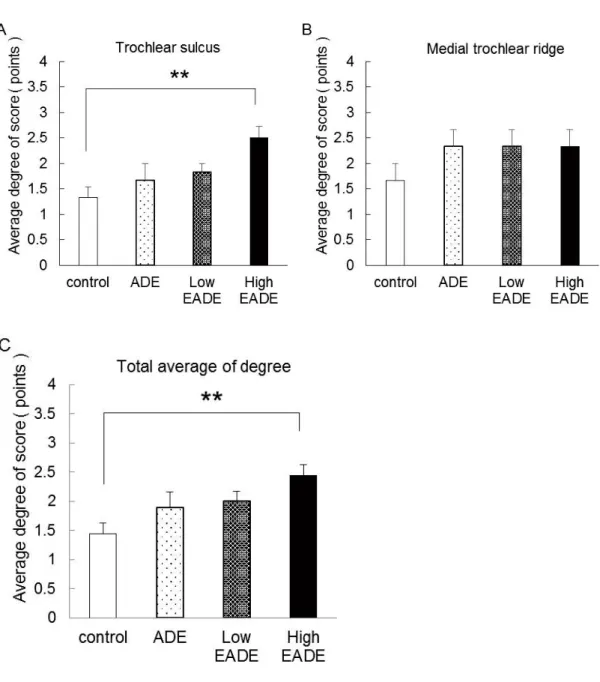
Figure 1 shows the average degree of restoration score for each group. In the control group, the degree of restoration score were lightly increased at trochlear sulcus (A) and the medial trochlear ridge (B). In the ADE group, the Low and High EADE group, the degree of restoration score was dose-dependently increased by 20-hydroxyecdysone and was significantly increased in the High EADE group compared to the control group in the trochlear sulcus (p < 0.01). Conversely, while the degree of restoration score was increased in the ADE, Low EADE, and High EADE groups in the medial trochlear ridge, there were no significant differences compared to the control group. Importantly, the average degree of restoration score, taken from pooling the data from the trochlear sulcus and medial trochlear ridge, was significantly increased in the High EADE group compared to the control group (p < 0.01), which was similar to that seen with the trochlear sulcus alone.
3.1.2 Effect of EADE on cartilage regeneration as assessed by histological HE staining
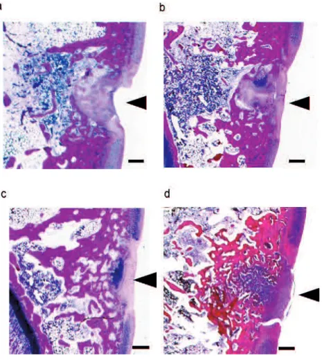
Figure 2 shows representative HE staining images of the trochlear sulcus 3 weeks after surgery. Tissue defects were still clearly visible in the control group (a) at the trochlear sulcus. In contrast, tissue regeneration was apparent in the cartilage matrix and subchondral matrix in the ADE (b), Low EADE (c), and High EADE (d) groups, with defective areas filled with proliferating fibroblasts, cartilaginous cells, and subchondral bone matrix. The surface of the wound area was covered with regenerated connective tissue; at a deeper zone, a lack of bone trabeculae, migration of mononuclear cells and neutrophils was observed. These tissues were surrounded by proliferating cells (fibroblasts and cartilaginous cells), giving the tissue an
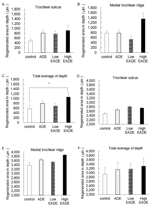
appearance that was almost identical to the formation of mature cartilaginous substrates. In the deeper zone, subchondral bone matrix was massively regenerated in ADE (b), Low EADE (c), and High EADE (d) groups. The cancellous bone structure was also somewhat regenerated in these groups, particularly in the High EADE group (d). Figure 3 shows the proportion of restoration depth in the cartilage matrix (A, B, C) and the subchondral bone matrix (D, E, F). The proportion of regenerated cartilage matrix was greater in the ADE, Low EADE, and High EADE groups compared to the control group in the trochlear sulcus. At the medial trochlear ridge, the restoration area in depth was more developed in the High EADE group, compared to the control group, and the average depth of the two areas combined was significantly higher in the High EADE group (p < 0.05). In contrast, the proportion of regenerated subchondral bone matrix in the trochlear sulcus and medial trochlear ridge, and the average combined depth, was greater in ADE, Low EADE, and High EADE groups compared to the control group.
3.1.3 Effect of EADE on cartilage regeneration as assessed by histological Safranin O and Alcian blue staining
To evaluate cartilaginous matrix regeneration more clearly, histological staining using Safranin O and Alcian blue was carried out. Figure 4 shows Safranin O staining of the trochlear sulcus (A) and Alcian blue staining of trochlear sulcus (B) with PGs in the cartilage matrix stained dark red. There was very little staining of the cartilage matrix in the control group in the trochlear sulcus (A-a). Conversely, the cartilage matrix was stained more strongly in the ADE (A-b), Low EADE (A-c), and High EADE (A-d) groups compared to the control group. In Alcian blue staining, the GAGs in the
cartilage matrix were stained blue. The stained areas and color strength were similar to what was observed with Safranin O staining (A). Strong staining was seen in the cartilage matrix in the trochlear sulcus in the ADE (B-b), Low EADE (B -c), and High EADE (B -d) groups compared to the control group (B -a). Figure 5 shows the results of image analysis of the cartilage matrix following Safranin O and Alcian blue staining. The regeneration areas were significantly increased in the Low and High EADE groups as seen with Safranin O (A) and Alcian blue (D) staining in the trochlear sulcus (p < 0.05), with the two staining methods resulting in almost identical output. This demonstrates that EADE increased the amount of GAGs and PGs in the cartilage matrix in the trochlear sulcus in a dose-dependent manner. In the medial trochlear ridge, the greatest level of staining with either method was observed in the High EADE group (B, E) and the average of the total stained area (that is, staining within the trochlear sulcus and the medial trochlear ridge combined) was significantly higher in the Low and High EADE groups compared to the control group (p < 0.05).
3.1.4 Effect of EADE on osteogenesis as assessed by histological HE staining
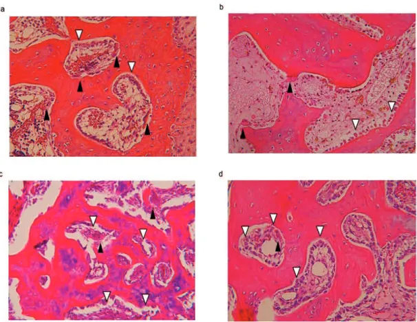
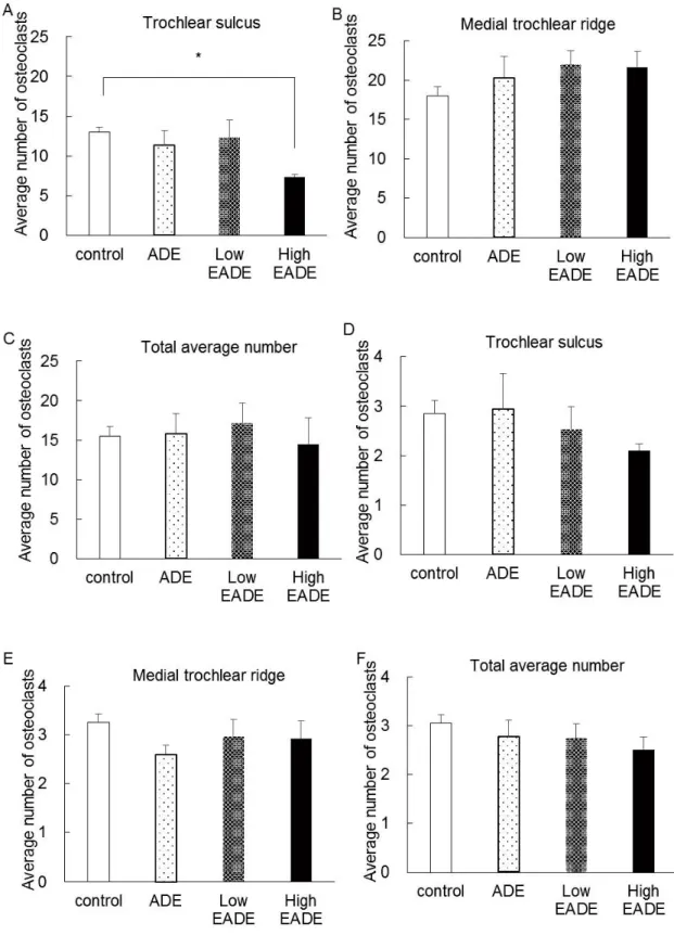
To estimate the effect of EADE on the balance of osteogenesis, osteoclasts and osteoblasts were counted in the subchondral bone following histological HE staining. Figure 6 shows the osteoclasts and osteoblasts in subchondral bone in HE staining. In control group, the osteoclasts were increased and little osteoblasts were observed. In the ADE group, the osteoblasts were observed and little osteoclasts were observed. In the Low EADE and the High EADE group, the osteoblasts were massively observed. Figure 7 shows the average number of osteoclasts (A, B, C) and we calculated the average number of osteoclasts per 100 counts of osteoblast
in the trochlear sulcus (D) and the medial trochlear ridge (E). The number of osteoclasts was significantly reduced in the High EADE group at the trochlear sulcus (A). However, the average number of osteoclasts was slightly increased in the ADE, Low EADE, and High EADE groups in the medial trochlear ridge (B). Interestingly, the average number of osteoclasts per 100 counts of osteoblast in the medial trochlear ridge (E) was slightly decreased in the ADE, Low EADE, and High EADE groups. These data show that osteogenesis was more highly activated at the medial trochlear ridge as a result of EADE treatment. Moreover, the average total number of osteoclasts per 100 counts of osteoblast for the trochlear sulcus and the medial trochlear ridge combined (F) was slightly decreased in the ADE, Low EADE, and High EADE groups. This suggests that EADE activates osteogenesis in subchondral bone.
3.2. Effect of EADE in vitro
3.2.1. Effect of EADE on chondrogenic differentiation in hMSCs
To determine whether chondrogenic differentiation was associated with cartilage matrix regeneration, we estimated MSC differentiation to chondrocytes following EADE treatment. Figure 8 shows the results of the cytochemical analysis carried out using Alcian blue staining after 3 weeks of hMSC culture. The hMSCs were cultured for 3 weeks with a normal growth medium or a chondrogenic differentiation medium containing the desired concentration of EADE. While undifferentiated MSCs have little extracellular matrix, chondrogenic differentiation results in the formation of cartilage with a typical extracellular matrix, composed of the PG aggrecan and other glycosaminoglycans. Aggrecan and other
glycosaminoglycans is used as an indicator of cartilage formation and can be detected via Alcian blue staining. Analysis showed that EADE dose-dependently increased aggrecan and other glycosaminoglycans -associated staining.
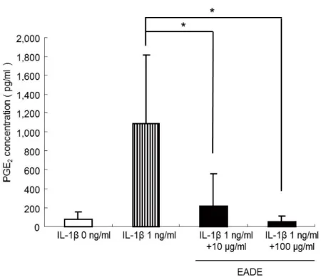
3.2.2 Effect of EADE on PGE2 production in chondrocytes
To evaluate its influence on osteogenesis, we measured the repressive effect of EADE on PGE2 production. Human chondrocytes were cultured in the absence or
presence of 1 ng/mL IL- 2
production was determined using the ELISA method (Figure 9). In the absence of IL-t of PGE2 was apparent in cultured cells. In
contrast, those cells cultured in the presence of IL- 2 in markedly
higher amounts than control cells. The effect of EADE on the IL- -induced production of PGE2 was also examined. Culture of cells
appeared to significantly block the stimulation of PGE2 production by
IL-concentration-dependent manner ( 0.05).
4. Discussion
In the present study, we evaluated whether concentration of the effective fraction of ADE (EADE), including 20-hydroxyecdysone, could be more effective in the treatment of OA and cartilage regeneration. In a cartilage injury model, the degree of restoration within the defective holes was dose-dependently increased by EADE administration (Figure 1). In particular, the cartilage matrix was strongly regenerated in the and High EADE groups ( 0.05) (Figure 2, 3). In addition, the regeneration
area was significantly enhanced in a dose-dependent manner in the Low and High EADE groups as seen following Safranin O and Alcian blue staining ( 0.05) (Figure 4, 5). The data obtained from Safranin O and Alcian blue staining were consistent with each other, and the extent of staining corresponded to the concentration of EADE, showing that the compound increases the amount of GAGs and PGs in cartilage matrix. Moreover, in our previous study, the number of osteoclasts was significantly decreased following administration of ADE or 20-hydroxyecdysone (Sawada et al., 2014), but the influence on bone metabolism was unclear. Bone metabolism is regulated by the balance of osteoclast and osteoblast cells. ADE has been shown to inhibit the differentiation of osteoclasts (Ono et al., 2008), while 20-hydroxyecdysone also has beneficial effects with respect to epiphyseal cartilage and trabecular bone in ovariectomized rats (Kapur et al., 2010). In the present study, the average number of osteoclasts per 100 counts of osteoblast in subchondral bone was decreased in the High ADE group compared to the control group (Figure 7). It is possible that EADE could influence not only the number of osteoclasts, but also, bone metabolism, and the regeneration of subchondral bone. As it has been reported that 20-hydroxyecdysone stimulates MSC osteogenic differentiation (Gao et al., 2008), its presence as an active component in EADE could stimulate MSCs and enhance osteogenesis in subchondral bone.
When cartilage is injured, natural healing tends to be slow and usually results in the formation of nonfunctional fibro-cartilage, while regeneration of hyaline cartilage seldom occurs (Tamai et al., 2002). However, the administration of GlcN does regenerate hyaline cartilage (Tamai et al., 2002). Experimental data have shown that the mechanism underlying this action is an association with chondroblast
activation (Hashida et al., 2003). Glucosamine promotes a chondrogenic phenotype in MSCs (Derfoul et al., 2007) and inhibits matrix met alloproteinase (MMP)-13 expression and matrix degradation (Derfoul et al., 2007). MMPs and
IL-shown to play a central role in the degradation of articular cartilage (Farahat et al., 1993, Campbell et al., 1990, Tetlow et al., 2001).
IL-cells in the arthritic synovium and chondrocytes (Nakamura et al., 2004), and enhances the production of various chemical mediators such as PGE2 (Campbell et al., 1990), nitric oxide (NO), and MMPs (Tetlow et al., 2001) from chondrocytes. To
more clearly delineate the mechanisms of the regeneration process following cartilage injury, the ability of EADE to induce differentiation of MSCs, and its anti-inflammatory effects in chondrocytes, were assessed. EADE stimulated PG production and induced in vitro chondrogenic differentiation of MSCs (Figure 8). Moreover, EADE significantly attenuated the stimulation of PGE2 production by
IL-in chondrocytes (Figure 10). It has been reported that 20-hydroxyecdysone suppresses IL- -induced catabolic gene expression in cartilage (Sheu et al., 2015), and that ADE inhibits expression of iNOS and NO production from macrophages (Ono et al., 2008). It has also been shown that PGE2 induces receptor activator of
nuclear factor kappa-B ligand (RANKL) expression by osteoblasts and directly enhances RANKL-induced osteoclastogenesis from precursors (Kotake et al., 2010). In addition, 20-hydroxyecdysone has been reported to stimulate MSC osteogenic differentiation (Gao et al., 2008). Therefore, 20-hydroxyecdysone, the active component in EADE, could suppress IL- -induced inflammation and PGE2-induced
RANKL expression. By this action, EADE could have an anti-inflammatory effect and suppress osteoclastogenesis.
References
Yoshimura, N., Muraki, S., Oka, H., Mabuchi, A., En-Yo, Y., Yoshida, M., Saika, A., Yoshida, H., Suzuki, T., Yamamoto, S., Ishibashi, H., Kawaguchi, H., Nakamura, K., Akune, T. (2009). Prevalence of knee osteoarthritis, lumbar spondylosis, and osteoporosis in Japanese men and women: the research on osteoarthritis/osteoporosis against disability study. J.Bone Miner.Metab, 27, 620-628.
Buckwalter, J.A., Martin, J., Mankin, H.J., (2000). Synovial joint degeneration and the syndrome of osteoarthritis. Instr Course Lect, 49, 481-489.
Buckwalter, J.A., Roughley, P.J., Rosenberg, L.C., (1994). Age-related changes in cartilage proteoglycans: quantitative electron microscopic studies. Microsc Res Tech, 28, 398-408.
Martin, J.A., Buckwalter, J.A., (2001). Roles of articular cartilage aging and chondrocyte senescence in the pathogenesis of osteoarthritis. Iowa Orthop J, 21, 1-7.
Reginster, J.Y., Deroisy, R., Rovati, L.C., Lee, R.L., Lejeune, E., Bruyere, O., Giacovelli, G., Henrotin, Y., Dacre, J.E., Gossett, C., (2001). Long-term effects of glucosamine sulphate on osteoarthritis progression: a randomised, placebo-controlled clinical trial. Lancet, 357, 251-256.
Nakamura, H., Shibakawa, A., Tanaka, M., Kato, T., Nishioka, K., (2004). Effects of glucosamine hydrochloride on the production of prostaglandin E2, nitric oxide and met alloproteases by chondrocytes and synoviocytes in osteoarthritis. Clin Exp Rheumatol, 22, 293-299.
Tamai, Y., Miyatake, K., Okamoto, Y., Takamori, Y., Sakamoto, H., Minami, S., (2002). Enhanced healing of cartilaginous injuries by glucosamine hydrochloride. Carbohydr Polym, 48, 369-378.
Naito, K., Watari, T., Furuhata, A., Yomogida, S., Sakamoto, K., Kurosawa, H., Kaneko, K., Nagaoka, I., (2010). Evaluation of the effect of glucosamine on an experimental rat osteoarthritis model. Life Sci, 86, 538-543.
Clegg, D.O., Reda, D.J., Harris, C.L., Klein, M.A. , J.R., Hooper, M.M., Bradley, J.D., Bingham, C.O., Weisman, M.H., Jackson, C.G., Lane, N.E., Cush, J.J., Moreland, L.W., Schumacher, H.R., Jr Oddis, C.V., Wolfe, F., Molitor, J.A., Yocum, D.E., Schnitzer, T.J., Furst, D.E., Sawitzke, A.D., Shi, H., Brandt, K.D., Moskowitz, R.W., Williams, H.J., (2006). Glucosamine, chondroitin sulfate, and the two in combination for painful knee osteoarthritis. N Engl J Med, 354, 795-808.
Ono, Y., Fukaya, Y., Imai, S., Yamakuni, T., (2008), Beneficial effects of Ajuga
decumbens on osteoporosis and arthritis. Biol Pharm Bull, 31, 1199-1204.
S., (2014). Oral administration of Ajuga decumbens extract has a synergetic effect with glucosamine on cartilaginous injury in a rabbit osteoarthritis model. J. Chitin Chitosan Sci, 2, 191-196.
Kapur, P., Wuttke, W., Jarry, H., Seidlova-Wuttke, D., (2010). Beneficial effects of beta-Ecdysone on the joint, epiphyseal cartilage tissue and trabecular bone in ovariectomized rats. Phytomedicine, 17, 350-355.
Mardones, R., Jofre, C.M., Minguell, J.J., (2015). Cell therapy and tissue engineering approaches for cartilage repair and/or regeneration. Int J Stem Cells, 8, 48-53.
Ham, O., Lee, C.Y., Kim, R., Lee, J., Oh, S., Lee, M.Y., Kim, J., Hwang, K.C., Maeng, L.S., Chang, W., (2015). Therapeutic potential of differentiated mesenchymal stem cells for treatment of osteoarthritis. Int J Mol Sci, 16, 14961-14978.
Johnson, K., Zhu, S., Tremblay, M.S., Payette, J.N., Wang, J., Bouchez, L.C., Meeusen, S., Althage, A., Cho, C.Y., Wu, X., Schultz, P.G., (2012). A stem cell-based approach to cartilage repair. Science, 336, 717-721.
Osaki, T., Kitahara, K., Okamoto, Y., Imagawa, T., Tsuka, T., Miki, Y., Kawamoto, H., Saimoto, H., Minami, S., (2012). Effect of fucoidan extracted from mozuku on experimental cartilaginous tissue injury. Mar Drugs, 13, 2560-2570.
in mouse mesenchymal stem cells and relieves osteoporosis. Biol Pharm Bull, 31, 2245-2249.
Hashida, M., Miyatake, K., Okamoto, Y., Fujita, K., Matsumoto, T., Morimatsu, F., Sakamoto, K., Minami, S., (2003). Synergistic effects of D-glucosamine and collagen peptides on healing experimental cartilage injury. Macromol Biosci, 3, 596-603.
Derfoul, A., Miyoshi, A.D., Freeman, D.E., Tuan, R.S., (2007). Glucosamine promotes chondrogenic phenotype in both chondrocytes and mesenchymal stem cells and inhibits MMP-13 expression and matrix degradation. Osteoarthritis Cartilage, 15, 646-655.
Farahat, M.N., Yanni, G., Poston, R., Panayi, G.S., (1993). Cytokine expression in synovial membranes of patients with rheumatoid arthritis and osteoarthritis. Ann Rheum Dis, 52, 870-875.
Campbell, I.K., Piccoli, D.S., Hamilton, J.A., (1990). Stimulation of human chondrocyte prostaglandin E2 production by recombinant human interleukin-1 and tumor necrosis factor. Biochim Biophys Acta, 1051, 310-318.
Tetlow, L.C., Adlam, D.J., Woolley, D.E., (2001). Matrix met alloproteinase and proinflammatory cytokine production by chondrocytes of human osteoarthritic cartilage: associations with degenerative changes. Arthritis Rheum, 44, 585-594.
Sheu, S.Y., Ho, S.R., Sun, J.S., Chen, C.Y., Ke, C.J., (2015). Arthropod steroid hormone (20-Hydroxyecdysone) suppresses IL- -induced catabolic gene expression in cartilage. BMC Complement Altern Med, 15, 1-8.
Kotake, S., Yago, T., Kawamoto, M., Nanke, Y., (2010) Effects of NSAIDs on differentiation and function of human and murine osteoclasts
Figures
Figure 1. The degree of restoration in the defective holes.
The degree of restoration in the defective holes was quantified using the following scoring system: 0 points: Less than 50 %; 1 point: 50 60 %; 2 points: 60 80 %; 3 points: 80 100 %. Scores for the degree of restoration of two holes in the trochlear sulcus (A), one hole in the medial trochlear ridge (B), and the average of the combined data for all holes (C). The degree of restoration in the defective holes was
calculated as the average of the scores obtained from subjects within each group. (A) The degree of restoration in the trochlear sulcus was dose-dependently increased by 20-hydroxyecdysone, with the High EADE group score being significantly higher than that of the control group. (B) The degree of restoration in the medial trochlear ridge was increased to a similar level within the ADE, Low EADE, and High EADE groups. (C) The average degree of restoration seen when the data from the trochlear sulcus and the medial trochlear ridge were pooled was also dose-dependently increased, and the score from the High EADE group was significantly higher compared to the control group. The data here were similar to the scores seen for the trochlear sulcus. **:
Figure 2. Histological HE staining 3 weeks after surgery.
Histological HE staining in the trochlear sulcus. Tissue defects (black arrowheads) were still clearly visible in the control group (a) at the trochlear sulcus. The holes were filled with proliferating fibroblasts, cartilaginous cells, and subchondral bone matrix in the ADE (b) and Low EADE (c) groups. It also appears that all holes were fully filled with cartilaginous matrix and subchondral matrix in the High EADE group (d). Bars = 500µm.
Figure 3. Restoration depth in the cartilage matrix and subchondral matrix (measured using HE staining).
The proportion of the regenerated area in depth at each part of the cartilage matrix (A), (B), (C) and the subchondral bone matrix (D), (E), (F). The cartilage matrix was regenerated to a greater extent in the ADE, Low EADE, and High EADE groups compared to the control at the trochlear sulcus (A); it showed enhanced regeneration in the High EADE group at the medial trochlear ridge (B); and it was significantly regenerated in the High EADE group when the total average depth was calculated (C). The subchondral bone matrix showed regeneration in the ADE, Low EADE, and High EADE groups in both areas and when the data were averaged (D), (E), (F), compared to the control group. *:
Figure 4. Histological Safranin O staining and Alcian blue staining 3 weeks after surgery.
Histological Safranin O staining in the trochlear sulcus (A) and Alcian blue staining in the trochlear sulcus (B). The proteoglycans in the cartilage matrix were darkly stained. Black arrowheads indicate the tissue-defective areas. At the trochlear sulcus (A), green and dark red staining was still weak in the control group, but the stained area appears to spread dose-dependently in the ADE (A-b), Low EADE (A-c), and High EADE (A-d) groups, compared to the control group (A-a). The defects were filled with collagen fibers in the control (A-a), ADE (A-b), Low EADE (A-c), and High EADE (A-d) groups and it seems that the cartilage matrix was strongly stained dark red in the High EADE group (A-d) only. The GAGs in the cartilage matrix were stained blue (B). The stained areas and intensity of color were similar to what was observed with Safranin O staining (A). The defects were filled with proteoglycan in the ADE (B-b), Low EADE (B-c), and High EADE (B-d) groups and it seems that the cartilage matrix was strongly stained blue in the High EADE group (B-d). Bars = 500µm.
Figure 5. Image analysis of the cartilage matrix following Safranin O and Alcian blue staining.
The proportion of pixels counted for the desired hue in Safranin O (A), (B), (C) and in Alcian blue (D), (E), (F) staining at each cartilage area (six areas in each group) was calculated. The increase in the stained area seen using either method, which represents enhanced PG and GAG content in the cartilage matrix, indicated significant dose-dependent regeneration in the Low ADE and High ADE groups compared to the control group at the trochlear sulcus (A), (D) and when the data were averaged for both areas (C), (F). It also appears that the stained area was greater in the High EADE group at the medial trochlear ridge (B), (E). *: 0.05, **:
Figure 6. Histological HE staining in subchondral bone 3 weeks after surgery. Histological HE staining in the trochlear sulcus and the enlarged photo in the subchondral bone. Black arrowheads indicate the osteoclasts and white arrowheads indicate the osteoblast. In control group (a), the osteoclasts were increased and little osteoblasts were observed. In the ADE group (b), the osteoblasts were observed and little osteoclasts were observed. In the Low EADE (c) and the High EADE (d) group, the osteoblasts were massively observed. At 200x magnification.
Figure 7. The average number of osteoclasts and the average number of osteoclasts per 100 counts of osteoblast.
The number of osteoclasts at the surface of the subchondral bone was recorded at the trochlear sulcus (A), and the medial trochlear ridge (B) at 10 random areas under the microscope, and the average total number was calculated (C). In addition, the number of osteoblasts at the subchondral bone was recorded and the average number of osteoclasts per 100 counts of osteoblast was calculated for each area (D), (E), (F). The number of osteoclasts was significantly reduced in the High EADE group in the trochlear sulcus (A). While the number of osteoclasts was increased in the ADE, Low EADE, and High EADE groups, compared to the control group, at the medial trochlear ridge (B), the balance of osteoblasts and osteoclasts was inclined towards osteogenesis in these groups, compared to the control (E). The average total number of osteoclasts per 100 counts of osteoblast was inclined towards osteogenesis in the ADE, Low EADE, and High EADE groups, compared to the control group (F). *: 0.05 versus Control using Dunnett s test.
Figure 8. Chondrocyte differentiation from MSCs.
The hMSCs were cultured for 3 weeks with normal growth medium (GM) or chondrogenic differentiation medium (CD) with the desired concentration of EADE. CD results in the formation of cartilage with a typical extracellular matrix consisting of the proteoglycan aggrecan. This is used as an indicator of cartilage formation and can be detected using Alcian blue staining at an absorbance of 630 nm. Absorbance was significantly increased by EADE in a concentration-dependent manner. *:
Figure 9. PGE2 levels in cytokine-stimulated chondrocytes.
Human chondrocytes were stimulated with
IL-(10 and 100 g/ml). The concentration of PGE2 was increased by
IL-Chapter II.
Evaluation of the efficacy of Ajuga decumbens extract supplement in individuals with knee discomfort associated with physical activity: a
randomized, double-blind, placebo-controlled study Abstract
The purpose of this study was to assess the efficacy and safety of the oral administration of Ajuga decumbens extract (EADE) supplement to individuals with knee discomfort associated with physical activity. A randomized, double-blind, placebo-controlled study was conducted using 48 subjects. The subjects were randomly allocated to an EADE diet group (oral administration of EADE-containing diet, n = 24) and a placebo group (n = 24), and the intervention was continued for 12 weeks. Finally, 22 subjects in the placebo group and 22 subjects in the EADE diet group assessed to be eligible for assessment of the efficacy of supplement. The knee function was assessed by changes in the scores of the Japanese Knee Osteoarthritis Measure (JKOM) questionnaire and the scores of the Japan Orthopedic Association (JOA) criteria as well as by analyzing the levels of type II collagen synthesis and degradation biomarkers (Procollagen II C-Terminal propeptide: PIICP, Degradation products of C-terminal telopeptides of type II collagen: CTX-II, C-terminus of the piece of type II collagen cleavage product: C2C, and matrix metalloproteinase-13: MMP13). Outcomes were measured at the baseline and 4, 8, and 12 weeks from the start of administration. The subscale II (Joint flexion/stiffness) of the JOA criteria was substantially improved in the EADE diet group compared with the placebo group at 8 and 12 weeks during the intervention. Moreover, in the subgroup analyses using subjects with less knee discomfort, the subscale II (Pain/stiffness) and IV (General
activities) scores of JKOM were significantly improved and total score was substantially improved in the EADE diet group compared with the placebo group. No adverse effects were identified for the administration of EADE. In conclusion, these observations suggested that the administration of EADE-containing diet was safe and improved the joint function (flexion and stiffness) and general activities in the subjects with mild knee discomfort. Thus, EADE could be a promising candidate as a functional food, which would be useful for the joint health.
1. Introduction
Knee osteoarthritis (OA) is characterized by the progressive destruction of articular cartilage and is the leading cause of pain and physical disability in the elderly (Yoshimura et al., 2009). Early-stage of OA limits the movement and causes the knee discomfort, thereby impairing the activities of daily life (ADL) and quality of life (QOL) (Yoshimura et al., 2009). The prevalence of radiographic knee OA in Japan is increasing and estimated to affect 30 million people in Japanese senior people over 50 years old (Yoshimura et al., 2009). According to the treatment guidelines for OA of the Knee Osteoarthritis Research Society International (OARSI), non-steroidal anti-inflammatory drugs, selective cyclooxygenase-2 inhibitors, and acetaminophen are listed as high level of evidence drugs for treatment of OA (McAlindon et al., 2014). However, these drugs enhance cartilage destruction, and promote OA (Xu et al., 2006, Ou 1989). Therefore, novel substances with a chondroprotective action have been sought. In our research, we focused on the extract of Ajuga decumbens (AD) as a candidate that exhibits a protective action on OA. AD is a natural herb and has long been used as a medicine for pain relief in Japan (Rashad et al., 1989, Herman
et al., 1984). Importantly, AD has been shown to have anti-inflammatory action on a
rat arthritis model (Ono et al., 2008). Moreover, AD promotes the osteoblastic differentiation of cultured osteoblasts (Ono et al., 2008) and the repair of articular cartilage and subchondral bone in a cartilage damaged model (Sawada et al., 2014). However, these beneficial effects on the joint have not been studied in humans. In this study, we investigated the effect of orally administered AD extract (EADE) on the subjects, who experienced the knee discomfort associated with physical activity but clinically did not need the treatment. Dietary supplement containing EADE was administered to the subjects, and the effect on the knee joint was evaluated using OA scores (the Japanese Knee Osteoarthritis Measure, JKOM and the Japan Orthopedic Association criteria, JOA). In addition, type II collagen degradation (Degradation products of terminal telopeptides of type II collagen: CTX-II and C-terminus of the piece of type II collagen cleavage product: C2C) and synthesis (Procollagen II C-Terminal propeptide: PIICP) markers and matrix metalloproteinase (MMP)-13 were analyzed to evaluate the effects of EADE on cartilage metabolism. Moreover, to make the effect of EADE more clear, subgroup analyses of the subjects with less knee discomfort (Kellgren Lawrence grades of 0 I and >75 points of JOA score) were performed.
2. Materials and Methods
2.1. Study Design
A prospective, randomized, double-blind, placebo-controlled and parallel-group comparative study was designed to assess the efficacy and safety of a diet supplemented with EADE. The study was conducted from November 2014 to June
2015 and involved one medical clinical service organization center under the control of one medical investigator in Japan. The protocol was submitted to and approved by the institutional ethics committee in October 2014 (20141030-2), and the study was conducted in accordance with the Declaration of Helsinki and Ethical Guidelines for Epidemiological Research (recognized by the Japanese Government in 2008). The objective of the study was explained to all the subjects and written informed consent was provided prior to enrollment in the study.
2.2. Subjects
Male and female Japanese subjects aged 40 80 years with knee discomfort associated with physical activity but without radiographic evidence of knee osteoarthritis (Kellgren and Lawrence (K/L) grades 0 II, mainly 0 I) were enrolled;
-- (Kellgren et al., 1957). The major exclusion criteria were the presence of gout or rheumatoid arthritis that may cause joint pain; suffering from any other injuries that may have required the use of anti-inflammatory or other medications or physiotherapy treatment by an orthopedist; previous surgical treatment of knee joints; routine use of dietary supplements or medicines containing EADE, hyaluronic acid, glucosamine, chondroitin sulfate, collagen peptides, or rich in calcium; need to undergo pharmacological treatments during the study period; performing hard exercise; history of osseous or articular diseases within the past year; diagnosis as having malignant tumor, hypertension, heart disease, kidney disease, thyroid disorder or other serious illness that may have required other physiotherapy treatment; pregnancy; nursing mothers or women of childbearing
potential; participation in another clinical study; and presence of any medical
study.
2.3. Study Interventions
The study diet was manufactured in the form of a 1.5 g powder. The ingredients were EADE (>1% 20-hydroxyecdysone), dextrin, hydroxypropyl methylcellulose, cellulose, calcium stearate, and palatinose. EADE was purchased from Matsuura Yakugyo Company. The amount of EADE was 10 mg in the 1.5 g EADE diet. The placebo diet comprised crystalline cellulose, dextrin and caramel pigment instead of EADE. The study supplement was wrapped in a single pack (1.5 g) and provided with a wafer, and the subject took the supplement after breakfast once a day with two cups of water for 12 weeks.
2.4. Randomization and Blinding
Because the symptoms, especially pain, vary according to sex in arthritis (Muraki et al., 2009), research coordinators created an allocation table for males and females, randomly assigned eligible subjects and granted allocation numbers to test diet. The allocation table was sealed until the end of the study. All of the research staff and subjects were blinded to the treatment allocation during the test period. Only the statistics experts and data monitoring committee were unblinded, but they had
2.5. Evaluation of Efficacy
The outcomes for the evaluation of efficacy were based on the changes in the subscale scores of the Japan Orthopedic Association criteria for osteoarthritic knees (JOA) (Koshino et al., 1988); the changes in the subscale scores of the Japanese Knee Osteoarthritis Measure (JKOM) (Akai et al., 2005); the levels of urinary CTX-II and serum C2C as type II collagen degradation biomarkers (Reijman et al., 2004, Poole et al., 2004); the levels of serum PIICP as a type II collagen synthesis biomarker (Nelson et al., 1998), the levels of serum MMP-13 (Wang et al., 2014), the ratio of CTX-II/PIICP, and the ratio of C2C/PIICP. These parameters were measured at the baseline and 4, 8, and 12 weeks. Serum and urine samples were stored at 80°C until measurement. After the end of study period, CTX-II, C2C, PIICP, and MMP-13 were measured on the same day using the respective ELISA assay kits; CTX-II, Urine CartiLaps® EIA, Immunodiagnostic Systems Ltd, Tyne&Wear, United Kingdom.; C2C, Collagen Type II Cleavage ELISA, IBEX Pharmaceuticals Inc, Quebec, Canada; PIICP, Enzyme-linked Immunosorbent Assay Kit For Procollagen II C-Terminal Propeptide, Uscn Life Science Inc, Houston, USA.; MMP-13, and Enzyme-linked Immunosorbent Assay Kit For Matrix Metalloproteinase 13, Uscn Life Science Inc, Houston, USA. Serum C2C and urinary CTX-II were used as markers for type II collagen degradation, and serum PIICP was used as a marker for type II collagen synthesis. The ratios of C2C/PIICP and CTX-II/PIICP were also analyzed to assess the changes in the balance of type II collagen degradation and synthesis (cartilage metabolism). The concentrations of CTX-II were corrected for urinary creatinine (Cre) and expressed as ng/mmol/Cre. Serum MMP-13 was also used as a marker for cartilage degradation, because MMP-13 is a collagenase that
contributes to cartilage degradation by cleaving type II collagen triple helix (Poole et
al., 2003).
The JOA is the criteria established by the Japanese Orthopedic Association (Akai et al., 2005), which is used by physicians for subjectively evaluating the treatment, based on the following subcategories; I) Pain/walking function; II) Pain/step-up and -down function; III) Joint flexion/stiffness; IV) Swelling; and V) Aggregated total symptoms. The first four subscales are rated from 0 to 30, from 0 to 25, from 0 to 35, and from 0 to 10, respectively. The maximum value indicates no symptoms or functional disability, and a score of 0 indicates a condition resulting in extreme difficulty to perform daily living tasks. The sum of the scores of these four subscales represents the score of the fifth subscale (Total symptoms score).
The JKOM is a self-answered, evaluation questionnaire that includes five subcategories: I) Pain was evaluated by a visual analog scale (VAS); II) Pain and stiffness during the past few days (8 questions); III) Activities of daily living during the past few days (10 questions); IV) General activities during the past month (5 questions); and V) General health conditions during the past month (2 questions). VAS values range from 0 (no pain) to 100 (pain that cannot be tolerated). The responses to each question (II-V subcategories) are assigned 1 to 5 points, with 1 point indicating good functional status and 5 points indicating the worst functional status. The JKOM score is higher in subjects with more pain and physical disability, and this evaluation modality is reported to be sufficiently reliable and valid for studying the clinical outcomes of knee OA (Braham et al., 2003). The outcome of JKOM has been shown to be closely correlated with that of other arthritis-related scales, such as the Western Ontario and McMaster Universities Arthritis Index