PAPER
Compressed Sensing Framework Applying Independent
Component Analysis after Undersampling for Reconstructing Electroencephalogram Signals ∗
Daisuke KANEMOTO†a),Member, Shun KATSUMATA††, Masao AIHARA†††,Nonmembers, andMakoto OHKI††††,Member
SUMMARY This paper proposes a novel compressed sensing (CS) framework for reconstructing electroencephalogram (EEG) signals. A fea- ture of this framework is the application of independent component analysis (ICA) to remove the interference from artifacts after undersampling in a data processing unit. Therefore, we can remove the ICA processing block from the sensing unit. In this framework, we used a random undersampling measurement matrix to suppress the Gaussian. The developed framework, in which the discrete cosine transform basis and orthogonal matching pur- suit were used, was evaluated using raw EEG signals with a pseudo-model of an eye-blink artifact. The normalized mean square error (NMSE) and correlation coefficient (CC), obtained as the average of 2,000 results, were compared to quantitatively demonstrate the effectiveness of the proposed framework. The evaluation results of the NMSE and CC showed that the proposed framework could remove the interference from the artifacts under a high compression ratio.
key words: EEG, compressed sensing, independent component analysis, random undersampling, artifact
1. Introduction
Electroencephalogram (EEG) signals are important bio- metric signals used to reveal signs of brain inflammation, epilepsy[1], sleep disorders, and Alzheimer’s disease[2].
Long-term wireless telemonitoring of EEG signals is re- quired for accurately tracing brain activity. In general EEG wireless monitoring systems, sensors in the sensing unit are placed on the scalp, and EEG signals are transmitted wire- lessly to a data processing unit, such as a workstation or a
Manuscript received May 1, 2020.
Manuscript publicized June 22, 2020.
†The author is with Division of Electrical, Electronic and Infor- mation Engineering, Osaka University, Suita-shi, 565-0871 Japan.
††The author is with Integrated Graduate School of Medicine, Engineering, and Agricultural Sciences, University of Yamanashi, Kofu-shi, 400-8511 Japan.
†††The author is with Graduate School of Medicine, University of Yamanashi, Kofu-shi, 400-8511 Japan.
††††The author is with Graduate Faculty of Interdisciplinary Re- search, University of Yamanashi, Kofu-shi, 400-8511 Japan.
∗This paper is based on “Framework of Applying Indepen- dent Component Analysis After Compressed Sensing for Elec- troencephalogram Signals” by D.KANEMOTO, S.KATSUMATA, M.AIHARA, and M.OHKI which appeared in Proceedings of 2018 IEEE Biomedical Circuits and Systems Conference, October 2018.
©2018 IEEE.
a) E-mail: [email protected] (Corresponding author)
DOI: 10.1587/transfun.2020EAP1058
PC, for analysis. The data exchange in wireless commu- nication requires large power consumption. Therefore, the energy efficiency of the sensing unit is important because its battery life is limited. Thus, a low-power sensing technique is required.
The compressed sensing (CS) theory, which is an at- tractive method for signal acquisition and compression[3], can enable the realization of a low-power sensing technique.
This theory suggests that signals can be recovered from fewer measurements than required in the conventional Nyquist sampling technique, if the signal is sparse in some transform domains. However, if interferences from artifacts (from eye blinking, muscle activity, etc.) occur during EEG monitor- ing, the compressed signals become less sparse. Particularly, impulse noise strongly affects the quality of signal recovery [4].
On the other hand, independent component analysis (ICA) [5] has proved to be an effective algorithm for re- moving artifacts [6]–[8]. In some other studies, ICA was also performed to effectively separate only the desired infor- mation from the measured EEG signals before compressing the signals in a sensing unit [9]–[11]. Thus, performing ICA to improve sparsity before applying compression can be also considered. However, to perform ICA before un- dersampling, a micro-processing unit (MPU) that performs calculations must be installed in the sensing unit. This how- ever this results in an increase in the power dissipation of the sensing unit owing to the additional implementation of the MPU. Therefore, it is not efficient to use ICA process- ing before undersampling for removing artifacts. Hence, we developed a new framework to remove artifacts from the compressed signals outside the sensing unit. The proposed framework does not require additional MPUs in the sensing unit to perform ICA.
This paper is organized as follows. Section 2 briefly de- scribes the CS theory and ICA. Section 3 presents the details of the new framework. Section 4 presents the calculation results obtained with this proposed framework. In Sect. 5, we discuss the difference in reconstruction accuracy between two frameworks: ICA after compression (proposed frame- work) and ICA before compression. Section 6 presents the conclusions of the study. This paper is based on international conference proceedings[12].
Copyright © 2020 The Institute of Electronics, Information and Communication Engineers
Here, y is a known vector, andΦ and Ψare known matrices. Θ=Φ Ψis called the sensing matrix. The rela- tionship betweenyandsis shown in Fig. 1 using an example withM=5,P=20. The color depth indicates the numeric size of each element. White elements indicate zeros. The vector sis sparse and there are many zero elements. The length of yis much smaller than the length ofs. Therefore, we can understand from Fig. 1 that Eq. (1) is underdetermined. The most original approach for solving this problem is to find the sparsest vector. A solution is given by the followingl0 minimization problem:
min||s||0 subject to y=Θs. (2)
It is well known that thel0minimization problem is NP- hard[13], and it cannot be used for practical applications.
Therefore, other reconstruction algorithms (e.g. basis pursuit (BP) [14], orthogonal matching pursuit (OMP) [15], and block sparse Bayesian learning (BSBL)[16]) are widely used for solving CS problems. In general, there is a trade-off between the reconstruction time and accuracy. For example, it was reported that BP is better than OMP in terms of the reconstruction accuracy. However, the reconstruction time of BP tends to be longer owing to calculation complexities[17].
It was reported that BSBL is a powerful reconstruction tool for compressed EEG signals[16]. However, reconstruction using BSBL is more time consuming than that using OMP.
Determining the basis for EEG signal is also important to realize the sparsest vector. In some studies, discrete cosine transform (DCT) basis[16], Gabor basis[18], and wavelet
Fig. 1 Relationship betweenyands.
a low-rate random undersampling analog-to-digital (A/D) converter, was developed[20].
The design of a data acquisition system in which CS is applied, requires specific consideration of the reconstruc- tion algorithm, basis, and measurement matrix that should be selected. For example, reconstruction algorithms have a trade-off relationship between the reconstruction time and re- construction accuracy. We also have to find a suitable sensing matrix, which can transform to the sparsest vector. There- fore, selecting the appropriate reconstruction algorithm, ba- sis, and measurement matrix depends on the application and type of the target signal. In this study, taking EEG mea- surement application as an example, we used OMP as the reconstruction algorithm and DCT as the basis to evaluate our proposed framework. The measurement matrix will be discussed later in this paper.
2.2 ICA for EEG Signals
From the ICA model, the measured L-channel EEG sig- nals D=[d1(t),· · ·,dL−1(t),dL(t)]T may be considered as a linear combination of L unknown underlying sourcesE
=[e1(t),· · ·,eL−1(t),eL(t)]T, with
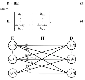
D=HE, (3)
where
H=
h11 · · · h1L
... . .. ...
h(L−1)1 · · · h(L−1)L hL1 · · · hL L
, (4)
Fig. 2 Linear mixture model.
Fig. 3 EEG measurement frameworks. (a) ICA before compression (b) ICA after compression (pro- posed framework) ©2018 IEEE.
is the unknown mixing matrix (Fig. 2). The goal of ICA is to estimateEfrom the givenD. We can calculate the estimated sources by obtaining the unmixing matrixLW, whereLW = H−1. In some EEG studies, ICA is used to remove artifacts [6]–[8]. However, in general, ICA relies on the assumption thatEis not Gaussian becauseLWbecomes unidentifiable[5].
Thus, it is difficult to separate the data, which are compressed using a Gaussian measurement matrix.
3. Proposed Framework
Figure 3 shows the existing framework (ICA before com- pression) and our proposed EEG measurement framework (ICA after compression) [12]. These frameworks are pri- marily composed of two parts: ICA processing part and CS part. Figure 3(a) shows the existing framework in which the desired information can be separated from the measured signals effectively before compression to reduce the amount of data[9]–[11]. However, a power consumption problem occurs if we add digital processing in the sensing unit such as that in ICA. Therefore, we developed a fundamentally new framework to solve this problem (Fig. 3(b)). In this frame- work, additional power-consuming processing components are not required in the sensing unit, owing to the application of ICA in the data processing unit. The details of both units in our proposed framework are given below.
3.1 Sensing Unit in Our Proposed Framework
Figure 4 shows the details of the sensing unit used in L- channel EEG measurements. In actual EEG measurements, artifacts are present in the measured signals. Here, xCh-i andaCh-iindicate the measured pure EEG signals and arti- facts ini-th channel, respectively. The length of each signal is presented as N. Initially, these signals are appropriately processed in an analog circuit block. Subsequently,Lran- dom sampling A/D converters perform undersampling of the signals,xCh-i +aCh-i, to achieveM data fromN data. The
Fig. 4 Sensing unit in our framework. ©2018 IEEE.
measurement matrix is presented at the top of Fig. 4. The random sampling technique introduced in [20] is used to compress the measured EEG signal in the sensing unit; this is to avoid the problem in which ICA is difficult to apply for separating signals following a Gaussian distribution[5].
In actual A/D converters, the input signals are quantized.
Thus, we have to determine the appropriate resolution for A/D converters. However, in this study, resolution was not considered, and we used the A/D converters as samplers to explain the principle of our proposed framework. In each row of theM×Nmeasurement matrix in the A/D converter, only one element is “1” and the others are all “0.” The compressed signals, ˙xCh-i+a˙Ch-i, are transferred to the data processing unit through a wireless transmitter.
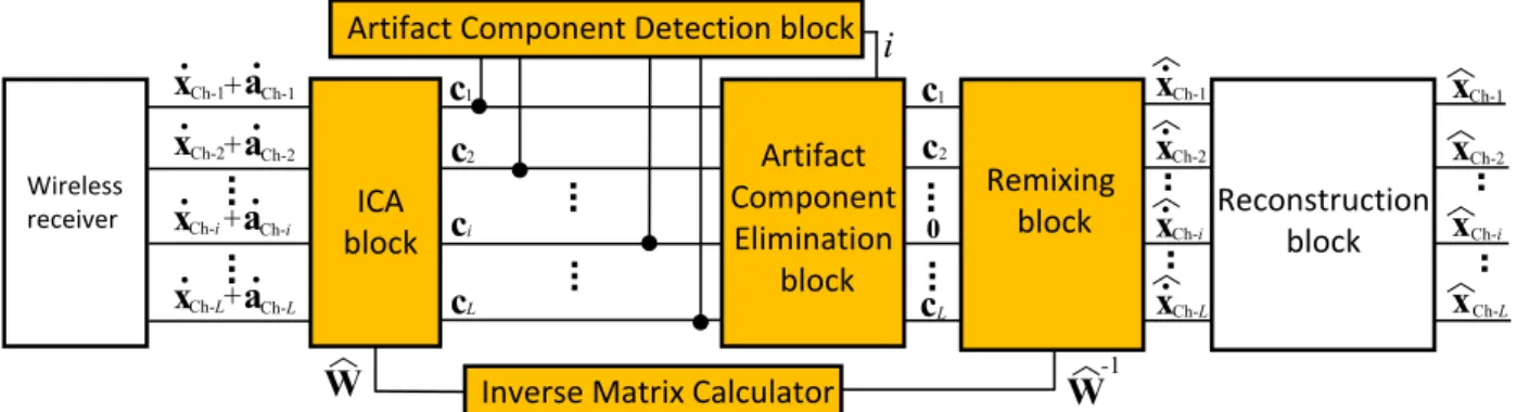
3.2 Data Processing Unit in Our Proposed Framework Figure 5 shows the block diagram of the data processing unit.
The compressed data, ˙xCh-i +a˙Ch-i, are received through a wireless receiver. Subsequently, ICA is performed in the ICA block to generate L components from c1 to cL, and the unmixing matrixLW. Next, the component representing an artifact is revealed in the artifact component detection block. We assumed that thei-th component indicated the artifact. Next, all data of thei-th component are reduced to 0 in the artifact component elimination block. Finally, the estimated EEG signal of each channel fromDxCh-1 toDxCh-L can be achieved using the compressed signals fromDx˙Ch-1to Dx˙Ch-Lin the CS reconstruction block.
4. Evaluation
We evaluated our framework using the measured EEG sig- nals. The measured raw EEG signals, which were recorded at 200 Hz in 16 channels (L=16) at a hospital, were used as the input EEG signals for the measurement frameworks. The time corresponding to one epoch was 3 s. The evaluation was performed using MATLAB software. The model encom- passed the EEG signal acquisition in the sensing unit to data reconstruction in the data processing unit. To focus on the verification of our framework operation, the analog circuit adopted an ideal model in all verification results. Eye-blink artifacts are the most common artifacts that easily interfere with EEG measurements. Therefore, we generated a pseudo- model of eye-blink artifacts to evaluate the extent to which these artifacts were eliminated in our proposed framework.
Several shapes of eye-blink artifact in EEG signals have been reported and the shapes differ by subject[21]. There- fore, we used a simple model of an impulse and triangular shaped eye-blink artifact (e.g.[22]) in this paper. It has also been reported that the period and magnitude of an eye-blink artifact depend on the subject. For example, it was reported that the distortion of the EEG caused by an eye-blink lasts longer than 100 ms[21]. The range is approximately between
50µV and 250µV of the magnitude, as shown in[22]. In
some papers, the received magnitude of the eye-blink arti- facts near the eye electrodes is larger than that at the other
Fig. 6 EEG signals (Original with artifact, only CS, and proposed frame- work). As an example,k=20 was used. ©2018 IEEE.
parts. Further, it is well known that eye-blink artifacts de- crease rapidly with an increase in distance from the eyes (e.g.
[23]).
Based on the above information and our EEG measure- ment experience, a pseudo-model of the eye-blink artifact was created and used for evaluation. The maximum volt- age of the pseudo-model of the eye-blink artifact was set as
150µV in the electrodes FP1 and FP2. The peaks of the
artifacts in F3, F4, F7, and F8 correspond to 75µV. In the other electrodes (C3, C4, P3, P4, T3, T4, T5, T6, O1, and O2), artifacts with a voltage of 15µV were added to the mea- sured EEG signals. In this evaluation, we used the principal component analysis (PCA)–ICA algorithm[24]in the ICA block. OMP was used in the reconstruction block, in which the sparse parameter k was set at 20 as an example. The DCT basis was used asΨ.
Figure 6 shows the examples of the original signal with the pseudo-model of the eye-blink artifacts and the recon- structed signal by using the proposed framework at FP1.
Figure 6 also shows the results obtained using only CS. The compression ratio (CR = N/M) is 4 during the evaluation using only CS and the proposed framework. A pseudo- model artifact was inserted from 0.5 s to 0.65 s in the original
Fig. 7 EEG signals (zoomed-in view of Fig. 6) ©2018 IEEE.
Fig. 8 NMSE with variouskvalues at FP1 and F3.
data. Using our framework, the artifact could be removed effectively when compared with that using only CS. Fig- ure 7 shows the zoomed-in view of Fig. 6 from 1.0 s to 1.8 s.
These results indicate successful reconstruction of the EEG waveform using our proposed framework.
To evaluate the performance of the proposed frame- work, we used two different performance indices. One is the normalized mean square error (NMSE) that normalizes the average of the square of the difference between the estimated and original values. It was calculated as follows:
NMSE= kx−xkˆ 22
kxk22 , (5)
wherexis the original EEG signal and ˆxis the reconstructed EEG signal. k · k2 denotes the Euclidean norm. Another performance index is the correlation coefficient (CC), which measures the similarity between two waveforms. It was calculated as
Fig. 9 CC with variouskvalues at FP1 and F3.
Fig. 10 NMSE with various CRs at FP1.
CC= Sxˆx
SxSxˆ, (6)
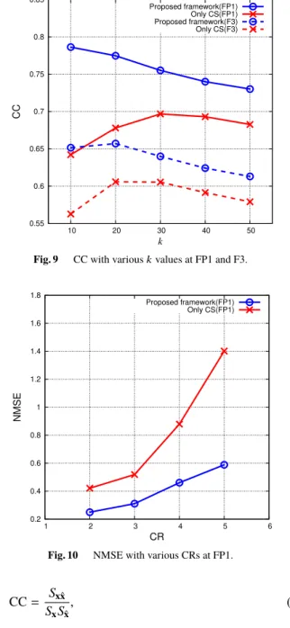
whereSxˆxis the covariance betweenxand ˆx, andSx andSxˆ are the standard deviations ofxand ˆx, respectively. We eval- uated the NMSE and CC at CR=4 with various values ofk ranging from 10 to 50. Figure 8 shows the calculated NMSE values at FP1 and P3. We prepared 10 types of measure- ment matrices in which the positions of “1” were different.
Therefore, we calculated the NMSE values by averaging the results 2,000 times (= 200 epochs × 10 times simulation results with 10 types of measurement matrices). In these results, we ignored the data from 0.5 s to 0.65 s, in which the results obtained using only CS were strongly affected by the artifacts; we did so to perform a fair comparison between the results obtained using the proposed framework and those obtained with only CS. We observed that the NMSE val- ues obtained using the proposed framework were lower than those obtained using only CS for any value ofk. For exam- ple, at k =20 shown in Fig. 8, the NMSE obtained using
5. Discussion
In the discussion presented up to Sect. 4, it was mentioned that the proposed framework is capable of signal reconstruc- tion owing to the suppression of the influence of artifacts, even if the MPU is not installed in the sensing unit. In this section, we discuss the differences in the reconstruction ac- curacy between the two frameworks: ICA after compression (proposed framework) and ICA before compression (e.g.[9]–
[11]). The most important difference in both frameworks is where the ICA is operated. In ICA before compression, the artifact component can be separated from uncompressed measurement EEG signals (i.e., fromxCh-i+aCh-itoxCh-iand aCh-i). This is a typical method that applies ICA to EEG sig- nals, and there are many papers which show artifact separa- tion successfully(e.g.[6]). On the other hand, in the proposed framework, ICA is applied to compressed measurement EEG signals (i.e., ˙xCh-i+a˙Ch-i=Φ(xCh-i+aCh-i)). Therefore, the reconstruction accuracy of the proposed framework depends on whether the ICA can separate compressed measurement EEG signals for independent components correctly. Based on the above discussion, we note that the measurement ma- trix is particularly important.
To separate independent components using ICA, the components obtained by decomposition should be statisti- cally independent and non-Gaussian[5]. When the random undersampling matrix is used as the measurement matrix, the original data are only thinned out, and the independence of the original data is not affected. In addition, as explained in Sect. 3.1, avoiding a Gaussian distribution of independent components can be also realized using random undersam- pling matrix as the measurement matrix, which is different from the Gaussian measurement matrix. From the above, we found that the compressed measurement EEG signals and artifacts can be separated in the proposed framework, theoretically.
Next, the difference between the existing ICA before compression framework, in which MPUs are required in the sensing unit, and the proposed framework was evaluated using CC. The evaluation was performed under the same conditions as described in Sect. 4 (i.e., using OMP as the reconstruction algorithm, PCA–ICA as the ICA algorithm,
Fig. 11 CC evaluation results by using OMP, PCA–ICA at FP1 (proposed framework (ICA after compression), ICA before compression framework, and only CS.
and averaging the results 2,000 times). Figure 11 shows the evaluation results with the proposed framework (ICA af- ter compression), ICA before compression framework, and only CS at FP1. From Fig. 11, it can be seen that up to CR = 3, a reconstruction accuracy of the proposed framework as high as ICA before compression framework can be achieved.
When CR = 4 or more, there are slight differences in the re- construction accuracy; however, it is clear that the proposed framework can recover with a higher accuracy than only CS, although the proposed framework does not need MPUs to operate ICA in the sensing unit. If a higher reconstruction accuracy is desired, then an additional signal processing can be accepted in the data processing unit; for example, apply- ing an additional method for removing artifacts efficiently [25]is also one solution.
6. Conclusion
We developed a new framework for reconstructing EEG sig- nals affected by artifacts. The sensing unit in our framework does not require ICA to suppress the artifacts, and the ICA block can be moved to the data processing unit. Therefore, we can remove the additional digital processing functions in the sensing unit. In this framework, we used the ran- dom undersampling measurement matrix in CS to perform ICA for suppressing the Gaussian. The proposed framework was evaluated on MATLAB using 3-s 16-channel raw EEG signals with a pseudo-model of the eye-blink artifact. In the evaluation, a combination of OMP as the reconstruc- tion algorithm, DCT basis, and the PCA–ICA algorithm was used. The NMSE and CC values were compared to quantita- tively evaluate the effectiveness of our proposed framework.
For example, the NMSE obtained using only CS was ap- proximately 0.88 although that obtained using the proposed framework was below 0.48 with CR=4 at FP1. The evalu- ation results showed that the framework could remove the
interference from artifacts under a high CR without using MPUs in the sensing unit.
Acknowledgments
This work was supported by JSPS KAKENHI Grant Number JP18K18023, Adaptable and Seamless Technology Transfer Program through Target-driven R&D (A-STEP) from Japan Science and Technology Agency (JST), and JKA foundation.
The authors would like to thank the staff at the University of Yamanashi Hospital for their helpful suggestions and tech- nical assistance with EEG data collection.
References
[1] H.R. Mohseni, A. Maghsoudi, and M.B. Shamsollahi, “Seizure de- tection in EEG signals: A comparison of different approaches,” Proc.
IEEE Engineering in Medicine and Biology Society, pp.6724–6727, Sept. 2006.
[2] T. Musha, H. Matsuzaki, Y. Kobayashi, Y. Okamoto, M. Tanaka, and T. Asada, “EEG markers for characterizing anomalous activities of cerebral neurons in NAT (neuronal activity topography) method,”
IEEE Trans. Biomed. Eng., vol.60, no.8, pp.2332–2338, Aug. 2013.
[3] D. Gangopadhyay, E.G. Allstot, A.M.R. Dixon, K. Natarajan, S.
Gupta, and D.J. Allstot, “Compressed sensing analog front-end for bio-sensor applications,” IEEE J. Solid-State Circuits, vol.49, no.2, pp.426–438, Feb. 2014.
[4] X. Zou, L. Feng, and H. Sun, “Robust compressive sensing of multi- channel EEG signals in the presence of impulsive noise,” Information Sciences, vol.429, pp.120–129, March 2018.
[5] A. Hyvärinen, J. Karhumen, and E. Oja, Independent Component Analysis, John Wiley & Sons, 2001.
[6] N.P. Castellanos and V.A. Makarov, “Recovering EEG brain signals:
Artifact suppression with wavelet enhanced independent component analysis,” J. Neurosci. Meth., vol.158, no.2, pp.300–312, Dec. 2006.
[7] S. Makeig, A.J. Bell, T. Jung, and T.J. Sejnowski, “Independent component analysis of electroencephalographic data,” Advances in Neural Information Processing Systems, pp.145–151, Aug. 1996.
[8] S. Çınar, N. Acır, “A novel system for automatic removal of ocular artefacts in EEG by using outlier detection methods and independent component analysis,” Expert Syst. Appl., vol.68, pp.36–44, Feb.
2017.
[9] B. Zhou, X. Wu, Z. Lv, L. Zhang, and C. Zhang, “Independent component analysis combined with compressed sensing for EEG compression in BCI,” Proc. International Conference on Information, Communications and Signal Processing, pp.1–4, Dec. 2015.
[10] B. Mijović, V. Matić, M. De Vos, and S. Van Huffel, “Independent component analysis as a preprocessing step for data compression of neonatal EEG,” Proc. IEEE Engineering in Medicine and Biology Society, pp.1–5, Aug. 2011.
[11] S. Fauvel, A. Agarwal, and R. Ward, “Compressed sensing and energy-aware independent component analysis for compression of EEG signal,” Proc. IEEE International Conference on Acoustics, Speech and Signal Processing, pp.973–977, May 2013.
[12] D. Kanemoto, S. Katsumata, M. Aihara, and M. Ohki, “Framework of applying independent component analysis after compressed sens- ing for electroencephalogram signals,” Proc. 2018 IEEE Biomedical Circuits and Systems Conference, pp.145–148, Oct. 2018.
[13] B.K. Natarajan, “Sparse approximate solutions to linear systems,”
SIAM J. Comput., vol.24, no.2, pp.227–234, April 1995.
[14] D. Donoho, “Compressed sensing,” IEEE Trans. Inf. Theory, vol.52, no.4, pp.1289–1306, April 2006.
[15] J.A. Tropp and A.C. Gilbert, “Signal recovery from random measure- ments via orthogonal matching pursuit,” IEEE Trans. Inf. Theory, vol.53, no.12, pp.4655–4666, Dec. 2007.
[16] Z. Zhang, T. Jung, S. Makeig, and B.D. Rao, “Compressed sensing of EEG for wireless telemonitoring with low energy consumption and inexpensive hardware,” IEEE Trans. Biomed. Eng., vol.60, no.1, pp.221–224, Jan. 2013.
[17] A.M. Abdulghani, A.J. Casson, and E. Rodriguez-Villegas, “Com- pressive sensing scalp EEG signals: Implementations and practical performance,” Med. Biol. Eng. Comput., vol.50, no.11, pp.1137–
1145, Nov. 2012.
[18] M. Mohsina and A. Majumdar, “Gabor based analysis prior formu- lation for EEG signal reconstruction,” Biomedical Signal Processing and Control, vol.8, no.6, pp.951–955, Nov. 2013.
[19] L. Lin, Y. Meng, J.P. Chen, and Z.B. Li, “Multichannel EEG com- pression based on ICA and SPIHT,” Biomedical Signal Processing and Control, vol.20, pp.45–51, April 2015.
[20] J. Laska, S. Kirolos, Y. Massoud, R. Baraniuk, A. Gilbert, M. Iwen, and M. Strauss, “Random sampling for analog-to-information con- version of wideband signals,” Proc. IEEE Dallas/CAS Workshop Design, Applications, Integration and Software, pp.119–122, Oct.
2006.
[21] I. Erkens and G.G. Molina, “Artifact detection and correction in Neurofeedback and BCI applications,” Philips Research, Tech. note TN-PR 2008/00409, 2008.
[22] W.D. Chang, H.S. Cha, K. Kim, and C.H. Im, “Detection of eye blink artifacts from single prefrontal channel electroencephalogram,”
Comput. Meth. Prog. Bio., vol.124, pp.19–30, Feb. 2016.
[23] V. Krishnaveni, S. Jayaraman, S. Aravind, V. Hariharasudhan, and K.
Ramadoss, “Automatic identification and removal of ocular artifacts from EEG using wavelet transform,” Measurement Science Review, vol.6, no.4, pp.45–57, 2006.
[24] K. Zhang and L. Chan, “ICA by PCA approach: Relating higher- order statistics to second-order moments,” Proc. International Con- ference on Independent Component Analysis and Signal Separation, pp.311–318, March 2006.
[25] S. Katsumata, D. Kanemoto, and M. Ohki, “Applying outlier de- tection and independent component analysis for compressed sensing EEG measurement framework,” Proc. 2019 IEEE Biomedical Cir- cuits and Systems Conference, pp.1–4, Oct. 2019.
Daisuke Kanemoto received his B.S. de- gree in Electronics and Information Science from Kyoto Institute of Technology, Kyoto, Japan, in 2004 and M.S. and Ph.D. degrees in Electronic Engineering from Osaka University, Osaka, Japan, in 2006, 2011. From 2010 to 2013, he was an assistant professor in the De- partment of Electronics, Graduate School of In- formation Science and Electrical Engineering, Kyushu University, Fukuoka, Japan. From 2013 to 2019, he was an assistant professor in the De- partment of Research Interdisciplinary Graduate School of Medicine and Engineering, University of Yamanashi, Yamanashi, Japan. From 2015 to 2017, he was a visiting assistant professor in the Department of Electri- cal Engineering, Stanford University, Stanford, USA. From 2019, he has been an associate professor with Graduate School of Engineering Osaka University, Osaka, Japan. His research interests include analog circuits, mixed-signal integrated circuits and compressed sensing. He is an IEEE member.
plinary Research, Graduate School, University of Yamanashi. His research interest is the cog- nitive neuroscience of frontal lobe functions in children and developmental disorders.
Makoto Ohki received his B.E. degree from University of Yamanashi, Kofu, Japan, in 1985, and M.E. and D.E. degrees from Tohoku Uni- versity, Sendai, Japan, in 1987 and 1990, re- spectively, all in electronic engineering. He is currently a professor with University of Yama- nashi, Kofu, Japan. His research interests in- clude multidimensional signal processing and adaptive signal processing.
![Figure 3 shows the existing framework (ICA before com- com-pression) and our proposed EEG measurement framework (ICA after compression) [12]](https://thumb-ap.123doks.com/thumbv2/123deta/5633781.1501446/3.892.461.819.437.790/figure-existing-framework-pression-proposed-measurement-framework-compression.webp)