Effect of Chemotherapy for Luminal A Breast Cancer
Naotaka Uchida,*† Takako Suda† and Kiyosuke Ishiguro†*Clinic of Surgery, Tottori Prefectural Kosei Hospital, Kurayoshi 682-0804, Japan and †Clinical Department of Mammoendocrinol-ogy, Tottori University Hospital, Yonago 683-8504, Japan
ABSTRACT
Background The addition of chemotherapy to en-docrine therapy for luminal A breast cancer generally provides little benefit. However, the least benefit of che-motherapy in all patients with luminal A breast cancer is controversial.
Methods This was a retrospective study of 140 pa-tients with luminal A breast cancer who underwent surgery at Tottori University Hospital between 2001 and 2010. Luminal A breast cancer was defined as positive for estrogen receptors and/or progesterone receptors and negative for human epidermal growth factor 2. Postop-erative endocrine therapy was given to all patients. The prognostic values of age, tumor size, presence of lym-phovascular invasion and lymph node status were evalu-ated. In addition, the prognostic value of chemotherapy for patients with identified risk factors affecting relapse-free survival and overall survival was evaluated.
Results Tumor size greater than 2 cm and positive lymph node status were factors significantly affecting relapse-free survival. There were no factors significantly affecting overall survival. There was no significant dif-ference in the relapse-free survival of patients with tu-mor size greater than 2 cm and/or positive lymph node status who either received chemotherapy or not. How-ever, the relapse event was earlier in patients with tumor size greater than 2 cm and positive lymph node status who did not receive chemotherapy than in those who re-ceived chemotherapy.
Conclusion Chemotherapy could provide little benefit to patients with luminal A breast cancer. However, che-motherapy may bring them longer relapse-free periods. Key words breast cancer; chemotherapy; luminal A Luminal A breast cancers that express estrogen recep-tors (ERs) and/or progesterone receprecep-tors (PRs) and are negative for human epidermal growth factor receptor 2 (HER2) expressions respond well to endocrine therapy and have a generally favorable prognosis. Patients with luminal A breast cancers are not so sensitive to paclitaxel- and doxorubicin-containing preoperative chemotherapy.1 Patients with node-positive luminal A
breast cancer gain little benefit from taxane therapy administered.2, 3 Proceedings from the 12th St. Gallen
Corresponding author: Naotaka Uchida, MD, PhD uchidana@pref.tottori.jp
Received 2013/01/09 Accepted 2013/03/08
Abbreviations: ER, estrogen receptor; HER2, human epidermal growth factor receptor 2; IHC, immunohistochemistry; PR, pro-gesterone receptor
International Breast Cancer Conference held in 2011 showed that the luminal A subtype was fairly unrespon-sive to chemotherapy and said that node positivity per se was not an indication for use of chemotherapy, although a large majority of physicians would use it if more than 3 axillary lymph nodes were involved.4 However, the
least benefit of chemotherapy in all patients with lumi-nal A breast cancer is controversial. The 11th St. Gallen Conference held in 2009 presented the relative indica-tions for using endocrine therapy alone or a combination of chemotherapy and endocrine therapy for luminal A breast cancer.5 The clinicopathological risk factors for
prognosis included lymph node status, tumor size, pres-ence of lymphovascular invasion, histological grade, and proliferative status. Therefore, it is essential to clarify the significance of chemotherapy for patients with these risk factors and luminal A breast cancer. In this report, we analyzed the significance of chemotherapy for patients with luminal A breast cancer who underwent surgery and received postoperative endocrine therapy.
SUBJECTS AND METHODS Patients
A total of 140 patients with initial luminal A primary invasive breast cancers who underwent surgery in our department at Tottori University Hospital between June 2001 and December 2010 were retrospectively investi-gated (Table 1). Men with breast cancer, women with in situ carcinoma, bilateral breast carcinoma or who un-derwent neoadjuvant chemotherapy or chemotherapy for another disease were excluded from the study. Informed consent was obtained from every patient. Patients were treated with breast-conserving surgery or mastectomy. All patients received post-operative endocrine therapy (Table 2). Most patients with breast-conserving surgery received radiotherapy to the whole breast after surgery. There were 24 patients who received chemotherapy (Ta-ble 2). Decisions regarding chemotherapy were made by the treating physician on the basis of patient preference
and risk factors. In principle, the chemotherapy regimen was as follows: FEC (fluorouracil 500 mg/m2, epirubicin
60 mg/m2 and cyclophosphamide 500 mg/m2) alone on
day 1 every 3 weeks for 4 cycles or FEC (fluorouracil 500 mg/m2, epirubicin 60 mg/m2 and cyclophosphamide
500 mg/m2) on day 1 every 3 weeks for 4 cycles
fol-lowed by docetaxel (60 mg/m2) on day 1 every 3 weeks
for 4 cycles or paclitaxel (80 mg/m2) on day 1 and 8
ev-ery 3 weeks for 4 cycles.
Histopathological and immunohistochemical eval-uations
All histopathological and immunohistopathological diagnoses were determined by several pathologists at our facilities or at another laboratory facility within our partnership. Surgical specimens were embedded in par-affin, sectioned and stained with hematoxylin and eosin. Luminal A subtype was defined as ER and/or PR posi-tive and HER2 negaposi-tive. If the Ki-67 labeling index and/
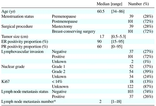
Table 1. Patients characteristics
Median [range] Number (%)
Age (yr) 60.5 [34–86]
Menstruation status Premenopause 39 (28%)
Postmenopause 101 (72%)
Surgical procedure Mastectomy 39 (28%)
Breast-conserving surgery 101 (72%)
Tumor size (cm) 1.7 [0.5–5.3]
ER positivity proportion (%) 90 [15–95]
PR positivity proportion (%) 60 [0–95]
Lymphovascular invasion Negative 37 (27%)
Positive 101 (72%)
Unkown 2 (1%)
Nuclear grade Grade 1 52 (37%)
Grade 2 54 (39%)
Unkown 34 (24%)
Ki67 < 14% 18 (13%)
Unknown 122 (87%)
Lymph node metastasis status Negative 103 (74%)
Positive 37 (26%)
Lymph node metastasis number* 2 [1–18]
* Number in 37 positive cases.
ER, estrogen receptor; PR, progesterone receptor.
Table 2. Adjuvant systemic therapy
Number (%)
Endocrine therapy [n = 140] Tamoxifen ± LH-RH agonist 48 (34%)
Aromatase inhibitor 75 (54%)
Tamoxifen ± LH-RH agonist Aromatase inhibitor 15 (10%)
Aromatase inhibitor Tamoxifen 1 (1%)
Toremifene citrate 1 (1%)
Chemotherapy [n = 24] Anthracyclin 19 (79%)
Anthracyclin + Taxane 5 (21%)
LH-RH, luteinizing hormone-releasing hormone.
or nuclear grade were determined, the luminal A subtype was also defined as low Ki-67 labeling in-dex (< 14%) and/or nuclear grade 1 or 2. ER and PR positivity was confirmed by immunohistochem-istry (IHC); greater than 10% of tumors cells staining positive was considered positive. HER2-negative-status was confirmed by IHC (with 0, 1+, 2+ scores indicat-ing no cells with stainindicat-ing, < 10% of cells with membrane staining, or > 10% of cells with low or me-dium membrane staining, respec-tively). Tumors that were 2+ by IHC were also examined by fluo-rescence in situ hybridization (with an amplification ratio < 2.0 indi-cating negative status). The Ki-67 labeling index was determined by IHC.
Statistics
Relapse-free survival was defined as the period from the date of operation to the date of the first confirma-tion of relapse (i.e., local relapse or metastasis) or death from any cause, whichever came first. Overall survival was defined as the period from the date of operation to the date of death from any cause. Relapse-free and overall survival periods were estimated with the Kaplan-Meier method and compared with the log-rank test and Wilcoxon test. The Mann-Whitney U test was used for comparisons of continuous outcomes, while the chi-square test, for comparisons of categorical variables. Risk factors affecting prognosis were estimated with univariate and multivariate analyses. Cox’s proportional-hazards model was applied in multivarate analysis. Dif-ferences were considered significant when the P value was < 0.05.
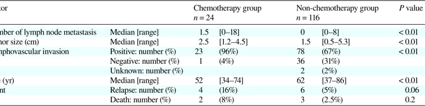
Table 3. Comparion between chemotherapy and non-chemotherapy groups
Factor Chemotherapy group Non-chemotherapy group P value
n = 24 n = 116
Number of lymph node metastasis Median [range] 1.5 [0–18] 0 [0–8] < 0.01
Tumor size (cm) Median [range] 2.5 [1.2–4.5] 1.5 [0.5–5.3] < 0.01
Lymphovascular invasion Positive: number (%) 23 (96%) 78 (67%) < 0.01
Negative: number (%) 1 (4%) 36 (31%)
Unknown: number (%) 2 (2%)
Age (yr) Median [range] 52 [34–74] 62 [37–86] < 0.01
Event Relapse: number (%) 4 (16%) 6 (5%) 0.06
Death: number (%) 2 (8%) 3 (2.5%) 0.2
RESULTS
Patient characteristics
Table 1 shows the characteristics of 140 luminal A breast cancer patients. The median patient age was 60.5 years (range: 34–86 years). Menstruation status of 39 (28%) patients was premenopausal and that of 101 (72%) patients was postmenopausal. There were 101 (72%) patients receiving breast-conserving surgery and 39 (28%) patients receiving mastectomy. The median tumor size was 1.7 cm (range: 0.5–5.3 cm), and 103 patients (74%) were free of axillary lymph node metastasis. In 37 patients with axillary lymph node metastasis, the median number of involved lymph nodes was 2 (range: 1–18). Median ER and PR positivity proportion of tu-mor cells in each tutu-mor were 90% (range: 15–95%) and 60% (range: 0–95%) respectively. There were 101 (72%) patients positive for lymphovascular invasion. There were 52 (37%) and 54 (39%) patients with nuclear-grade tumors of 1 and 2, respectively. The median follow-up period was 40 months (range: 3–113 months).
Adjuvant systemic therapy
Table 2 lists the post-operative systemic therapies. All patients received some type of endocrine therapy. A total of 48 (34%) patients received tamoxifen and/or a lutein-izing hormone-releasing hormone agonist, and 75 (54%) patients received an aromatase inhibitor. There were 15 (10%) patients who changed from tamoxifen and/or lu-teinizing hormone-releasing hormone agonist to the aro-matase inhibitor for the following reasons: change from
pre- to post-menopausal in 9 patients, patient preference to continue endocrine therapy after 5 years of treatment with 1 type of therapy in 3, thrombosis in 1, uterine en-dometrial hypertrophy in 1, and uterine cancer in 1 pa-tients. One patient changed from the aromatase inhibitor to tamoxifen because of joint pain.
A total of 24 patients received chemotherapy. Che-motherapy was performed more frequently to patients with metastases involving several lymph nodes, large tumor size, positive lymphvascular invasion and younger age (Table 3). Relapse events were 4 (16%) in the che-motherapy group and 6 (5%) in the non-cheche-motherapy group, and intergroup difference was not significant (P = 0.06) (Table 3). Death events, 2 (8%) in the chemothera-py group and 3 (2.5%) in the non-chemotherachemothera-py group, showed no significance (P = 0.20) (Table 3). Of 24 chemotherapy-based patients, 19 (79%) patients received anthracycline alone, and the remaining 5 (21%) received anthracycline and taxane (Table 2). Chemotherapy that included taxane was adopted more frequently in patients with metastases involving several lymph nodes: median number of involved nodes (range) was 1 (0–13) when the agent was anthracycline, and 11 (3–18) when taxane was coadministered.
Risk factors associated with relapse and death Relapse or death risk factors were investigated by uni-variate analysis among positive lymph node status, tu-mor size > 2 cm, positive lymphovascular invasion and age < 40 years old. As the result, positive lymph node status and tumor size > 2 cm were significant relapse
Table 4. Relapse-free survival and overall survival in 140 patients with luminal A breast cancer (univariate analysis) Relapse-free survival Overall survival
Univariate analysis Univariate analysis
Hazard 95% Confi- P Hazard 95% Confi- P ratio dence interval value ratio dence interval value Lymph node status (positive versus negative) 8.08 2.02–32.2 < 0.01 4.68 0.66–32.7 0.11 Tumor size (> 2 cm versus ≤ 2 cm) 3.68 1.01–13.3 0.04 1.73 0.26–11.3 0.56 Lymphovascular invasion (positive versus negative) 1.98 0.43–9.04 0.37 1.57 0.21–11.5 0.65
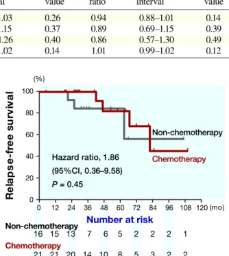
Fig. 3. Relapse-free survival in patients with tumor sizes > 2 cm and axillary lymph node metastasis. There is no difference be-tween groups undergoing chemotherapy or not. But chemotherapy brings a longer relapse-free period.
Fig. 1. Relapse-free survival in patients with tumor sizes > 2 cm. There is no difference between groups undergoing chemotherapy or not.
Fig. 2. Relapse-free survival in patients with axillary lymph node metastasis. There is no difference between groups undergoing chemotherapy or not.
risk factors (Table 4). Meanwhile, there were no signifi-cant death risk factors (Table 4). In multivariate analysis, there were no significant relapse and death risk factors (Table 5).
Relapse-free survival
The relapse-free survival rates of patients with significant risk factors were estimated for patients with or without chemotherapy. In 44 patients with tumor size > 2 cm, 16 patients received chemotherapy. The 5-year relapse-free survival rate of the patients undergoing chemotherapy was estimated at 78.7%, whereas for those not undergo-ing chemotherapy it was estimated at 86.6%. The differ-ence was not significant (P = 0.40) (Fig. 1).
In 37 patients with positive lymph node status, 21 patients received chemotherapy. The 5-year relapse-free survival rate of the patients undergoing chemotherapy was estimated at 82.5%, whereas for those not undergo-ing chemotherapy it was estimated at 83.3%. The differ-ence was also not significant (P = 0.45) (Fig. 2).
In 20 patients with positive lymph node status and tumor size > 2 cm, 14 patients received chemotherapy. The 5-year relapse-free survival rate of the patients un-dergoing chemotherapy was estimated at 78.7%, whereas
for those not undergoing chemotherapy it was estimated at 33.3%. The difference were also not significant on log-rank analysis (P = 0.21), but significant on Wilcoxon’s analysis (P = 0.02) (Fig. 3). These suggest earlier relapse
Table 5. Relapse-free survival and overall survival in 140 patients with luminal A breast cancer (multivariate anal-ysis)
Relapse-free survival Overall survival Multivariate analysis Multivariate analysis
Hazard 95% Confidence P Hazard 95% Confidence P
ratio interval value ratio interval value
Lymph node status (/1 involved node) 0.95 0.88–1.03 0.26 0.94 0.88–1.01 0.14
Tumor size (/1 cm) 0.88 0.68–1.15 0.37 0.89 0.69–1.15 0.39
Lymphovascular invasion (positive versus negative) 0.84 0.56–1.26 0.40 0.86 0.57–1.30 0.49
were more frequent in non-chemotherapy group than chemotherapy group.
DISCUSSION
Using complementary DNA microarrays representing 8102 human genes, Perou et al. defined 4 breast cancer subtypes (luminal/ER, HER2-overexpressing, basal-like and normal), based on gene expression patterns.6 Carley
et al. subsequently classified 5 subtypes based on immu-nohistochemical staining of various markers as follows: luminal A (ER+ and/or PR+, HER2–), luminal B (ER+ and/or PR+, HER2+), HER2 (ER–, PR–, HER2+), basal-like (ER–, PR–, HER2–, CK5/6+ and/or HER1+) and an unclassified type, and also demonstrated the applicabil-ity of this system to past classification systems based on gene microarray analysis.7 The clinical application of
gene expression profiling is limited because the proce-dure is technically difficult and expensive. Therefore, the consensus of the 12th St. Gallen Conference defined luminal A breast cancer as ER+ and/or PR+, HER2– tumors with a Ki-67 labeling index of < 14%.4 However,
there was a lack of complete consensus on the threshold indication for administering chemotherapy to patients with luminal A breast cancer.4 It has been reported that
Oncotype DX recurrence scores were high in 13% of pa-tients with luminal A breast cancer.8 Among
tamoxifen-treated patients, patients with a low or intermediate recurrence score on the 21-gene assay benefited from tamoxifen, whereas patients with a high recurrence score did not benefit.9 However, the 21-gene assay has
also demonstrated that chemotherapy provided a large benefit when the recurrence score was high.10 These
re-sults suggest that chemotherapy is beneficial for patients with potentially malignant luminal A breast cancer.
The value of chemotherapy for patients with large luminal A breast tumors is unclear. The 21-gene recur-rence score assay has shown that there is a significant as-sociation between increased tumor size and high recur-rence score.10 Tumor size was prognostic for local and
distant relapse.11, 12 These findings suggest that the larger
the tumor the greater the association of gene expression with malignant potential and that chemotherapy may lead to a better prognosis. However, in our study, chemo-therapy had no beneficial effects on relapse in patients with tumors larger than 2 cm.
The value of chemotherapy for patients with lymph node involvement of luminal A breast cancer is also not clear. Generally speaking, the number of axillary lymph node metastases is the most reliable prognostic factor and an important indication for adjuvant therapy. The addition of a paclitaxel protocol after the completion
of anthracycline treatment has improved the disease-free and OS of patients with node-positive early breast cancer.13, 14 However, patients with luminal A breast
cancer benefit less from treatment with a taxane when administered after anthracycline, even if lymph nodes are involved.2, 3 In our study, chemotherapy had no
ben-eficial effects on relapse in patients with lymph node in-volvement. The effects on relapse in patients with large tumor size and lymph node involvement were also not beneficial. Multivariate analysis revealed that endocrine therapy alone sufficiently removed relapse and death risk from patients with luminal A breast cancer.
The following reasons could explain why, in our study, patients with larger tumors and lymph node in-volvement did not benefit from chemotherapy. First, mi-croarray analysis has shown that nodal status and tumor size cannot be correlated with biologically distinct dis-eases.15 These findings suggest that biological
character-istics, not node positivity, are indications for the use of chemotherapy.4 Therefore, in our study, luminal A breast
cancer in patients with larger tumors and lymph node involvement may exhibit lower malignant potential. Sec-ond, the chemotherapy regimen used in this study was at the lower limit of standard regimens in regard to dosages (epirubicin: 60–100 mg/m2, docetaxel: 60–100 mg/m2,
paclitaxel: 80–100 mg/m2). A less-efficacious regimen
might be the reason that we did not detect a significant effect in the chemotherapy group. Third, the size of this study is a limitation. Our study is retrospective, and the chemotherapy group contained 24 patients. We cannot draw a conclusion using such a small dataset. Fourth, unknown cases regarding nuclear grade and/or Ki-67 could affect the results. In our study, nuclear grade in 34 patients was unknown because the grading is gener-ally used in patients with invasive ductal carcinoma and there is no consensus to decide nuclear grade in patients with special types of breast cancer. Ki-67 in 122 patients was unknown because it was introduced in May 2009, and the confidence for specimens embedded in paraf-fin and preserved for long time is not established. Ki-67 and/or nuclear grade are important factors to distinguish luminal A and luminal B (HER2 negative type) in the 12th St. Gallen International Breast Cancer Conference held in 2011. Among them, the cases belonging to lu-minal B (HER2 negative type) could be included and it may influence the result in patients with large tumor size and lymph node involvement.
In conclusion, chemotherapy could provide little benefit to patients with luminal A breast cancer, even those with high relapse risk factors including large tumor size and many positive nodes. However, chemotherapy may bring patients longer relapse-free periods.
Prospec-tive studies and additional subject recruitment are neces-sary to draw definitive conclusions.
The authors declare no conflict of interest. REFERENCES
1 Rouzier R, Perou CM, Symmans WF, Ibrahim N, Cristofanilli M, Anderson K, et al. Breast cancer molecular subtypes re-spond differently to preoperative chemotherapy. Clin Cancer Res. 2005;11:5678-85. PMID: 16115903.
2 Hayes DF, Thor AD, Dressler LG, Weaver D, Edgerton S, Cowan D, et al. Cancer and Leukemia Group B (CALGB) In-vestigators. HER2 and response to paclitaxel in node-positive breast cancer. N Engl J Med. 2007;357:1496-506. PMID: 17928597.
3 Hugh J, Hanson J, Cheang MC, Nielsen TO, Perou CM, Dumontet C, et al. Breast cancer subtypes and response to docetaxel in node-positive breast cancer: use of an immuno-histochemical definition in the BCIRG 001 trial. J Clin Oncol. 2009;27:1168-76. PMID: 19204205.
4 Goldhirsch A, Wood WC, Coates AS, Gelber RD, Thürlimann B, Senn HJ. Panel members. Strategies for subtypes--dealing with the diversity of breast cancer: highlights of the St. Gal-len International Expert Consensus on the Primary Therapy of Early Breast Cancer 2011. Ann Oncol. 2011;22:1736-47. PMID: 21709140.
5 Goldhirsch A, Ingle JN, Gelber RD, Coates AS, Thürlimann B, Senn HJ. Panel members; Thresholds for therapies: high-lights of the St Gallen International Expert Consensus on the primary therapy of early breast cancer 2009. Ann Oncol. 2009;20:1319-29. PMID: 19535820.
6 Perou CM, Sørlie T, Eisen MB, van de Rijn M, Jeffrey SS, Rees CA, et al. Molecular portraits of human breast tumours. Nature. 2000;406:747-52. PMID: 10963602.
7 Carey LA, Perou CM, Livasy CA, Dressler LG, Cowan D, Conway K, et al. Race, breast cancer subtypes, and survival
in the Carolina Breast Cancer Study. JAMA. 2006;295:2492-502. PMID: 16757721.
8 Ademuyiwa FO, Thorat MA, Jain RK, Nakshatri H, Badve S. Expression of Forkhead-box protein A1, a marker of luminal A type breast cancer, parallels low Oncotype DX 21-gene re-lapse scores. Mod Pathol. 2010;23:270-5. PMID: 19946260. 9 Sparano JA, Paik S. Development of the 21-gene assay and its
application in clinical practice and clinical trials. J Clin Oncol. 2008;26:721-8. PMID: 18258979.
10 Paik S, Tang G, Shak S, Kim C, Baker J, Kim W, et al. Gene expression and benefit of chemotherapy in women with node-negative, estrogen receptor-positive breast cancer. J Clin On-col. 2006;24:3726-34. PMID: 16720680.
11 Sanpaolo P, Barbieri V, Genovesi D. Prognostic value of breast cancer subtypes on breast cancer specific survival, distant metastases and local relapse rates in conservatively managed early stage breast cancer: a retrospective clinical study. Eur J Surg Oncol. 2011;37:876-82. PMID: 21824742.
12 Paik S, Shak S, Tang G, Kim C, Baker J, Cronin M, et al. A multigene assay to predict relapse of tamoxifen-treated, node-negative breast cancer. N Engl J Med. 2004;351:2817-26. PMID: 15591335.
13 Mamounas EP, Bryant J, Lembersky B, Fehrenbacher L, Sedlacek SM, Fisher B, et al. Paclitaxel after doxorubicin plus cyclophosphamide as adjuvant chemotherapy for node-pos-itive breast cancer: results from NSABP B-28. J Clin Oncol. 2005;23:3686-96. PMID: 15897552.
14 Henderson IC, Berry DA, Demetri GD, Cirrincione CT, Goldstein LJ, Martino S, et al. Improved outcomes from adding sequential Paclitaxel but not from escalating Doxoru-bicin dose in an adjuvant chemotherapy regimen for patients with node-positive primary breast cancer. J Clin Oncol. 2003;21:976-83. PMID: 12637460.
15 Sotiriou C, Neo SY, McShane LM, Korn EL, Long PM, Jazaeri A, et al. Breast cancer classification and prognosis based on gene expression profiles from a population-based study. Proc Natl Acad Sci U S A. 2003;100:10393-8. PMID: 12917485.