Yonago Acta Medica 2018;61:008–018 Original Article
Corresponding author: Yasuyuki Hasegawa, MD, PhD hasechi@med.tottori-u.ac.jp
Received 2017 November 27 Accepted 2017 December 21
Abbreviations: BLT1, leukotriene B4 receptor subtype 1; COX, cy-clooxygenase; EPA, eicosapentaenoic acid; ELISA, enzyme-linked immunosorbent assay; DHA, docosahexaenoic acid; FBS, fetal bo-vine serum; IL, interleukin; mPGES-1, microsomal prostaglandin E synthase-1; MMP, matrix metalloproteinase; NFATc1, nuclear factor of activated T cells c1; OPG, osteoprotegerin; PBS, phosphate-buff-ered saline; PGE2, prostaglandin E2; PUFA, polyunsaturated fatty acid; RANKL, receptor activator of nuclear factor-κB ligand; RvE1, resolvin E1; TNF, tumor necrosis factor; TRAP, tartrate-resistant acid phosphatase
Resolvin E1 Inhibits Osteoclastogenesis and Bone Resorption by Suppressing
IL-17-induced RANKL Expression in Osteoblasts and RANKL-induced Osteoclast
Differentiation
Yoshihiro Funaki, Yasuyuki Hasegawa, Ryota Okazaki, Akira Yamasaki, Yuriko Sueda, Akihiro Yamamoto, Masaaki Yanai, Takehito Fukushima, Tomoya Harada, Haruhiko Makino and Eiji Shimizu
Division of Medical Oncology and Molecular Respirology, Department of Multidisciplinary Internal Medicine, School of Medicine, Tottori University Faculty of Medicine, Yonago 683-8504, Japan
ABSTRACT
Background Resolvin E1 (RvE1) derived from the ω-3 polyunsaturated fatty acid eicosapentaenoic acid is known to be a potent pro-resolving lipid mediator that prevents chronic inflammation and osteoclastogenesis. We investigated the inhibitory effects of RvE1 on osteo-clastogenesis and bone resorption to clarify its therapeu-tic potential for rheumatoid arthritis (RA).
Methods Receptor activator of nuclear factor-κB li-gand (RANKL)-induced osteoclast differentiation was assessed with tartrate-resistant acid phosphatase stain-ing. RANKL-induced bone resorption was assessed by the measurement of pit formation using calcium phosphate-labeled fluorescent polyanionic molecules in RAW264.7 cells as osteoclast precursors. The effects of RvE1 on the RANKL-induced mRNA expression of os-teoclast-specific genes and transcriptional factors such as c-fos and nuclear factor of activated T cells c1 (NFATc1) in RAW264.7 cells were measured by quantitative re-al-time PCR. The distribution of NFATc1 induced by RANKL was evaluated by immunofluorescence stain-ing in RAW264.7 cells. To analyze the mechanism of the inhibitory effect of RvE1 on osteoclastogenesis, we measured IL-17-induced RANKL mRNA expression in MC3T3-E1 osteoblast cells treated with RvE1 using quantitative real-time PCR and determined the level of
prostaglandin E2 (PGE2) production by enzyme-linked
immunosorbent assay.
Results RvE1 significantly suppressed
RANKL-in-duced osteoclast differentiation and bone resorption. RvE1 inhibited the RANKL-induced mRNA expression of osteoclast-specific genes along with the transcription factors NFATc1 and c-fos. Moreover, NFATc1 translo-cation from the cytoplasm to the nucleus of RAW264.7 cells was suppressed following RvE1 treatment. RvE1 also inhibited IL-17-induced RANKL mRNA
expres-sion and PGE2 production in MC3T3-E1 cells.
Conclusion RvE1 inhibited osteoclastogenesis and bone resorption by suppressing RANKL-induced NFATc1 and c-fos expression in osteoclasts and IL-17-induced RANKL expression through the autocrine
ac-tion of PGE2 in osteoblasts. Our data suggest RvE1 as a
new therapeutic target of RA.
Key words interleukin-17; osteoblasts; osteoclasts; RANK ligand; resolvin E1
The bone undergoes continuous remodeling to achieve a balance between bone formation and resorption medi-ated by osteoblasts and osteoclasts, respectively. Rheu-matoid arthritis (RA) is a chronic inflammatory auto-immune disease characterized by progressive synovial inflammation and destruction of the joint cartilage and
bone.1 In particular, RA involves a breakdown of
met-abolic balance in the bone, and is characterized by an increase in bone resorption that results in impaired bone
formation.2 This imbalance is caused by the increase
of various inflammatory cytokines, including receptor
activator of nuclear factor-κB ligand (RANKL) and
its competitive inhibitor osteoprotegerin (OPG), in the
inflammatory tissue.3, 4 Thus, anti-inflammatory drugs
are most commonly prescribed to treat the symptoms of RA. However, recent evidence points to the beneficial effects of natural compounds on reducing inflammatory cascades, providing new options for RA treatment.
For example, a diet enriched in ω-3 polyunsaturated
fatty acids (PUFAs) was found to reduce joint stiffness
in the morning and the number of tender joints,5 and
also decreased the levels of inflammatory cytokines in
RvE1 inhibits osteoclastogenesis and bone resorption
interleukin (IL)-1β, and IL-6.6, 7 Indeed, several
random-ized clinical studies have revealed that dietary
supple-mentation with ω-3 PUFAs is efficacious in reducing
joint pain, the duration of morning stiffness, the number of tender or swollen joints and non-steroidal
anti-in-flammatory drug usage in RA patients.8, 9 However, the
mechanisms of these clinical effects of ω-3 PUFAs have
remained unclear. Recent discoveries demonstrate that ω-3 PUFAs such as eicosapentaenoic acid (EPA) and docosahexaenoic acid (DHA) can be enzymatically con-verted in vivo to novel bioactive lipid mediators termed specialized pro-resolving mediators, including resolvins, protectins and maresins, which promote the resolution of inflammation and have more potent effects than their lipid precursors.10
One of the specialized pro-resolving mediators derived from EPA, resolvin E1 (5S, 12R, 18R-trihy-droxy-6Z, 8E, 10E, 14Z, 16E-eicosapentaenoic acid; RvE1), was originally identified in exudates of the
mu-rine dorsal air pouch, an acute inflammation model.11
Recent findings demonstrated that RvE1 had protective effects in periodontal disease,12 peritonitis,13 asthmatic
airway inflammation,14 bacterial pneumonia and acute
lung injury in vivo.15 Accordingly, we speculated that the
same bone-protective effects noted in periodontal dis-ease might be applicable to RA. There have been only a few reports on the regulation of RvE1 for bone
resorp-tion and osteoclast differentiaresorp-tion,16 and the effects and
mechanism of RvE1 on bone remodeling are still not fully understood. Therefore, in this study, we originally investigated the effects of RvE1 on the inflammation and signaling pathways in osteoclasts and osteoblasts to provide a foundation for RvE1 as a new therapeutic ap-proach in RA treatment. Our results are the first to indi-cate that RvE1 interacts with osteoclasts and osteoblasts via inhibition of RANKL production in osteoblasts.
RANKL is a member of the TNF superfamily of cy-tokines and is known to induce osteoclastogenesis from monocytes or macrophages. The binding of RANKL to its receptor RANK, expressed on osteoclast precursors, induces the expression and/or activation of transcription factors, including nuclear factor of activated T cells c1 (NFATc1) and c-fos, which have been shown to be es-sential for osteoclast differentiation.17, 18
Pro-inflammatory molecules play a significant, but primarily indirect, role in osteoclast regulation as they
act through modulating RANKL and OPG.19 TNFα,
IL-1, IL-6, IL-11 and IL-17 act on osteoblasts and bone marrow stromal cells to increase RANKL and/
or decrease OPG expression in osteoblasts.20, 21 IL-17 is
a pro-inflammatory cytokine induced by a subset of T
helper 17 cells.3, 22 In osteoblasts, RANKL is induced
by prostaglandin E2 (PGE2), which is in turn strongly
induced by IL-17; however, this PGE2-induced
upregula-tion of RANKL expression could be inhibited by NS398,
a selective cyclooxygenase-2 (COX-2) inhibitor.3, 23 Thus,
IL-17 indirectly induces osteoclastogenesis via PGE2
-in-duced RANKL expression in osteoblasts and has been implicated in the promotion of the pathogenesis of RA. Based on this background, in this study, we focused on the potential effects of RvE1 on mediating IL-17-induced osteoclastogenesis to alleviate the inflammation associated with RA. We further examined the potential mechanism underlying the effects of RANKL, IL-17 and/or RvE1 in RAW264.7 and mouse MC3T3-E1 cells as osteoclasts and osteoblasts, respectively. We exam-ined the effect on osteoclastogenesis by determining the expression of matrix metalloproteinase-9 (MMP-9) and cathepsin K, which are highly expressed in osteoclastic cells and considered as markers of mature osteoclasts
that play important roles in osteolysis.24 PGE2
produc-tion tends to be induced by endogenous COX-2 and microsomal PGE synthase-1 (mPGES-1). Therefore, the COX-2 and mPGES-1 mRNA expression levels were examined with real-time PCR, and the production
level of PGE2 was examined with an enzyme-linked
immunosorbent assay (ELISA) in MC3T3-E1 cells to understand the mechanism of the inhibitory effects of RvE1 on osteoclastogenesis. Osteoblasts are involved in osteoclast regulation by expressing RANKL on their
membranes or releasing it as a soluble factor.2 We
there-fore further verified the mRNA expression of RANKL and its competitive inhibitor OPG in MC3T3-E1 cells by real-time PCR. Together, these in vitro results could pro-vide a foundation for the clinical application of RvE1 in maintaining the balance of bone metabolism and in the treatment or prevention of RA.
MATERIALS AND METHODS Cell culture and reagents
RAW264.7 cells were purchased from American Type Culture Collection (Manassas, VA) and were used as osteoclastic cells. Mouse calvarial cells (MC3T3-E1) were purchased from the RIKEN BioResource Center (Tsukuba, Japan) and were used as osteoblastic cells. RAW264.7 cells were cultured in Dulbecco’s minimal essential medium (D-MEM; Wako Pure Chemicals, Osaka, Japan) containing 10% fetal bovine serum (FBS), 100 µg/mL penicillin and 100 µg/mL
streptomy-cin. MC3T3-E1 cells were cultured in α-MEM (Wako
Pure Chemicals) containing 10% FBS, 100 µg/mL penicillin and 100 µg/mL streptomycin. Both cell lines
were cultured at 37 °C in a humidified 5% CO2
Y. Funaki et al.
soluble RANKL (Oriental Yeast, Tokyo, Japan) and RvE1 (Toronto Research Chemicals, Toronto, Canada). MC3T3-E1 cells were cultured with or without IL-17 (Pepro Tech, Rocky Hill, NJ) and RvE1.
Cell proliferation assay
Cell viability was assessed using Cell Counting Kit-8 (CCK-8; Dojindo, Kumamoto, Japan). RAW264.7 and MC3T3-E1 cells were seeded in 96-well plates at 5.0 ×
103 cells/well in D-MEM or MEM-α each containing
10% FBS. Following incubation for 24 h, the cells were treated with 0, 50, 100 or 200 nM RvE1. After 1 h, the cells were treated with 100 ng/mL RANKL (RAW 264.7 cells) or 50 ng/mL IL-17 (MC3T3-E1 cells). After 24 h, 10 µL CCK-8 solution was added to the culture and the cells were further incubated in the dark at 37 °C for 2 h. The plate was then read using a Sunrise micro-plate analyzer (Tecan, Mannedorf, Switzerland) at 450 nm with a reference at 600 nm. The number of surviving cells was quantifi ed by measuring the absorbance at this wavelength.
Tartrate-resistant acid phosphatase (TRAP) stain-ing
RAW264.7 cells were plated in 24-well microplates at a density of 1.0 × 10⁴ cells/mL and left overnight to settle. Conditioned medium containing 50 or 100 ng/mL sol-uble RANKL was then added to the cells and cultured for 6 days. On day 7 of culture, the cells were fi xed and stained using a TRAP staining kit (Wako Pure Chemi-cals) according to the manufacturer’s instructions. The number of osteoclast-like cells per well was then count-ed (TRAP-positive cells with more than three nuclei). Each experiment was performed in duplicate.
Quantitative real-time PCR
Total RNA was isolated from cultured cells using the RNeasy Plus mini kit (Qiagen, Valencia, CA) according to the manufacturer’s instructions. The mRNA was reverse-transcribed into cDNA using the Super Script VILO Master Mix (Invitrogen, Carlsbad, CA) and the resultant cDNA was subjected to real-time PCR using the TaqMan Fast Advanced Master Mix (Applied Biosystems, Carlsbad, CA). Specific primers (Table 1) were purchased from Applied Biosystems (Foster City, CA). PCR was performed using the TaKa-Ra PCR Thermal Cycler Dice system (Takara Bio, Kusatsu, Japan) under the following conditions: initial holding at 25 °C for 10 min and then 42.0 °C for 60 min and fi nally 85.0 °C for 5 min. Real-time PCR was performed on a ViiA7 Real-Time PCR system (Applied Biosystems) for 40 cycles of 95 °C for 1 s and 60 °C for 20 s. The ex-pression levels of NFATc1, c-fos, cathepsin K, MMP-9, RANKL, OPG, COX-2 and mPGES-1 were normalized to that of glyceraldehyde 3-phosphate dehydrogenase (GAPDH). All real-time PCR experiments were per-formed in triplicated and analyzed by the comparative 2-∆∆Ct relative quantifi cation meth od.
Immunofl uorescence staining
RAW264.7 cells were cultured in glass chamber slides (Lab-Tek II Chamber Slide w/Cover RS Glass Slide Sterile; Nalge Nunc) for 24 h with or without RANKL plus RvE1. The glass chamber slides were then removed, washed in phosphate-buffered saline (PBS), fi xed with 4% paraformaldehyde for 20 min, permeabilized with 0.5% TritonX-100 for 1 h, incubated with bovine serum albumin for 1 h to block non-specifi c binding and then incubated with mouse NFATc1 monoclonal antibody (di-luted 1:50; Santa Cruz Biotechnology, Dallas, TX) over-night at 4 °C. The cells were washed again in PBS and incubated with Alexa Fluor 488 goat anti-mouse second-ary antibody (diluted l:2000; Life Technologies, Carls-bad, CA) for 1 h. The nuclei of cells were stained using the blue fluorescent dye 4’,6-diamidino-2-phenylindole in Vectashield mounting medium (Vector Laboratories, Burlingame, CA). Images were obtained with a confo-cal microscope (TCS-SP2 confoconfo-cal microscope; Leica, Wetzlar, Germany).
Bone resorption assay
The bone resorption assay was performed using Bone Table 1. Primers for real-time PCR
Genes Assay IDa RefSeq Exon
Boundary Product length (bp)
NFATc1 Mm00479445_m1 NM_00116409.1 7-8 75 c-fos Mm00487425_m1 NM_010234.2 1-2 59 Cathepsin K Mm00484039_m1 NM_007802.4 7-8 73 MMP-9 Mm00442991_m1 NM_013599.3 5-6 80 RANKL Mm00441906_m1 NM_011613.3 2-3 66 OPG Mm00435454_m1 NM_008764.3 4-5 71 COX-2 Mm00478374_m1 NM_011198.3 5-6 80 mPGES-1 Mm00452105_m1 NM_022415.3 2-3 87 GAPDH Mm99999915_m1 NM_001289726.1 2-3 107
COX-2, Cyclooxygenase-2; GAPDH, glyceraldehyde-3-phosphate dehydrogenase; MMP-9, matrix metalloproteinase-9; mPGES-1, microsomal prostaglandin E synthe-tase-1; NFATc1, nuclear factor of activated T cells c1; OPG, osteoprotegerin; RANKL, receptor activator of nuclear factor κB ligand. a: TaqMan Gene Expression Assay (Applied Biosystems)
RvE1 inhibits osteoclastogenesis and bone resorption
Resorption Assay Kit 24 (PG Research, Kodaira,
Ja-pan).25 First, 0.5 mL fluorescein amine-labeled
chon-droitin polysulfate (Bone Resorption Assay FACS) was added to each well of a calcium phosphate-coated 24-well plate (Bone Resorption Assay 24) and incubated at
37 °C in a humidifi ed 5% CO2 atmosphere for 2 h under
a light-shielded condition. After the incubation, each well of the 24-well plate was washed with 1 mL PBS
twice and then 1 mL MEM-α without phenol red (Wako
Pure Chemicals) containing 10% FBS was added. The RAW264.7 cells were inoculated into each well at a
density of 1.0 × 10⁴ cells/mL and allowed to attach for 4
h befo re being treated with 50 or 100 nM RvE1 for 1 h, followed by incubation with 10–200 ng/mL RANKL for 6 days without a medium change. RvE1 and RANKL were added at the same dose after 3 days. After 6 days, 100 µL of the culture supernatant from each well was harvested into a 96-well plate and mixed with 50 µL of 0.1 N NaOH (Bone Resorption Assay Buffer). The fl
u-orescence intensity of the culture supernatant was mea-sured using a fluorescence plate reader (Infinite F500, Tecan, Mannedorf, Switzerland) with an excitation wavelength of 485 nm and emission wavelength of 535 nm. The remaining plates were washed with PBS and treated with 5% sodium hypochlorite for 5 min. After washing the plates with water and drying them, the pit area was photographed by a fluorescence microscope (BZ-8100, Keyence, Osaka, Japan).
ELISA
The amount of PGE2 in the culture medium was
deter-mined using a commercially available ELISA kit (Enzo Life sciences, Farmingdale, NY) according to the man-ufacturer’s instructions, and the data were converted to pg/mL. Finally, duplicate assays were performed on each sample, and the absorbance at 405 nm was record-ed.
Control
RANKL 50 ng/mL
RANKL 100 ng/mL
RvE1 (–)
RvE1 100 nM
(A) Fig. 1 50 100 150 0 RANKL (ng/mL) TRAP posi@ve MN Cs / well ** ** RvE1 (–) RvE1 100 nM Fig. 1 (B) 0 50 100 50 100 150 0 RANKL (ng/mL) TRAP posi@ve MN Cs / well ** ** RvE1 (–) RvE1 100 nM Fig. 1 (B) 0 50 100Fig. 1. Effects of RvE1 on the number of TRAP-positive osteo-clast-like RAW264.7 cells.
RAW264.7 cells were cultured in conditioned medium contain-ing 50 or 100 ng/mL RANKL with or without 100 nM RvE1 for 6 days. Osteoclast-like cells were stained by TRAP on day 7 of culture. Representative microscope images are shown. Original magnifi cation, × 100 (A). The number of TRAP-positive multi-nucleated cells in the conditioned medium treated with 100 nM RvE1 was signifi cantly reduced compared to groups treated with both 50 and 100 ng/mL RANKL (B). Data are expressed as the mean ± SD (n = 3). **P < 0.01 versus RvE1 (–) in the presence of 50 or 100 ng/mL RANKL. MNC, multinucleated cell; RANKL, receptor activator of nuclear factor-κB ligand; RvE1, resolvin E1; TRAP, tartrate-resistant acid phosphatase.
(A)
Y. Funaki et al. Fig. 2 (A) Vehicle control RANKL 10 ng/mL RANKL 10 ng/mL + RvE1 100 nM Ve h ic le R 1 0 R 1 0 0 R 2 0 0 0 .0 0 .5 1 .0 1 .5
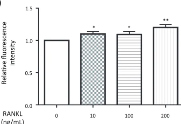
flu o re s c e n c e in te n s ity re la tiv e R 0 n ly O n e -w a y A N O V A d a ta Fig. 2 10 100 200 0.0 0.5 1.0 * ** (B) Rela@ve fluorescence intensity * RANKL (ng/mL) 1.5 0 Fig. 2 50 100 RANKL (10 ng/mL) ** ** (C) Rela@ve fluorescence intensity ** ** 0.0 0.5 1.0 1.5 0 RvE1 (nM) (A) (B) (C)
Fig. 2. Effects of RvE1 on osteoclastic bone resorption in RAW264.7 cells. RAW264.7 cells were cultured with 50 or 100 nM RvE1 in the presence of different doses (10, 100, 200 ng/mL) of RANKL for 6 days. Resorption pits were observed on day 7 in the conditioned medium treated with 10 ng/mL RANKL, whereas no resorption pit was observed in the medium without RANKL. However, the area of resorption pits was decreased by treatment with 100 nM RvE1. Original magnifi cation, × 200 (A). The levels of fl uorescence intensity, representing the activity of osteoclast generation, of the supernatant in the presence of 10, 100 and 200 ng/mL RANKL were determined (B). The levels of fluorescence intensity of the supernatant treated with 50 or 100 nM RvE1 in the presence of 10 ng/mL RANKL were deter-mined (C). Data are expressed as the mean ± SD (n = 3). *P < 0.05, **P < 0.01 versus control (B). **P < 0.01 versus RANKL (C). RANKL, receptor activator of nuclear factor-κB ligand; RvE1, resolvin E1.
Statistical analysis
The experimental data were analyzed using Graphpad Prism 6 (Graphpad Software, San Diego, CA). All ex-periments were conducted separately at least three times, and all data are presented as the mean ± SD. Statistical-ly significant differences were assessed by anaStatistical-lysis of variance (followed by Bonferroni multiple comparisons test) or Student’s t-test with P-values < 0.05 considered signifi cant.
RESULTS
RvE1 reduced the number of TRAP-positive osteo-clast-like RAW264.7 cells
The CCK-8 assay confi rmed that any effects of RvE1 on osteoclastogenesis would not be due to the cytotoxicity of this compound, since no change in cell viability was noted in RAW264.7 cells and MC3T3-E1 cells treated with RvE1 (data not shown).
TRAP-positive osteoclast-like cells were observed
when RAW264.7 cells were cultured in the presence RANKL, and the number of positive cells increased in a dose-dependent manner. However, the number of TRAP-positive cells decreased in the conditioned medi-um treated with both 100 nM RvE1 and RANKL (Fig. 1A), representing a statistically significant reduction compared to those detected with 50 and 100 ng/mL RANKL alone (Fig. 1B).
RvE1 suppressed osteoclastic bone resorption in RAW264.7 cells
To assess the effects of RvE1 on bone resorption, RAW264.7 cells were cultured with or without RvE1 in the presence of different doses (10, 100 and 200 ng/mL) of RANKL. Resorption pits were observed in the con-ditioned medium of RAW264.7 cells treated with 10 ng/ mL RANKL, which were not detected in the medium without RANKL (Fig. 2A). The fl uorescence intensity, representing the activity of osteoclast generation,
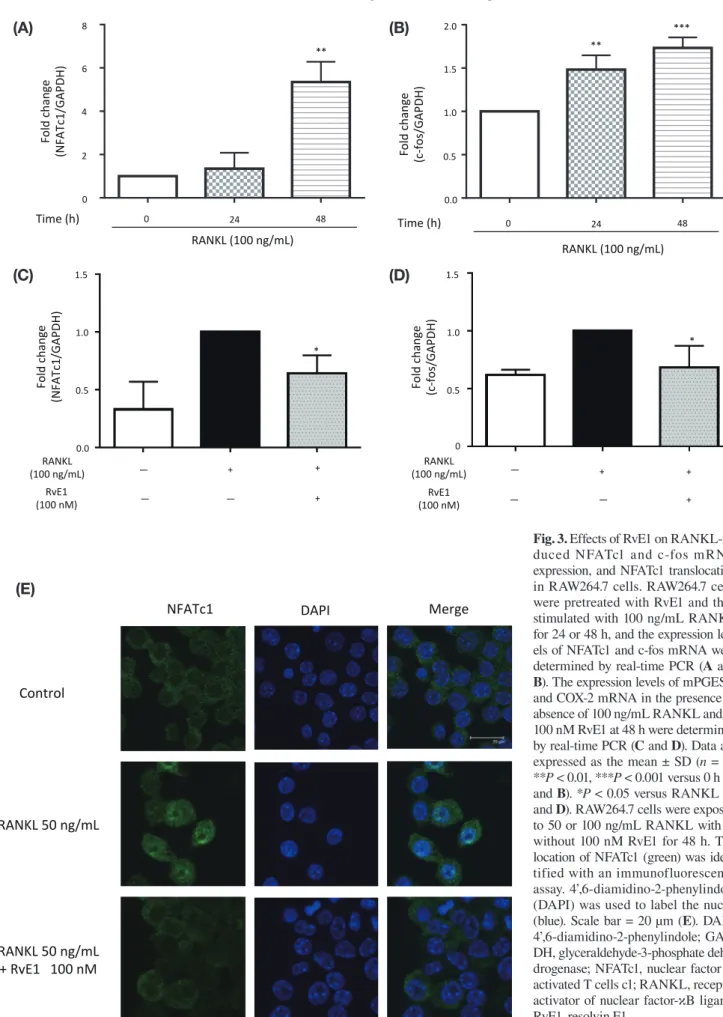
sig-RvE1 inhibits osteoclastogenesis and bone resorption C o n tr o l 2 4 h 4 8 h 0 2 4 6 8 N F A T c 1 2 4 ,4 8 h tim e c o u rs e O n e -w a y A N O V A d a ta (A) 8 4 2 0 Fold change (N FATc1/GAPDH) 6 0 24 48 ** RANKL (100 ng/mL) Fig. 3 Time (h) C o n tr o l 2 4 h 4 8 h 0 .0 0 .5 1 .0 1 .5 2 .0 c -fo s 2 4 ,4 8 h tim e c o u rs e O n e -w a y A N O V A d a ta (B) 2.0 1.0 0.5 0.0
Fold change (c-‐fos
/GAPDH) *** ** 1.5 Fig. 3 0 24 48 RANKL (100 ng/mL) Time (h) (C) Fold change (N FATc1/GAPDH) 1.0 0.5 0.0 1.5 * Fig. 3 RANKL (100 ng/mL) RvE1 (100 nM) ― ― + + + ― (D)
Fold change (c-‐fos
/GAPDH) 1.0 0.5 0 1.5 * Fig. 3 RANKL (100 ng/mL) RvE1 (100 nM) ― ― + + + ― (A) (C) (B) (D)
Fig. 3. Effects of RvE1 on RANKL-in-duced NFATc1 and c-fos mRNA expression, and NFATc1 translocation in RAW264.7 cells. RAW264.7 cells were pretreated with RvE1 and then stimulated with 100 ng/mL RANKL for 24 or 48 h, and the expression lev-els of NFATc1 and c-fos mRNA were determined by real-time PCR (A and B). The expression levels of mPGES-1 and COX-2 mRNA in the presence or absence of 100 ng/mL RANKL and/or 100 nM RvE1 at 48 h were determined by real-time PCR (C and D). Data are expressed as the mean ± SD (n = 3). **P < 0.01, ***P < 0.001 versus 0 h (A and B). *P < 0.05 versus RANKL (C
and D). RAW264.7 cells were exposed
to 50 or 100 ng/mL RANKL with or without 100 nM RvE1 for 48 h. The location of NFATc1 (green) was iden-tified with an immunofluorescence assay. 4’,6-diamidino-2-phenylindole (DAPI) was used to label the nuclei (blue). Scale bar = 20 µm (E). DAPI, 4’,6-diamidino-2-phenylindole; GAP-DH, glyceraldehyde-3-phosphate dehy-drogenase; NFATc1, nuclear factor of activated T cells c1; RANKL, receptor activator of nuclear factor-κB ligand; RvE1, resolvin E1.
Fig. 3
NFATc1 DAPI Merge
Control
RANKL 50 ng/mL
RANKL 50 ng/mL + RvE1 100 nM
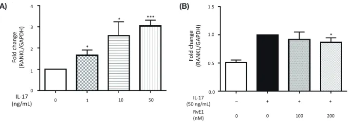
Y. Funaki et al. Fig. 4 1500 500 0 Fold change (MMP9/GAPDH) 1000 ** (A) ** 0 24 48 RANKL (100 ng/mL) Time (h) 72 Fig. 4 (B) 150 50 0 Fold change (Cathepsin K /GAPDH) 100 ** * 0 24 48 RANKL (100 ng/mL) Time (h) 72 Fig. 4 ** 0 100 200 300 400 Fold change (MMP9/GAPDH) RANKL (100 ng/mL) 50 ** (C) * RvE1 (nM) – + + + + 100 200 0 0 Fig. 5 (A) Fold change (RAN KL/GAPDH) 0 1 2 3 4 *** * * 1 10 50 IL-‐17 (ng/mL) 0 Fig. 4 (D) * 0 5 10 15 Fold change (Cathepsin K /GAPDH) ** RANKL (100 ng/mL) 50 RvE1 (nM) – + + + + 100 200 0 0 Fig. 5 (B) Fold change (RAN KL/GAPDH) 0.0 0.5 1.0 1.5 * IL-‐17 (50 ng/mL) RvE1 (nM) – + + + 100 200 0 0 (A) (C) (A) (B) (D) (B)
Fig. 4. Effects of RvE1 on RANKL-induced MMP-9 and cathepsin K mRNA expression in RAW264.7 cells.RAW264.7 cells were pre-treated with RvE1 and then stimulated with 100 ng/mL RANKL for 24 or 48 h, and the mRNA expression levels of MMP-9 and cathep-sin K were determined by real-time PCR (A and B). The mRNA expression levels of MMP-9 and cathepsin K in the presence or absence of 100 ng/mL RANKL and/or 100 nM RvE1 at 48 h were determined by real-time PCR (C and D). Data are expressed as the mean ± SD (n = 3), *P < 0.05, **P < 0.01 versus 0 h (A and B). *P < 0.05, **P < 0.01 versus RANKL (C and D). GAPDH, glyceraldehyde-3-phosphate dehydrogenase; MMP-9, matrix metalloproteinase-9; RANKL, receptor activator of nuclear factor-κB ligand; RvE1, resolvin E1.
Fig. 5. Effect of RvE1 on IL-17-induced RANKL mRNA expression in MC3T3-E1 cells. MC3T3-E1 cells were cultured in the presence of 1, 10 and 50 ng/mL IL-17, and the mRNA expression level of RANKL was determined by real-time PCR (A). The cells were also cultured with 50, 100 and 200 nM RvE1 in the presence of 50 ng/mL IL-17. The expression levels of RANKL mRNA in the presence or absence of 50 ng/mL IL-17 and/or 100 nM RvE1 at 24 h were determined by real-time PCR. The value of the fold change in mRNA lev-els of IL-17 alone was normalized as 1. (B). Data are expressed as the mean ± SD (n = 3), *P < 0.05, ***P < 0.01 versus control (A). *P < 0.05 versus IL-17 alone (B). GAPDH, glyceraldehyde-3-phosphate dehydrogenase; IL, interleukin; RANKL, receptor activator of nuclear factor-κB ligand; RvE1, resolvin E1.
RvE1 inhibits osteoclastogenesis and bone resorption
nifi cantly increased compared to that of the control in a RANKL concentration-dependent manner (Fig. 2B). However, co-treatment with 100 nM RvE1 decreased the area of the resorption Pits (Fig. 2A) and signifi cantly reduced the fl uorescent intensity compared to that of the vehicle control (Fig. 2C).
RvE1 suppressed RANKL-induced NFATc1 and c-fos mRNA expression and NFATc1 nuclear trans-location in RAW264.7 cells
The NFATc1 and c-fos mRNA levels of RAW264.7 cells treated with 100 ng/mL RANKL increased in a time-de-pendent manner with a signifi cant increase at 48 h, and at both 24 and 48 h, respectively, compared to 0 h (Figs. 3A and B). However, co-treatment with 100 nM RvE1 signifi cantly reduced the RANKL-induced NFATc1 and c-fos mRNA expression levels compared to those of the vehicle control (Figs. 3C and D). The immunofluores-cence assay showed that NFATc1 was mainly localized in the cytoplasm in the control cells, but was distinctly translocated to the nucleus after treatment with 50 ng/ mL RANKL for 48 h; however, the nuclear immunos-taining intensity was reduced in the presence of 100 nM RvE1 (Fig. 3E).
RvE1 reduced RANKL-induced MMP-9 and cathepsin K mRNA expression in RAW264.7 cells
The mRNA expression levels of MMP-9 and cathepsin K signifi cantly increased at 48 and 72 h in RAW264.7 cells treated with 100 ng/mL RANKL compared to 0 h (Figs. 4A and B). RvE1 significantly reduced RANKL-induced MMP-9 and cathepsin K mRNA ex-pression in a dose-dependent manner compared to the vehicle control (Figs. 4C and D).
RvE1 reduced IL-17-induced RANKL mRNA ex-pression in MC3T3-E1 cells
After culturing the cells with IL-17 (1, 10 or 50 ng/mL) for 24 h, the RANKL mRNA level was significantly increased in a dose-dependent manner (Fig. 5A). Pre-treatment with RvE1 (100 or 200 nM) for 24 h tended to reduce the IL-17-induced RANKL mRNA level in a dose-dependent manner with signifi cant downregulation observed following treatment with 200 nM RvE1 (Fig. 5B). Similarly, the OPG mRNA level was decreased after IL-17 treatment in a dose-dependent manner, and OPG expression was upregulated with RvE1 pretreat-ment (data not shown). Therefore, RvE1 improved the
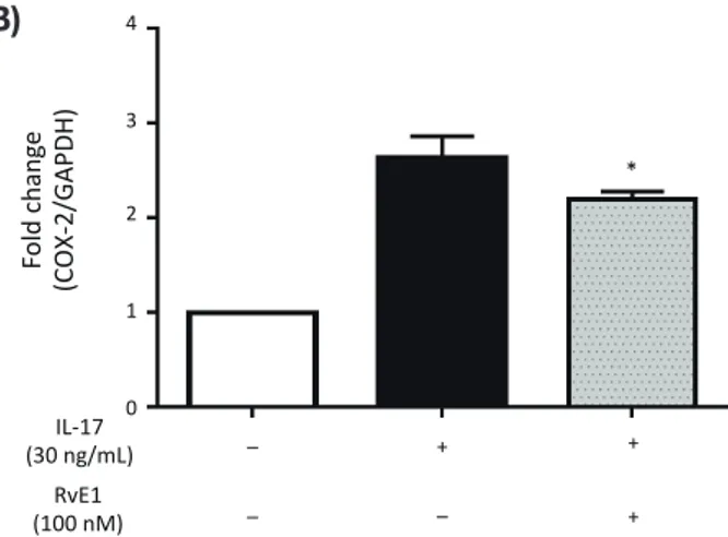
Fig. 6 100 0 200 300 PGE 2 (pg /mL) 400 C o n tr o l IL -1 7 :1 0 IL -1 7 + R v E 1 0 0 n M 0 1 0 0 2 0 0 3 0 0 4 0 0 P G E 2 c o n tro l+ h a s e g a w a O K O n e -w a y A N O V A d a ta (A) IL-‐17 (30 ng/mL) RvE1 (100 nM) – – + + + – C o n tr o l IL -1 7 :3 0 n g /m L IL -1 7 + R v E 0 .0 0 .5 1 .0 1 .5 2 .0 2 .5 m P G E S -1 n = 5 C o n tro l= 1 O K O n e -w a y A N O V A d a ta 2.5 2.0 1.5 1.0 0.5 0.0 Fold change (mPGES-‐1/GAPDH) Fig. 6 (C) * IL-‐17 (30 ng/mL) RvE1 (100 nM) – – + + + – Fold change (COX-‐2/GAPDH) 3 2 1 0 4 Fig. 6 (B) * IL-‐17 (30 ng/mL) RvE1 (100 nM) – – + + + – (A) (C) (B)
Fig. 6. Effects of RvE1 on IL-17-induced PGE2 production and mRNA expression of COX-2 and mPGES-1 in MC3T3-E1 cells.MC3T3-E1 cells were cultured with 30 ng/mL IL-17 for 24 h after pretreatment with 100 nM RvE1 and the PGE2 pro-duction level was determined by ELISA. Data are expressed as the mean ± SD (n = 2 for control, n = 3 for 10 ng/mL IL-17 and IL-17 + 100 nM RvE1); P = 0.075 versus IL-17 (A). MC3T3-E1 cells were cultured with or without 100 nM RvE1 in the pres-ence of 30 ng/mL IL-17 for 24 h, and the mRNA expression levels of COX-2 (B) and mPGES-1 (C) were determined by real-time PCR. Data are expressed as the mean ± SD (n = 3 for COX-2, n = 5 for mPGES-1). *P < 0.05 versus IL-17. COX-2, cyclooxygenase-2; GAPDH, glyceraldehyde-3-phosphate dehy-drogenase; IL, interleukin; mPGES-1, microsomal prostaglan-din E synthase-1; PGE2, prostaglandin E2; RvE1, resolvin E1.
Y. Funaki et al.
imbalance in the RANKL/OPG ratio, which is likely to induce an inhibitory effect on bone resorption.
RvE1 reduced IL-17-induced PGE2 production and
the mRNA expression levels of COX-2 and mPG-ES-1 in MC3T3-E1 cells
The ELISA results showed that pretreatment of MC3T3-E1 cells with RvE1 inhibited the stimulatory
effect of IL-17 on PGE2 production; the PGE2
produc-tion levels in the presence of 100 nM RvE1 tended to be reduced compared to those of cells treated with the vehicle control (Fig. 6A). RvE1 treatment also signifi-cantly reduced the IL-17-induced expression of COX-2 and mPGES-1 mRNA compared to the vehicle control group (Figs. 6B and C).
DISCUSSION
RvE1 is a member of the E series of resolvins that are
biosynthesized from EPA.11 Recent reports identified
an-other member of the E series of resolvins, called resolvin
E2 (RvE2) and resolvin E3 (RvE3).26, 27 Although Barden
and colleagues28 showed that synovial fluid RvE2 was
negatively associated with pain score in humans, the effects of RvE2 and RvE3 on osteoclasts and osteoblasts were not fully understood. Thus, we investigated RvE1 as a therapeutic agent for RA. With respect to bone me-tabolism, RvE1 was shown to inhibit osteoclast growth and bone resorption.16 Gao and colleagues29 reported that RvE1 has a direct bone-preserving function via chemo-kine-like receptor 1, which mediates bone preservation in osteoblasts. Moreover, a recent study indicated that RvE1 influenced pain, a major symptom of RA, showing simultaneous anti-inflammatory and analgesic properties in experimental models closely related to translational
sites in humans.30 These studies have demonstrated the
good therapeutic potential of RvE1 in many chronic in-flammatory diseases. Indeed, RvE1 (Rx-10001) and its synthetic analog (Rx-10045) are currently under clinical trials for the relief of chronic dry eyes.31 The aim of our study was to clarify the effect of RvE1 on osteoclasto-genesis and determine its feasibility in the development of a new therapeutic approach to RA.
We showed that RvE1 reduced the number of TRAP-positive osteoclast-like cells, which is consistent with a previous report indicating that RvE1 markedly decreased the number of differentiated osteoclasts in-duced by macrophage colony-stimulating factor and
RANKL in primary osteoclast cultures.16
Recently, a new assay method of evaluating the bone resorption activity in RAW264.7 cells was reported based on the measurement of pit formation using calci-um phosphate labeled with fluorescent polyanionic
mole-cules.25 With this method, we found that RvE1 decreased
the bone resorption activity in RAW264.7 cells, in line with a previous report showing that RvE1 diminished
dentin resorption in vitro.16 These results in RAW264.7
cells suggest that RvE1 could inhibit RANKL-induced osteoclastogenesis and bone resorption.
We further found that RvE1 significantly inhibited RANKL-induced NFATc1 and c-fos mRNA expres-sion and the nuclear translocation of NFATc1 from the cytoplasm in RAW264.7 cells, providing a potential mechanism underlying this inhibitory effect of RvE1 on osteoclastogenesis. A previous study showed that RvE1 attenuated the RANKL-induced nuclear translocation
of the p50 subunit of NF-κB.16 Zhu and colleagues32
reported another mechanism of RvE1 in osteoclasts in bone marrow cells of C57BL/6 mice, in which RvE1 inhibits osteoclast fusion through binding NFATc1 to the pivotal fusion protein DC-STAMP, thereby suppressing RANKL-induced NFATc1 nuclear translocation. Anoth-er report showed that Bioaggregate, a laboratory-synthe-sized and calcium silicate-based nanoparticulate cement, also inhibited the translocation of NFATc1 and c-fos from the cytoplasm to the nucleus in osteoclast precursor cells, which was determined with immunofluorescence analysis.33
Our data indicated that RvE1 inhibited RANKL-in-duced osteoclastogenesis through suppressing the mRNA expression of NFATc1 and c-fos. NFATc1 is known as a master transcription factor that plays a key role in regulating the expression of several osteo-clast-specific genes such as TRAP, cathepsin K and MMP-9, which are involved in regulating osteoclast
differentiation.18 Our results suggested that RvE1
sig-nificantly inhibited RANKL-induced cathepsin K and MMP-9 mRNA expression by decreasing the transloca-tion of NFATc1 from the cytoplasm to the nucleus.
We further showed that RvE1 might correct the im-balance of RANKL and OPG expression in osteoblasts induced by IL-17 using MC3T3-E1 cells.
PGE2 is well known to be the key mediator of
in-flammation, pain and joint destruction in RA, and its production is an important target of anti-inflammatory
drugs. PGE2 is produced by the conversion of
arachi-donic acid to prostaglandin H2 by COX-1/COX-2, with
the subsequent conversion of prostaglandin H2 to PGE2
by mPGES-1, which is the terminal enzyme in the PGE2
production process at the sites of inflammation, and has
an important role in the pathogenesis of RA.34, 35 PGE2
has several pro-inflammatory effects, including increas-ing vascular permeability, vasodilation, blood flow and local pyrexia, along with potentiation of pain caused by
stim-RvE1 inhibits osteoclastogenesis and bone resorption
ulates osteoclast differentiation through the prostanoid receptors EP4 and EP2 by inducing RANKL production
and inhibiting OPG expression.36–38 OPG is a soluble
de-coy receptor for RANKL that prevents it from binding to RANK. RANKL interacts with its receptor, which is expressed on osteoclast precursors, to induce the
differentiation and activation of osteoclasts.21 A recent
study showed that EPA and DHA inhibit PGE2-induced
RANKL expression in MC3T3-E1 cells.39 Similarly, we
showed that RvE1 inhibits the IL-17-induced COX-2 and
mPGES-1 mRNA expression followed by PGE2
produc-tion in MC3T3-E1 cells. These results might indicate that RvE1 indirectly inhibits osteoclastogenesis by re-ducing IL-17-induced COX-2 and mPGES-1 expression in osteoblasts, suggesting that RvE1, like EPA and DHA, has an anti-inflammatory effect in RA.
We did not examine the effects of RvE1 on the
ex-pression of PGE2 receptors in osteoblasts, such as EP4
receptor. However, recent reports showed that PGE2
en-hanced osteoclast formation through EP4 receptor acti-vation on osteoblasts.40, 41 In addition, the effects of RvE1 on the leukotriene B4 receptor subtype 1 (BLT1)
ex-pressed on osteoclasts, identified as receptors for RvE1,42
should be further investigated, as a previous study showed that BLT1 mediates the actions of RvE1 on
os-teoclasts.16 Although the major effect of PGE2 on bone
resorption is generally considered to occur indirectly via upregulation of RANKL and inhibition of OPG
ex-pression in osteoblastic cells,43 further study is needed
to determine whether the EP4 receptor and BLT1 are therapeutic targets of RvE1. The mechanisms of inflam-mation in RA are complex. In RA, many
pro-inflamma-tory cytokines including TNF-α, 1β, 6, and
IL-17 increase RANKL expression, leading to an increased osteoclast differentiation and subsequent bone erosions. Several of these cytokines also act synergistically with
RANKL in promoting osteoclast differentiation.44
Fur-ther studies are needed to assess the anti-inflammatory effect of RvE1 in RA.
In conclusion, our results suggest that RvE1 inhibits osteoclastogenesis and bone resorption by suppressing RANKL-induced osteoclast differentiation. The mecha-nism of action was determined to occur by the downreg-ulation of NFATc1 and c-fos in osteoclasts and suppres-sion of IL-17-induced RANKL expressuppres-sion through the
autocrine action of PGE2 in osteoblasts. These findings
suggest the potential of RvE1 as a new therapeutic ap-proach to RA, providing the foundation for further pre-clinical and pre-clinical investigations.
Acknowledgments: This research was partly performed at the Re-search Center for Bioscience and Technology, Tottori University. We thank Katsumi Higaki (Division of Functional Genomics,
Research Center for Bioscience and Technology, Tottori Universi-ty, Yonago, Japan) for helpful scientific discussions and technical assistance.
The authors declare no conflict of interest. REFERENCES
1 McInnes IB, Schett G. The pathogenesis of rheumatoid arthri-tis. N Engl J Med. 2011;365:2205-19. PMID: 28617323. 2 Corrado A, Maruotti N, Cantatore FP. Osteoblast Role in
Rheumatic Diseases. International journal of molecular sci-ences. 2017;18. PMID: 28617323.
3 Kotake S, Udagawa N, Takahashi N, Matsuzaki K, Itoh K, Ishiyama S, et al. IL-17 in synovial fluids from patients with rheumatoid arthritis is a potent stimulator of osteoclastogene-sis. J Clin Invest. 1999;103:1345-52. PMID: 10225978. 4 Walsh NC, Crotti TN, Goldring SR, Gravallese EM.
Rheu-matic diseases: the effects of inflammation on bone. Immunol Rev. 2005;208:228-51. PMID: 16313352.
5 Kremer JM, Bigauoette J, Michalek AV, Timchalk MA, Lininger L, Rynes RI, et al. Effects of manipulation of dietary fatty acids on clinical manifestations of rheumatoid arthritis. Lancet. 1985;1:184-7. PMID: 2857265.
6 Caughey GE, Mantzioris E, Gibson RA, Cleland LG, James MJ. The effect on human tumor necrosis factor alpha and in-terleukin 1 beta production of diets enriched in n-3 fatty acids from vegetable oil or fish oil. Am J Clin Nutr. 1996;63:116-22. PMID: 8604658.
7 Trebble T, Arden NK, Stroud MA, Wootton SA, Burdge GC, Miles EA, et al. Inhibition of tumour necrosis factor-alpha and interleukin 6 production by mononuclear cells following dietary fish-oil supplementation in healthy men and response to antioxidant co-supplementation. The British journal of nu-trition. 2003;90:405-12. PMID: 12908901.
8 Calder PC. Session 3: Joint Nutrition Society and Irish Nu-trition and Dietetic Institute Symposium on ‘NuNu-trition and autoimmune disease’ PUFA, inflammatory processes and rheumatoid arthritis. Proc Nutr Soc. 2008;67:409-18. PMID: 18847518.
9 Goldberg RJ, Katz J. A meta-analysis of the analgesic ef-fects of omega-3 polyunsaturated fatty acid supplementation for inflammatory joint pain. Pain. 2007;129:210-23. PMID: 17335973.
10 Serhan CN. Pro-resolving lipid mediators are leads for resolu-tion physiology. Nature. 2014;510:92-101. PMID: 24899309. 11 Serhan CN, Clish CB, Brannon J, Colgan SP, Chiang N,
Gronert K. Novel functional sets of lipid-derived mediators with antiinflammatory actions generated from omega-3 fatty acids via cyclooxygenase 2-nonsteroidal antiinflammatory drugs and transcellular processing. J Exp Med. 2000;192:1197-204. PMID: 11034610.
12 Hasturk H, Kantarci A, Ohira T, Arita M, Ebrahimi N, Chiang N, et al. RvE1 protects from local inflammation and osteoclast- mediated bone destruction in periodontitis. FASEB J. 2006;20:401-3. PMID: 16373400.
13 Schwab JM, Chiang N, Arita M, Serhan CN. Resolvin E1 and protectin D1 activate inflammation-resolution programmes. Nature. 2007;869-74. 17568749.
14 Aoki H, Hisada T, Ishizuka T, Utsugi M, Kawata T, Shimizu Y, et al. Resolvin E1 dampens airway inflammation and hyperre-sponsiveness in a murine model of asthma. Biochem Biophys
Y. Funaki et al. Res Commun. 2008;367:509-15. PMID: 18190790.
15 Seki H, Fukunaga K, Arita M, Arai H, Nakanishi H, Taguchi R, et al. The anti-inflammatory and proresolving mediator resolvin E1 protects mice from bacterial pneumonia and acute lung injury. J Immunol. 2010;184:836-43. PMID: 20007539. 16 Herrera BS, Ohira T, Gao L, Omori K, Yang R, Zhu M, et
al. An endogenous regulator of inflammation, resolvin E1, modulates osteoclast differentiation and bone resorption. Br J Pharmacol. 2008;155:1214-23. PMID: 18806821.
17 Grigoriadis AE, Wang ZQ, Cecchini MG, Hofstetter W, Felix R, Fleisch HA, et al. c-Fos: a key regulator of osteoclast-mac-rophage lineage determination and bone remodeling. Science. 1994;266:443-8. PMID: 7939685.
18 Takayanagi H, Kim S, Koga T, Nishina H, Isshiki M, Yoshida H, et al. Induction and activation of the transcription factor NFATc1 (NFAT2) integrate RANKL signaling in terminal differentiation of osteoclasts. Developmental cell. 2002;3:889-901. PMID: 12479813.
19 Gyurko R, Van Dyke TE. The role of polyunsaturated ω-3 fatty acid eicosapentaenoic acid-derived resolvin E1 (RvE1) in bone preservation. Crit Rev Immunol. 2014;34:347-57. PMID: 24941160.
20 Kong YY, Boyle WJ, Penninger JM. Osteoprotegerin ligand: a regulator of immune responses and bone physiology. Immu-nology today. 2000;21:495-502. PMID:11071528.
21 Boyle WJ, Simonet WS, Lacey DL. Osteoclast differentiation and activation. Nature. 2003;423:337-42. PMID: 12748652. 22 Moseley TA, Haudenschild DR, Rose L, Reddi AH.
Interleu-kin-17 family and IL-17 receptors. Cytokine & growth factor reviews. 2003;14:155-74. PMID: 12651226.
23 Suda T, Takahashi N, Udagawa N, Jimi E, Gillespie MT, Martin TJ. Modulation of osteoclast differentiation and func-tion by the new members of the tumor necrosis factor receptor and ligand families. Endocrine reviews. 1999;20:345-57. PMID: 10368775.
24 Boeyens JC, Deepak V, Chua WH, Kruger MC, Joubert AM, Coetzee M. Effects of omega3- and omega6-polyunsaturated fatty acids on RANKL-induced osteoclast differentiation of RAW264.7 cells: a comparative in vitro study. Nutrients. 2014;6: 2584-601. PMID: 25010555.
25 Miyazaki T, Miyauchi S, Anada T, Imaizumi H, Suzuki O. Evaluation of osteoclastic resorption activity using calcium phosphate coating combined with labeled polyanion. Analyti-cal biochemistry. 2011;410:7-12. PMID: 21078287.
26 Tjonahen E, Oh SF, Siegelman J, Elangovan S, Percarpio KB, Hong S, et al. Resolvin E2: identification and anti-inflamma-tory actions: pivotal role of human 5-lipoxygenase in resolvin E series biosynthesis. Chem Biol. 2006;13:1193-202. PMID: 17114001.
27 Isobe Y, Arita M, Matsueda S, Iwamoto R, Fujihara T, Nakanishi H, et al. Identification and structure determination of novel anti-inflammatory mediator resolvin E3, 17,18-dihy-droxyeicosapentaenoic acid. J Biol Chem. 2012;287:10525-34. PMID:22275352
28 Barden AE, Moghaddami M, Mas E, Phillips M, Cleland LG, Mori TA. Specialised pro-resolving mediators of inflamma-tion in inflammatory arthritis. Prostaglandins Leukot Essent Fatty Acids. 2016;107:24-9. PMID: 27033423.
29 Gao L, Faibish D, Fredman G, Herrera BS, Chiang N, Serhan CN, et al. Resolvin E1 and chemokine-like receptor 1 medi-ate bone preservation. J Immunol. 2013;190:689-94. PMID: 23241890.
30 Fonseca FC, Orlando RM, Turchetti-Maia RM, de Francischi
JN. Comparative effects of the omega3 polyunsaturated fat-ty acid derivatives resolvins E1 and D1 and protectin DX in models of inflammation and pain. Journal of inflammation research. 2017;10:119-33. PMID: 28919798.
31 Hessen M, Akpek EK. Dry eye: an inflammatory ocular dis-ease. Journal of ophthalmic & vision research. 2014;9:240-50. PMID: 25279127.
32 Zhu M, Van Dyke TE, Gyurko R. Resolvin E1 regulates osteoclast fusion via DC-STAMP and NFATc1. FASEB J. 2013;27:3344-53. PMID: 23629863.
33 Zhang J, Zhu L, Yan P, Peng B. Effect of BioAggregate on Re-ceptor Activator of Nuclear Factor-Kappa B Ligand-induced Osteoclastogenesis from Murine Macrophage Cell Line In Vitro. J Endod. 2015;41:1265-71. PMID: 25975181.
34 Jakobsson PJ, Thoren S, Morgenstern R, Samuelsson B. Identification of human prostaglandin E synthase: a microso-mal, glutathione-dependent, inducible enzyme, constituting a potential novel drug target. Proc Natl Acad Sci U S A. 1999;96:7220-5. PMID: 10377395.
35 Samuelsson B, Morgenstern R, Jakobsson PJ. Membrane pros-taglandin E synthase-1: a novel therapeutic target. Pharmacol Rev. 2007;59:207-24. PMID: 17878511.
36 Suzawa T, Miyaura C, Inada M, Maruyama T, Sugimoto Y, Ushikubi F, et al. The role of prostaglandin E receptor sub-types (EP1, EP2, EP3, and EP4) in bone resorption: an analy-sis using specific agonists for the respective EPs. Endocrinolo-gy. 2000;141:1554-9. PMID: 10746663.
37 Nukaga J, Kobayashi M, Shinki T, Song H, Takada T, Takiguchi T, et al. Regulatory effects of interleukin-1beta and prostaglandin E2 on expression of receptor activator of nucle-ar factor-kappaB ligand in human periodontal ligament cells. J Periodontol. 2004;75:249-59. PMID: 15068113.
38 Liu XH, Kirschenbaum A, Yao S, Levine AC. Cross-talk between the interleukin-6 and prostaglandin E(2) signaling systems results in enhancement of osteoclastogenesis through effects on the osteoprotegerin/receptor activator of nuclear factor-{kappa}B (RANK) ligand/RANK system. Endocrinol-ogy. 2005;146:1991-8. PMID: 15618359.
39 Poulsen RC, Wolber FM, Moughan PJ, Kruger MC. Long chain polyunsaturated fatty acids alter membrane-bound RANK-L expression and osteoprotegerin secretion by MC3T3-E1 osteoblast-like cells. Prostaglandins Other Lipid Mediat. 2008;85:42-8. PMID: 18077200.
40 Mano M, Arakawa T, Mano H, Nakagawa M, Kaneda T, Kaneko H, et al. Prostaglandin E2 directly inhibits bone-re-sorbing activity of isolated mature osteoclasts mainly through the EP4 receptor. Calcif Tissue Int. 2000;67:85-92. PMID: 10908419.
41 Sakuma Y, Tanaka K, Suda M, Yasoda A, Natsui K, Tanaka I, et al. Crucial involvement of the EP4 subtype of prostaglandin E receptor in osteoclast formation by proinflammatory cyto-kines and lipopolysaccharide. J Bone Miner Res. 2000;15:218-27. PMID: 10703923.
42 Arita M, Bianchini F, Aliberti J, Sher A, Chiang N, Hong S, et al. Stereochemical assignment, antiinflammatory properties, and receptor for the omega-3 lipid mediator resolvin E1. J Exp Med. 2005;201:713-22. PMID: 15753205.
43 Blackwell KA, Raisz LG, Pilbeam CC. Prostaglandins in bone: bad cop, good cop? Trends in endocrinology and metab-olism: TEM. 2010;21:294-301. PMID: 20079660.
44 Brennan FM, McInnes IB. Evidence that cytokines play a role in rheumatoid arthritis. J Clin Invest. 2008;118:3537-45. PMID: 18982160.