Considering the Better Donor-side in Reconstruction of Composite Radius After
Resection of Spindle Cell Sarcoma Using Free Fibular Osteocutaneous Flap:
A Case Report
Shunjiro Yagi,* Yoshiko Suyama,* Kohei Fukuoka,* Maki Morita,* Kensaku Yamaga† and Hideki Nagashima† *Department of Plastic and Reconstructive Surgery, Tottori University Hospital, Yonago 683-8504, Japan, and †Division of Orthopedic Surgery, Department of Sensory and Motor Organs, School of Medicine, Tottori University Faculty of Medicine, Yonago 683-8503 Japan
ABSTRACT
Reconstruction of bone and soft-tissue defects in the forearm is a surgery that often proves unsuccess-ful. Free fibular osteocutaneous flaps are a useful material for reconstruction that enable simultaneous reconstruction of bone, skin, and soft tissues. However, in free fibular osteocutaneous flaps, the fibula, skin, and vascular pedicle are tightly bound together by the posterior intermuscular septum and the perforators that pass through the septum, giving the disadvantage of a low degree of freedom when setting these structures in place. We take into account the 3-dimensional structure of the free skin flap when selecting which lower leg to use as the donor. We report here the case of a 61-year-old man with defects in the radius, skin, and soft tissues after resection of spindle cell carcinoma of the right forearm, which was reconstructed using a free fibular osteocutaneous flap harvested from the left lower leg. Two years postoperatively, recovery has been unevent-ful with no complications. Donor-side selection of free fibular osteocutaneous flap is an important factor for safely completing composite radius reconstruction.
Key words donor side; fibular flap; forearm; microsur-gery; reconstruction
Advances in microsurgery mean that free fibular os-teocutaneous flaps are now a useful material for use in forearm reconstruction after injury and malignant tumor resection.1–7 However, these materials confer several
disadvantages, including a high risk of partial necrosis due to unstable blood supply from the perforators to the skin and the fact that only a limited amount of skin can be harvested. In addition, in free fibular osteocutaneous
flaps, the fibula, skin, and vascular pedicle are tightly bound together by the posterior intermuscular septum and the perforating branches that pass through, leading to the disadvantage of a low degree of freedom when setting these structures in place. This also makes such flaps more difficult to use compared to another flaps.
The selection of which lower leg to use as the donor site involves a number of considerations, such as whether to use the leg on the same side as the defect, the non-dominant leg, or a leg without vascular lesions. The preferences and experience of the surgeon are often major factors. By taking into account the 3-dimensional (3D) structure of the fibular osteocutaneous flap when selecting the leg to use as the donor, we fix the skin flap in the best possible way for safe reconstruction. We here report our surgical strategy in detail for reconstruction with a free fibular osteochondral flap after resection of soft-tissue carcinoma of the forearm.
PATIENT REPORT
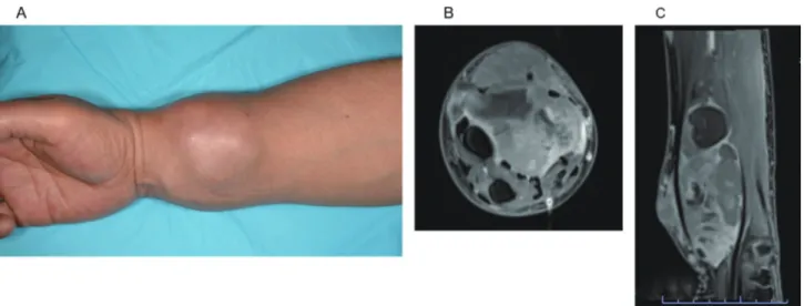
A 61-year-old man presented with a mass that had suddenly increased in size about 1 year previously. Computed tomography (CT) revealed a soft-tissue mass measuring 6 × 4.5 cm2 touching the radius on the
cranial side of the right forearm. Magnetic resonance imaging (MRI) depicted a tumor involving the flexor carpi radialis, brachioradialis, and radial artery. The tumor was also touching the skin on the flexor side (Fig. 1). The pathological diagnosis from needle biopsy was spindle cell sarcoma. After chemotherapy with adria-mycin and ifosfamide, wide resection of the tumor and reconstruction with a fibular osteocutaneous flap were planned.
Surgery Tumor resection
A longitudinal skin incision was made from the antero-lateral side of the forearm with an oval patch of skin measuring 12 × 13 cm2. Wide resection of tumor was
then performed according to the surgical plan. In fact, the radius, palmaris longus, flexor digitorum superfi-cialis (FDS), flexor digitorum profundus (FDP), flexor
Patient Report Yonago Acta Medica 2020;63(2):127–131 doi: 10.33160/yam.2020.05.003
Corresponding author: Shunjiro Yagi, MD, PhD yagishun68@gmail.com
Received 2020 February 5 Accepted 2020 April 3 Online published 2020 April 22
Abbreviations: 3D, 3-dimensional; CT, computed tomography; FCU, flexor carpi ulnaris; FDP, flexor digitorum profundus; FDS, flexor digitorum superficialis; FPL, flexor pollicis longus; MRI, magnetic resonance imaging; MP, metacarpophalangeal
pollicis longus (FPL), brachioradialis, extensor carpi radialis brevis, extensor carpi radialis longus, flexor carpi radialis, pronator quadratus muscle, extensor pollicis brevis, abductor pollicis longus, interosseous membrane, median nerve, radial nerve, and radial artery were resected together with tumor. The radius was cut 13 cm proximal to the wrist. The muscles, nerves, and radial artery were cut at the level of the carpometacar-pal joint on the distal side and at 14 cm proximal to the wrist on the proximal side (Fig. 2).
To reconstruct the function of flexion in the fingers, cut ends of residual FDSs were each resected at a length of about 5 cm, and resected FDSs were sutured to the cut end of residual FDPs and FPL. Sutured FDSs were bundled in a cluster, then sutured to the residual flexor carpi ulnaris (FCU) for bridging between FDPs and
FCU. Further, to reconstruct the function of the op-ponens pollicis, the extensor indicis proprius was cut at the level of metacarpophalangeal (MP) joint and routed subcutaneously around the ulnar side of the forearm and across the palm to the abductor pollicis brevis tendon in the region of the thumb MP joint according to Burkhalter’s procedure.8
Decision on fibula harvesting side and reconstruction
In consideration of the 3D structure of the free fibular osteocutaneous flap, the left lower leg was selected as the donor.
If the flap had been taken from the right leg, the vascular pedicle would have to pass either over or under the fibula to use the radial artery as the recipient vessel. This would have risked placing the vascular pedicle
Fig. 1. Preoperative clinical photograph (A) and MRI (B and C). The tumor is seen touching the radius. The flexor carpi radialis, brachio-radialis tendon, and radial artery and vein run through the tumor, which has reached the skin on the flexor side.
Fig. 2. Intraoperative photograph showing the defect after tumor resection. The cut surface of the radius is indicated by an arrow, the radial artery and its accompanying vein by an asterisk, and the cephalic vein by two asterisks (left). Design of free fibular osteocutaneous flap on left leg (right).
Donor-side selection of fibular flap
under compression, and would also have brought the intermuscular septum and its perforators to the radial side, making it impossible for the skin flap to cover the skin defect effectively. Forcible skin closure would place the perforating branches under unnecessary strain, increasing the risk of skin necrosis (Fig. 3).
We therefore harvested the fibular osteocutaneous flap as a 20cm length of the fibula and 21 × 9 cm2 skin
island from the left lower leg. At the same time, we harvested the left sural nerve and reconstructed the median nerve. Lunate bone was decorticated for wrist arthrodesis and the vascularized fibula was grafted into the excision site of the radius and fixed with a locking plate after bending. The locking plate was fixed to the residual radius on the proximal side and to the third metacarpal bone on the distal side. The ulnar head was fixed to the carpal bones with a cannulated screw to
achieve stability. The radial artery and its accompany-ing vein as well as the cephalic vein were selected as the recipient vessels, and anastomosed to the vascular pedicle of the free fibular osteocutaneous flap under microscopic observation. The skin defect on the right forearm was covered with the skin flap. The skin defect on the lower leg was closed with a split-thickness graft from the left thigh.
Outcomes
The postoperative course was uneventful, with no complications such as infection. The right arm was immobilized for 1 month with a splint from the elbow to the wrist. The patient was discharged after postopera-tive chemotherapy. As of the time of writing, 2 years postoperatively, the transferred fibular osteocutaneous flap took completely. And good synostosis and slight movement of the fourth and fifth fingers has been
Fig. 3. Cross-sections of the right forearm and left lower leg (top row). If a free fibular osteocutaneous flap (dotted line) is harvested from the left lower leg, the perforating branches are not placed under unnecessary strain and the skin flap can effectively cover the skin defect. Cross-sections of the right forearm and right lower leg (bottom row). If a free fibular osteocutaneous flap (dotted line) is harvested from the right lower leg, the peroneal artery must pass either over the plate or under the fibula to reach the radial artery (dashed lines and ar-rows). The skin flap is displaced to the radial side and thus cannot effectively cover the skin defect.
achieved. Range of motion for the right elbow ranges from 0° extension to 140° of flexion (Fig. 4).
DISCUSSION
Since Taylor first reported using a fibular osteocutane-ous flap for limb reconstruction, free fibular osteo-cutaneous flaps have been used in a variety of ways, including limb bone reconstruction and mandibular reconstruction in the head and neck region.1 Fibular
osteocutaneous flaps are useful because they contain an extended piece of long bone and are raised attached to a skin flap from the lateral lower leg, meaning that they can be used to reconstruct skin and soft-tissue defects at the same time. On the other hand, a key disadvantage is a susceptibility to partial necrosis of the skin island as a complication. According to Wei,2 1 or
2 reliable perforators can cover more than 10 × 21 cm2
of skin. In our patient, the skin defect measured 12 cm width. In consideration of the decrease in resected soft tissues, we harvested the fibular osteocutaneous flap with a skin island of almost the maximum width size of 9 cm in order to cover the skin defect. However, as the fibula, skin island, and vascular pedicle of the free fibular osteocutaneous flap are bound closely together
by the intermuscular septum and the perforators, there is little freedom for manipulation, and suturing the skin island forcibly into position would place the perforators under unnecessary strain, increasing the risk of partial necrosis of the skin island. If the fibular osteocutaneous flap had been harvested from the other leg, the skin island would have been displaced to the radial side by the width of the lateral surface of the fibula, and sutur-ing the skin defect without placsutur-ing it under strain would have been difficult. The selection of the appropriate fibula harvesting site and the setting of fibular osteocu-taneous flap on the skin defect are thus important.
The bilateral cut surfaces of the fibular osteocuta-neous flap are mirror images of each other and differ 3-dimensionally. We have previously reported that donor side selection is important when a free fibular osteocutaneous flap is used for mandibular reconstruc-tion.9 We therefore created an algorithm for determining
the side from which the fibular osteocutaneous flap should be harvested, based on dividing the mandibular reconstruction method to be used into 4 types according to the positions of the skin island and vascular pedicle. A plate is fixed to the lateral surface of the fibula to prevent damage to the vascular pedicle of the fibular
Fig. 4. Photograph (left) and postoperative X-ray image (center and right) at 2 years postoperatively. The skin flap has survived and synostosis has been achieved.
Donor-side selection of fibular flap
osteocutaneous flap and its perforators. Another impor-tant consideration is not placing the perforators under unnecessary strain. Choosing the appropriate donor side for the skin flap is also important because the position of the skin defect that can be covered varies depending on the position of the intermuscular septum.
In the reconstruction of bone, skin, and soft-tissue defects of the forearm, the donor side must be decided according to the following: 1) whether the radius or ulna is to be reconstructed; 2) whether the skin to be recon-structed is on the flexor or dorsal side; and 3) the choice of recipient vessel. In this case, we decided on the donor side because the skin defect constituted almost the entire skin on the flexor side, the bone defect was in the radius, and the recipient vessels were the radial vessels. If the flap had been taken from the inadequate side, it would have been impossible for the skin flap to cover the skin defect effectively.
In conclusion, 3D characteristics for both the fibula and the defects should be taken into consideration in donor side selection of a free fibular osteocutaneous flap. We suppose that selecting the proper donor side of the fibular osteocutaneous flap enables safer composite forearm reconstruction.
The authors declare no conflict of interest.
REFERENCES
1 Taylor GI, Miller GDH, Ham FJ. The free vascularized bone graft. A clinical extension of microvascular techniques. Plast Reconstr Surg. 1975;55:533-44. DOI: 10.1097/00006534-197505000-00002, PMID: 1096183
2 Wei FC, Chen HC, Chuang CC, Noordhoff MS. Fibular osteoseptocutaneous flap: anatomic study and clinical application. Plast Reconstr Surg. 1986;78:191-9. DOI: 10.1097/00006534-198608000-00008, PMID: 3523559 3 van Alphen NA, Houdek MT, Steinmann SS, Moran SL.
Combined composite osteofasciocutaneous fibular free flap and radial head arthroplasty for reconstruction of the elbow joint. Microsurgery. 2014;34:475-80. DOI: 10.1002/ micr.22228, PMID: 24459031
4 Houdek M, Wagner E, Wyles C, Nanos G III, Moran S. New options for vascularized bone reconstruction in the upper extremity. Semin Plast Surg. 2015;29:020-9. DOI: 10.1055/ s-0035-1544167, PMID: 25685100
5 Leckenby JI, Grobbelaar AO, Aston W. The use of a free vascularised fibula to reconstruct the radius following the resection of an osteosarcoma in a paediatric patient. J Plast Reconstr Aesthet Surg. 2013;66:427-9. DOI: 10.1016/ j.bjps.2012.08.007, PMID: 22947773
6 Sato R, Hamada Y, Hibino N, Nishisho T, Tonogai I, Endo K, et al. Restoration of the active forearm rotation using vascularized free fibular graft and radial head arthroplasty for a wide defect of the proximal radius. J Hand Surg Asian Pac Vol. 2017;22:531-4. DOI: 10.1142/S0218810417720406, PMID: 29117845
7 Kamei Y, Nakayama B, Toriyama K, Hyodo I, Yagi S, Sugiura H, et al. Combined fibular osteocutaneous and omental flaps. Plast Reconstr Surg. 2007;119:1499-504. DOI: 10.1097/01.prs.0000256065.62382.89, PMID: 17415244 8 Burkhalter W, Christensen RC, Brown P. Extensor indicis
proprius opponensplasty. The Journal of Bone & Joint Surgery. 1973;55:725-32. DOI: 10.2106/00004623-197355040-00005, PMID: 4283744
9 Yagi S, Kamei Y, Torii S. Donor side selection in man-dibular reconstruction using a free fibular osteocutaneous flap. Ann Plast Surg. 2006;56:622-7. DOI: 10.1097/01. sap.0000205776.18090.9b, PMID: 16721074