INTRODUCTION
Video-assisted thoracoscopic surgery (VATS) is currently the first choice of treatment for broncho-genic cyst (BC). Problems associated with cyst ex-cision by VATS include adhesion to and peeling from the surrounding mediastinal organs, damage to mediastinal organs, incomplete excision of the cyst, recurrence from residual material, and transi-tion to thoracotomy. Among cases encountered at our facility since 1988, we report 2 recent cases of bronchogenic cyst in which satisfactory results were obtained by two different methods of treatment for strong adhesion to the trachea.
CASES
Case 1 : A 25-year-old female was referred to our
hospital because of an abnormal shadow on chest X-ray. Chest computed tomography (CT) demon-strated a tumor shadow 2 cm in diameter on the right side of the trachea at the upper level, 3 cm from the carina. T2-weighted magnetic resonance imaging (MRI) demonstrated a high-intensity tu-mor on the right side of the trachea (Figure 1a). The patient was strongly suspected as having broncho-genic cyst. She presented no symptoms, and labo-ratory studies revealed no notable findings. Surgery was performed by thoracoscopy through the 7th in-tercostal space at the midaxillary line. This revealed a whitish tumor 2 cm in diameter which was en-wrapped in the mediastinal pleura at the cephalic portion of the azygous vein and posterior to the su-perior vena cava (SVC) (Figure 2a). Two additional ports were inserted and we started to resect the tumor thoracoscopically. The cystic wall was sepa-rated from adjacent organs using sharp and dull de-tachment procedures, the cystic wall was subse-quently damaged, and a yellow-white, viscous fluid was released. As the cyst was adhered strongly to the right side of the trachea (Figure 2b), the surgi-cal procedure was changed to a limited thora-cotomy from thoracoscopic surgery. The sharp
sepa-CASE REPORT
Two cases of bronchogenic cyst with severe adhesion to
the trachea
Mitsuteru Yoshida, Kazuya Kondo, Hiroaki Toba, Koichirou Kenzaki, Shoji Sakiyama,
and Akira Tangoku
Department of Oncological and Regenerative Surgery, Institute of Health Biosciences, The Univer-sity of Tokushima Graduate School, Tokushima, Japan
Abstract : We experienced two bronchogenic cysts with severe adhesion to the trachea. Both cysts were located on the right side of the trachea. The surgical procedure was changed to thoracotomy from thoracoscopic surgery due to strong adhesion to the trachea. In case 1, sharp separation of the cystic wall from the trachea led to tracheal wall damage. In case 2, the cystic wall except a portion which was adhered to the trachea was re-sected. The inner side of the residual cystic wall underwent dull curettage to remove the epithelial component. There were no postoperative complications in either case. J. Med. Invest. 54 : 187-190, February, 2007
Keywords : bronchogenic cyst, adhesion, VATS
Received for publication November 20, 2006 ; accepted December 19, 2006.
Address correspondence and reprint requests to Mitsuteru Yoshida, Department of Oncological and Regenerative Surgery, Institute of Health Biosciences, The University of Tokushima Graduate School, Kuramoto-cho, Tokushima 770-8503, Japan and Fax : +81-88-633-7143/7144.
The Journal of Medical Investigation Vol. 54 2007 187
ration of the cystic wall from the tracheal wall led to tracheal wall damage (Figure 3a). The damaged site was sutured with an absorbent thread, and was covered with the fifth intercostal muscle. Histopa-thological findings demonstrated that the cyst wall contained cartilage tissue, pseudostratified ciliated columnar epithelium, bronchial glands, and smooth muscle. A diagnosis of bronchogenic cyst was made. Bronchoscopy on the 18th post operative day re-vealed the suture line at the repair site of the tra-chea (Figure 2c). There were no postoperative com-plications.
Case 2 : A 28-year-old female was referred to our hospital because of an abnormal shadow on chest X-ray. CT demonstrated an irregular tumor 4 cm in diameter on the anterior of the trachea at the level of the carina. T2-weighted MRI demonstrated a high-intensity tumor (Figure 1b). The patient was strongly
suspected of having bronchogenic cyst. She had no symptoms, and laboratory studies were devoid of notable findings. Three ports were inserted. A whitish, 4 cm-diameter tumor enwrapped in medi-astinal pleura was located at the cephalic portion of the azygous vein and posterior to the SVC. Although tumor resection was started thoracoscopically, the cystic wall was adhered strongly to the tracheal wall, as in case 1. The surgical procedure was thus changed to a limited thoracotomy. The cystic wall except a portion which was adhered strongly to the tracheal wall was resected. The inner side of the residual cystic wall which was adhered to the tra-cheal wall, underwent dull curettage to remove the epithelial component (Figure 3b). Microscopically, the cyst wall contained ciliated epithelium, bronchial glands, and cartilage. A diagnosis of bronchogenic cyst was made. There were no postoperative com-plications.
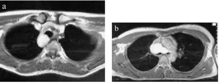
Figure 1. Preoperative MRI image of each case.
(a) Case 1 : T2-weighted MRI image demonstrating a high-intensity, cystic tumor 2 cm in diameter approximately 3 cm from the carina on the right and towards the head.
(b) Case 2 : T2-weighted MRI image demonstrating a high-intensity, wavy cystic tumor.
Figure 2. Perioperative findings in case 1.
(a) A tumor was present in the dorsum of the SVC. It was spherical with a smooth surface. (b) The tumor was adhered powerfully to the right side of the trachea.
(c) Bronchoscopy on postoperative hospital Day 18 revealed the formation of granulation tissue at the repair site (right side of the cartilaginous portion of the trachea) , but there were no postoperative complications, and progress was good.
M. Yoshida, et al. Bronchogenic cyst with severe adhesion to the trachea
DISCUSSION
The respiratory organs and esophagus have the foregut as their primordium. During gestational week 7, this duct lengthens vertically, leading to the separation of the respiratory system anteriorly and the pharyngoesophageal system posteriorly. Separation of a portion of these structures will result in a mediastinal bronchogenic cyst or esophageal cyst. According to 1995 statistics of the Japanese Association for Thoracic Surgery, mediastinal cysts comprise 14.1% of all mediastinal tumors, and the frequency of bronchogenic cysts is the highest among mediastinal cystic disease. The sites of occur-rence are the paratrachea, tracheal carina, pulmo-nary hilum, and paraesophagus (1) .
The first choice of treatment for bronchogenic cysts is surgical resection. Now, because of the re-cent dissemination of the thoracoscopic procedure, thoracoscopic removal or thoracoscopic-assisted re-moval is the method of first choice (1, 2). A thora-coscopic procedure is of course superior for reduc-ing postoperative pain, the number of inpatient days, and for cosmetic reasons. However, surgeons have experienced some cases of bronchogenic cyst strongly adherent to various adjacent organs, as we have presented.
We experienced two bronchogenic cyst cases with severe adhesion to the trachea among 14 cases of bronchogenic cysts in our hospital. These two bron-chogenic cysts were located in the right paracheal region, and were adhered strongly to the tra-cheal wall. The cystic wall and tratra-cheal wall were “shared”. In both cases, we first selected a thora-coscopic procedure, but had to convert to limited thoracotomy because of strong cystic adhesion to the trachea. In the fist case, we could not separate the cystic wall from the tracheal wall, and subse-quently damaged the latter. The damaged site was sutured with an absorbent thread, and was cov-ered with the fifth intercostal muscle. In the second case, the strongly adhesive portion of the cystic wall was left attached to the tracheal wall so as not to damage the tracheal wall, and the inner side of the residual cystic wall underwent dull curettage to re-move the epithelial component. Neither case had postoperative complications.
Kanemitsu, et al. reported one case of incomplete resected cyst wall because of tight adhesion to the trachea in 11 mediastinal bronchogenic cyst cases (3). Ribet, et al. also reported one case of injury to the trachea and 2 cases of incomplete resected cyst wall in 43 mediastinal bronchogenic cyst cases (4). Recently, Martinod et al. reported thoracoscopic
ex-Figure 3. Methods of treatment (a) Case 1
The tracheal wall and cystic wall were shared, and the bronchial wall was consequently damaged during separation and was sutured with an absorbent thread. The site was then covered with the fifth intercostal muscle.
(b) Case 2
The cystic wall on the tracheal side was spared, and the interior of the cystic wall underwent dull curettage to remove the epithelial component.
cision of 20 mediastinal bronchogenic cysts. In 7 patients, they had to convert from thoracoscopy to thoracotomy. Eleven (55%) bronchogenic cysts had strong adhesions to vital structures, and a portion of the cyst wall was left in place in 5 cases. There were no late complications and no cyst recurrence (5).
We suggest two reasons for severe adhesion to the trachea. One reason is incomplete separation between the cystic wall and the trachea during em-bryogenesis. The other reason is that infection is caused because a fine fistula is present between the trachea and the cyst. As a result, acute inflamma-tion is caused and the cyst adheres to the trachea.
We think that the reason for adhesion to the tra-chea is the former one because our two cases were asymptomatic.
There are various bronchogenic cysts ranging from cases with easy resection using thoracoscopy to cases which are difficult to resect because of strong cystic adhesion to the neighboring organs. In these difficult cases, including the 2 presented here, conversion to a supplementary limited thora-cotomy is strongly advised. Also, a small patch of the cystic wall should be left in place after destruc-tion of the mucosal lining to avoid recurrence.
REFERENCES
1. Fujimoto T, Yamanaka A, Hirai T : Application of thoracoscopic surgery handicraft for medi-astinal cysts. Nippon Kokyuu Gekagakkaishi 13 (7)(in Japanese) : 15-19, 1999
2. Urschel J, Horan TA : Mediastinoscopic treat-ment of mediastinal cysts. Ann Thoracic Surg 58 : 1698-1701, 1994
3. Kanemitsu Y, Nakayama H, Asamura H, Kondo H, Tsuchiya R, Naruke T : Clinacal features and management of bronchogenic cysts. Report of 17cases. Surgery Today Jpn J Surg 29 : 1201-1205, 1999
4. Michel ER, Marie CC, Bernard G : Broncho-genic cysts of the mediastinu- m. J Thorac Car-diovasc Surg 109 : 1003-1010, 1995
5. Martinod E, Pons F, Azorin J, Mouroux J, Dahan M, Faillon JM, Dujon A, Lajos PS, Riquet M, Jancovici R : Thoracoscopic exci-sion of mediastinal bronchogenic cysts : Re-sults in 20 Cases. Ann Thorax Surg 69 : 1525-1528, 2000
M. Yoshida, et al. Bronchogenic cyst with severe adhesion to the trachea