Introduction
Dayak ethnic group is the indigenous people of Kalimantan Island and mainly live in a remote area. Dayak ethnic group in West Kalimantan is divided into 151 sub-ethnic groups and their languages are classified into 168
groups (Alloy et al. 2008). Since their residential areas are isolated from other villages, they depend on their environment, especially, the forest. It functions as a place to meet their basic needs because they have the knowledge how to utilize natural resources. Especially, plants have been used as medicine to treat diseases. Therefore, medicinal plants are known as their
Regulatory effects of five medicinal plants used by Dayak
Uud Danum in West Kalimantan Indonesia on the
delayed-type hypersensitivity and the inflammation of human colon
epithelial cells
Yeni Mariani
1,2*, Fathul Yusro
1,2, Yuko Konishi
3, Takahiro Taguchi
1,
Akira Tominaga
11 Division of Human Health and Medical Science, Graduate School of Kuroshio Science, Kochi University, Okoh-Cho, Kohasu, Nankoku, Kochi 783-8505 Japan
2 Faculty of Forestry Tanjungpura University, Pontianak-Indonesia
3 Life and Functional Material Section, Science Research Center, Kochi University, Kohasu, Nankoku, Kochi 783-8505 Japan
Abstract
We evaluated the plants used to treat inflammatory diseases by Dayak Uud Danum by examining the anti-allergic activity and the preventive effects on the damage of colon epithelial cells. The plant species examined are Tekeriho (Callicarpa longifolia Lam), Penahan (Myrmeconauclea strigosa Merr.), Tebelion (Eusideroxylon zwageri Teijsm & Binn.), Kerokak (Scoparia dulcis L.) and Bungur (Lagerstroemia speciosa (L.) Pers.). Anti-inflammatory activities of methanol extracts from leaves of these plants were analyzed in terms of delayed-type hypersensitivity against 2, 4, 6-trinitrochlorobenzene using BALB/cAJc mice and in vitro model of intestinal inflammation using FPCK-1-1 human colon epithelial cells. The yield percentage of methanol extract ranges from 4.33 to 8.99%. Extracts from C. longifolia, M. strigosa, E. zwageri, and S. dulcis have suppressed delayed-type hypersensitivity at both 24 and 48 hours after challenge. These extracts also suppressed the migration of eosinophils to the site of inflammation. L. speciosa extract suppressed the delayed-type hypersensitivity only at 48 hours after challenge. M. strigosa extract showed the preventive effect in the damage model of colon epithelial cells by inhibiting the decrease of transepithelial electrical resistance. Methanol extract of M. strigosa stimulated FPCK1-1 cells to produce mucopolysaccharides and that of C. longifolia induced FPCK1-1 cells to produce IL-22. The production of mucopolysaccharides by FPCK-1-1 cells may explain, in part, the preventive effects of the plant extracts on the damage of human colon epithelial cells. It is suggested that leaves from five medicinal plants used by Dayak Uud Danum have the potential as anti-inflammatory agents. Key words: Medicinal plants, inflammation, delayed-type hypersensitivity: DTH, intestinal epithelial cells
Received June 17, 2016; Accepted August 27, 2016. *Corresponding author e-mail: yeni.mariani81@gmail.com
Research Paper
local wisdom and survival knowledge related to environment. Nowadays, the utilization of medicinal plants is facing several threats due to the scarcity of this knowledge and forest condition. Generally, this knowledge of the utilization of medicinal plants is not well documented since it is orally transferred from generation to generation. While younger generation accepts new culture from outside of their village, the knowledge of medicinal plants is fading. Forest conversion also contributed to the decreasing number of medicinal plant species. Approximately 21.51% of 9,125,486 ha of forest that functions as the main habitat of medicinal plants in West Kalimantan have been lost since 2010 (Sardana et al. 2011). Therefore, to conserve the knowledge of medicinal plants, it is necessary for the Dayak people to use them continuously to treat diseases.
The documentation of medicinal plants utilization of several Dayak sub-ethnic groups is already reported (Diba et al. 2013, Yusro et al. 2014). Dayak Uud Danum means a Dayak sub-ethnic group who lives in the upstream areas of Ambalau and Serawai river of Sintang Regency. In Dayak Uud Danum community, people use 95 species of medicinal plants (Mariani, unpublished data). Among of these medicinal plants, Tekeriho (Callicarpa longifolia Lam.), Penahan (Myrmeconauclea strigosa Merr.), Tebelion (Eusideroxylon zwageri Teijsm & Binn.), Kerokak (Scoparia dulcis L.) and Bungur (Lagerstroemia speciosa (L.) Pers.) have been used to treat diseases such as diarrhea, fever and allergy as the decoction of leaves by hot water. These diseases often caused by infections induce inflammation. Therefore, these medicinal plants are suggested to have anti-inflammatory activity.
Inflammation is one of process of natural protection to maintain the homeostasis of a living organism against tissue injury, infection or irritation (Mubashir et al. 2013). Although inflammation is necessary to protect hosts from infection and maintain homeostasis, it should be under control.
Cytokines are the proteins released by cells at the site of inflammation to regulate the inflammatory reaction (Zhang and An 2007). Some cytokines have a role to up-regulate the inflammation and called pro-inflammatory cytokines, while others have a role to down-regulate the inflammation and called anti-inflammatory cytokines (Dinarello 2000). So, the regulation of the pro-inflammatory cytokines is one way to control an inflammation.
Inflammation is one of causes of diarrhea (Brijesh et al. 2011, Hodges and Gill 2010) and plays an important role in the allergic disease such as delayed-type hypersensitivity (DTH) (Tominaga et al. 2011, Yoshino et al. 2010). Diarrhea is difficult to be fully recovered in a patient with an inflammatory bowel disease (Binder 2009). Prolonged inflammation of intestine may lead to the increasing risk of colon cancer (Kaser et al. 2010). Not only in cutaneous skin
infection, but also in patients bearing transplanted tissues and tumors, DTH must be considered.
Since both IFN-γ and TNF-α are involved in the inflammation of DTH and colon epithelial cells (Bruewer et al. 2003 and Tominaga et al. 2010), it is useful to examine the anti-inflammatory effects of medicinal plants on DTH and the inflammation of colon epithelial cells.
Among five medicinal plants we chose from Dayak Uud Danum, four of them were already investigated by several scientists and some of their biological activities are clarified. C. longifolia has bacterial, diabetic, analgesic, anti-pyretic, anti-fungal, and anti-inflammatory activities (Soni et al. 2014, Yadav et al. 2012a, Yadav et al. 2012b). E. zwageri has anti-melanogenesis activity (Arung et al. 2009). S. dulcis has anti-diabetic (Pari and Latha 2004, Perumai et al. 2014), while L. speciosa has oxidant (Unno et al. 1997), anti-gout (Unno et al. 2004), and anti-microbial (Nasrin et al. 2012) activities. Thus, except for M. strigosa, the activities of their plant extracts were reported. However, anti-inflammatory activities using a model of colon epithelial cell damage and DTH are not yet reported.
In this report, anti-inflammatory properties of medicinal plants were examined by using two approaches. Anti-inflammatory effects of plant extracts on colon epithelial cells were evaluated using FPCK-1-1 colon epithelial cells established from a tubular adenoma in a male familial polyposis coli patient. Regulatory effects of medicinal plants on allergic inflammation were analyzed in a delayed-type hypersensitivity response against 2,4,6-trinitrochlorobenzene using BALB/cAJc mice by measuring the suppression of ear swelling after the challenge with antigen. The leaves were selected for these experiments because this is the part of plants used by Dayak Uud Danum and they allegedly have the anti-inflammatory effects.
We found that all of the medicinal plants have anti-inflammatory activities equivalent to hydrocortisone except L. speciosa in the DTH assay. Extracts of all plants except L. speciosa suppressed the increment of ear thickness and inhibited the migration of eosinophils to the site of inflammation. Although the extract of L. speciosa suppressed the ear swelling in the DTH response 48 hours after challenge, the degree of suppression was much weaker at 24 hours after challenge compared with other plant extracts. Among these plants extracts M. strigosa extract showed preventive effect on the inflammation of colon epithelial cells.
Materials and Methods
Animals
from CLEA Japan, Inc. (Osaka, Japan) and used at the age of 8 weeks. All experiments were conducted under the guidelines of animal experiments of Kochi University.
Cell lines
FPCK-1-1 cells are precancerous colon epithelial cells derived from a male patient with familial adenomatous polyposis (Kawaguchi et al. 1991). THP-1 cells (Human monocytic leukemia) were purchased from Health Science Research Resources Bank, Japan Health Science Foundation, Osaka, Japan (Tsuchiya et al. 1982). Both FPCK-1-1 and THP-1 cells were maintained in Dulbecco’s-modified Eagle Medium (DMEM-high glucose) supplemented with 8% FCS, 20 U/ml penicillin, 50μg/ml kanamycin in 5% CO2at 37 C for several days. FPCK-1-1 cells were sub-cultured on Transwell permeable membrane with pores of 0.4μm and area 1.1 cm2 pre-coated with an equimolar mixture of collagen types I and III (3493, Corning, Ithaca, USA).
Chemicals
Picryl chloride (PCl; 2, 4, 6-trinitrochlorobenzene) was purchased from Nacalai Tesque (Kyoto, Japan). Cyclophosphamide (CY) was purchased from Shionogi Pharmaceutical Co. (Osaka, Japan). Hydrocortisone was purchased from Sigma-Aldrich (St. Louis, MO, USA).
Plant Materials and Extraction
Sample Collection
All samples described below were medicinal plants species used by Dayak Uud Danum and collected from Ambalau District of Sintang Regency, West Kalimantan Province, Indonesia: Tekeriho (C. longifolia), Penahan (M. strigosa), Tebelion (E. zwageri), Kerokak (S. dulcis), and Bungur (L. speciosa) (Fig. 1). After being dried in the air for 2 weeks, 40 grams of dried leaves were milled with an electric grinder (Oster, Sunbeam Products, Inc.) to obtain fine powder. Voucher specimen of all plants samples were deposited in the Laboratory of Wood Technology, Tanjungpura University Pontianak to identify the scientific name of each plant.
Extraction
Thirty grams of powdered leaves were extracted with 100 ml of methanol (99.7%) by using soxhlet extractor (Yamato Water Bath BS660, Yamato Scientific Co. Ltd) for 1 hour at 70 C, the extraction was repeated three times followed by the filtration with Whatman filter paper No. 2. The filtrated samples were evaporated with a vacuum rotary evaporator (Eyela No-1000, Tokyo, Japan) with a speed of 5 rpm at 40 C. The evaporated samples were dried for one day in a wind dryer (Pierce, Reacti-Therm and Reacti-Vap, Thermo Fisher Scientific Inc., Waltham, MA). Drying process was continued with a vacuum dryer (Ettas, AVO-250NB, Active Co. Saitama, Japan) for one day and the final residue was obtained. The percentage of the residues was determined at this point (Fig. 2).
Preparation of plant extracts
All dry methanol extracts were grinded in a mortar and suspended in distilled water (DW) at a concentration of 50 mg/ml. Then, these samples were homogenized with a Polytron (Kinematica, Luzern, Switzerland). The homogenized Fig. 1. Medicinal plants of Dayak Uud Danum used in this
study to analyze the anti-inflammatory effects.
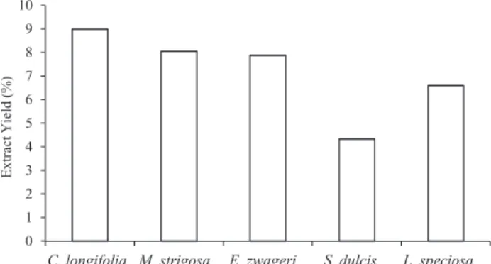
Fig. 2. Yield percentage of methanol extracts from plant leaves. Extractions were performed as described in Materials and Methods.
extracts were heated at 75 C for 30 minutes and left at room temperature overnight, then heated at 75 C again. Samples were stored at 4 C before use.
Immunization and Challenge
Two days before being immunized with PCl, mice were injected with cyclophosphamide subcutaneously (150 mg/kg in distilled water) at the nape of the neck. This injection was to remove proliferating immunosuppressive cells (Satoh et al. 1997). Abdominal coat hairs were removed and mice were immunized by painting abdominal skin with 0.05 ml of 7% PCl in ethanol: acetone (3:1). This process of immunization was repeated on the next day. Five mg of each plant extract suspended in a volume of 0.1 ml was orally administered to each mouse by using a polyethylene capillary every other day for two weeks after immunization. In one group, each mouse was orally administered with 0.5 mg hydrocortisone suspended in a volume of 0.1 ml distilled water every other day for two weeks after immunization. Mice of a positive control group (immunized and challenged) were administered with the same volume of distilled water. Two weeks after administration mice were challenged by painting 0.02 ml of 1% of PCl in acetone: olive oil (1:4) on each ear lobe. Ear thickness of each mouse was measured by using a dial thickness gauge (Peacock G-1M, Ozaki Mfg. Co. Ltd., Tokyo) before and after challenge.
Tissue Eosinophil Counts
Mice were sacrificed 48 hours after the challenge. The ear lobe of each mouse was removed and fixed with 8% paraformaldehyde in a 0.2 M Phosphate buffer with pH 7.2, and embedded in paraffin. Next, the sections of ears were stained with a hematoxylin/eosin solution. At the level of magnification of 400x the number of eosinophils was counted and expressed as number of cells in the observed area. The number of eosinophils was counted in 10 squares of 100 μm x 50μm of ear sections from each group.
Co-culture system to treat colon epithelial cells
FPCK-1-1 monolayer cells cultured at a density of 2 x 10⁵cells/insert (Transwell permeable membrane) in 12 wells plate (Corning 3513). THP-1 cells were cultured at density of 1 x 10⁵/well of 12 wells plate in the presence of phorbol 12-myristate 13-acetate (PMA 20 nM) for one day. After five days, the inserts with FPCK-1-1 cells were transferred into the wells where the THP-1 cells were cultured with PMA (lower chamber). The co-culture was maintained for three days to monitor the transepithelial electrical resistance of the FPCK-1-1 monolayer cells. In other co-culture, the methanol extracts ofC. longifolia, M. strigosa, E. zwageri, S. dulcis and L. speciosa were added to the upper chamber (insert) of the co-culture system at the time when co-culture started. The final concentration of each sample (1μg/ml) was determined by culturing NIH3T3 murine fibroblast cells in the presence various concentrations of each plant extract.
Measurement of transepithelial electrical resistance
(TER)
The TER of FPCK-1-1 monolayer cells was determined by measuring the electrical resistance between the lower chamber (well) and the upper chamber (filter insert) using a Millicell-ERS voltmeter with a MERSSTX01 electrode (Millipore, Bedford, MA). Two hours before measurement the medium in the transwell was changed and the electrode was soaked in 70% ethanol then rinsed with sterile DMEM and the temperature was maintained close to 37 C. The measurement of TER was repeated four times and the mean value was calculated. The TER value of FPCK-1-1 monolayer cells at the time of starting a co-culture was expressed as 100 % (TER value for FPCK-1-1 monolayer cells was 222 − 267 Ω x cm2 in this experiment).
Staining of mucopolysaccharide on the
FPCK-1-1 cell monolayer cells on the filter membrane
FPCK-1-1 monolayers cells were stained with Alcian blue (Muto Pure Chemicals Co., Ltd, Tokyo, Japan) according to manufacturer’s protocol.
IL-22 measurement in the supernatant
IL-22 released in the supernatant from FPCK-1-1 cells in the upper chamber was measured according to the manufacturer’s protocol using a Human IL-22 Quantikine ELISA kit (R&D System, Inc. Minneapolis, MN).
Results and Discussion
Methanol extraction yield from leaves
Among all the medicinal plants used by Dayak Uud Danum community to treat diseases such as diarrhea, fever and allergy, C. longifolia, M. strigosa, E. zwageri, S. dulcis and L. speciosa are most commonly used. Another reason to choose these five plants is that all these plants species are abundant and easy to find.
According to Departemen Pertanian (1976) wood chemical components can be classified into three levels (extractive content: < 2% lower, 2-4% moderate and > 4%
higher). From present results, all the plant species are categorized into higher level of extractive content after the methanol extraction with a yield ranged from 4.33%-8.99% (Fig. 2). The highest yield was from C. longifolia followed by M. strigosa, E. zwageri, L. speciosa, and S. dulcis.
The extractive yield content levels vary among the plant species. In general, plant extractive content ranges from less than 1% to more than 10%, while it is about 20% in a tropical wood (Tsoumis 1991). The different part of plants like woods, leaves, barks, roots and branches give differences in yield and chemical compound type. Generally, extraction from barks will give higher extractive yield followed by leaves, roots and stems (Sjostrom 1981).
All extracts used in this study derived from leaves, it assumed that higher extractive yield content of all plants related to the presence of chlorophyll. In the extraction process, the usage of polar solvent will dissolve chlorophyll resulting in the higher yield (Harborne 1996). Not only because of its high efficiency in extraction, methanol is generally used to acquire the intracellular components, because methanol penetrates the cell membrane expeditiously (Silva et al. 1998). As a polar solvent, methanol, ethanol and water are very effective to isolate bioactive compounds (Filho 2006). From methanol extraction, generally some chemical compounds such as oils, fats, waxes, alkaloids, flavones, polyphenols, tannins, saponins, glycosides aglycones are obtained (Filho 2006, Houghton and Raman 1998).
Alkaloids, flavonoids, condensed tannins, gallotannins, flavones, flavonols, flavanones, anthocyanidins, isoflavonoids, coumarins, isoquinolines, indoles, diterpenes, saponins, sterols, terpenoids and essential oils are chemical compounds from plants secondary metabolites known for their anti-inflammatory activity (Mohammed et al. 2014).
Except the methanol extract from M. strigosa leaves, some of the bioactive compounds of others plants are already identified. Methanol extracts from M. strigosa leaves may also contain useful bioactive compounds. It is also highly possible that other plant extracts may have unknown function. Since we perform functional assays that are never tried, we can expect that we will find new functions of these plant extracts.
Five plant extracts described above were applied to the following two functional assays: A delayed-type hypersensitivity that is characterized by eosinophilia and a prevention of damage of human colon epithelial cells caused by activated macrophage-like cells.
The effects of methanol extracts from leaves on
DTH in BALB/cAJc mice
At first, we investigated the anti-inflammatory effect of methanol extracts from five medicinal plant species of Dayak
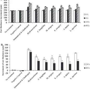
Uud Danum using the DTH response against picryl chloride (PCl). Plant extracts were orally administered for two weeks after immunization till one day before challenge and the ear thickness was measured 4, 24, and 48 hours after challenge. PCl is a hapten that usually used in in vivo model of T cell-mediated immunity assays to induce the DTH. As shown in Fig. 3, there were significant differences in ear thickness between a negative (immunized but not challenged) and a positive control (immunized and challenged). All mice except a negative control and normal mice exhibited inflammatory symptoms after being challenged with PCl that were recognized as ear thickness and active behaviors of mice such as scratching ears.
At 24 hours (h) after challenge, the increment of ear thickness of mice in a positive control was 148 μm in average. Mice administered with C. longifolia and M. strigosa extracts
Fig. 3. Effect of methanol extracts from five medicinal plants of Uud Danum on DTH response. DTH response was elicited against PCl as described in Materials and Methods. Hydrocortisone was administered orally at a dose of 0.5 mg/0. 1ml DW/mouse. Plants extracts were administered orally at a dose of 5 mg/0. 1ml DW/mouse. All reagents were administered every other day for two weeks after immunization. Increment of ear thickness was measured before (0h) and after challenge (4, 24 and 48 h). Results are shown as the mean ± SE (n = 5). Panel A: Ear thickness of each group after the challenge with PCl. Panel B: Increment of ear thickness was shown by subtracting the ear thickness before challenge from the ear thickness of each mouse. *Asterisks indicate that there are significant differences between positive control group and other groups (P < 0.01, Tukey-HSD, one-way ANOVA)
showed lower increment of ear thickness (78μm and 86μm, respectively) than those with hydrocortisone (96μm), followed by S. dulcis, E. zwageri, and L. speciosa (97μm, 98μm, and 122μm, respectively).
At 48 h after challenge, the increment of ear thickness of mice in a positive control was 129.5μm in average. Interestingly, S. dulcis extract strongly suppressed the increment of ear thickness (44.5μm) followed by extracts from M. strigosa, E. zwageri, C. longifolia and L. speciosa (50μm, 56.5μm, 57.5 μm, and 65μm, respectively). The increment of ear thickness of mice administered with hydrocortisone was 66μm.
As shown in Fig. 3, all methanol extracts had the anti-inflammatory activity to suppress the DTH response efficiently. At 24 hours after challenge, C. longifolia extract showed the highest suppression the DTH response (47.3%) followed by extracts from M. strigosa, E. zwageri, and S. dulcis with suppression of 41. 9%, 33. 8% and 34. 5%, respectively (% suppression = 1 - (the increment of ear thickness of each group/the increment of ear thickness of a positive control) x 100). At 48 h after challenge, S. dulcis extract showed the highest suppression (65.6%) followed by extracts from M. strigosa, E. zwageri and C. longifolia (61.4%, 56.4% and 55. 6%, respectively). L. speciosa showed the lowest suppression on 24 h after challenge (17.6%), but at 48 h after challenge it finally suppressed about 50% of the increment of ear thickness (49. 8%) of a positive control. At 24 h after challenge only one group of mice treated with L. speciosa did not show significant difference with a positive control group. But at 48 h after challenge, all groups of plant extracts showed significant difference with a positive control group and higher level of suppression than that of hydrocortisone. The levels of suppression of ear swelling at 24 and 48 hours after challenge in the group administered with hydrocortisone were 35.14% and 49.03%, respectively (Fig. 3).
DTH or Type IV allergy is characterized by the activation of T cells and macrophages to elicit ear swelling (Hou et al. 2006, Yoshino et al. 2010). Usually, the symptoms of DTH related to contact dermatitis. The activation of T lymphocytes and macrophages induced the production of pro-inflammatory cytokines such as IFN-γ and IL-17 (Ishii et al. 2010, Tominaga et al. 2010, 2011, Yoshimoto et al. 2000).
IFN-γ is engaged not only in the DTH but also in the host defense by killing tumors and intracellular microorganisms. As a potent macrophage activator IFN-γ also has a role as an immuno-regulatory cytokine. IL-17 is also engaged in the DTH, the host defense against bacterial infection, and the angiogenesis. In the DTH against PCl, both anti-IFN-γ and anti-IL-17 antibodies partially inhibit the ear swelling (Tominaga et al. 2010).
So, it is suggested that the down-regulation of the level of these two pro-inflammatory cytokines can be a target of these
plant extracts to suppress the DTH response. These results suggest that medicinal plants used by Dayak Uud Danum can be applied to reduce the inflammatory response in which T lymphocytes and macrophages are involved.
Medicinal plants have been used to treat diseases caused by the malfunction of the immune system. Many reports have been published to prove that chemical compounds from medicinal plants have immunosuppressive effects (Amirghofran 2012). Currently, it is well known that secondary metabolites from natural products such as medicinal plants have a broad spectrum of biological activities, and the anti-inflammatory activity is one of them. Flavonoid group, one group of secondary metabolites, has anti-inflammatory activity and is used to develop a new type anti-inflammatory agent (Kim et al. 1993).
Recently, some of the plant species in this study are identified to have bioactive compounds through phytochemical screening. C. longifolia leaves are reported to have alkaloid, phenolic, flavonoid, saponin and steroid compounds (Erwin et al. 2015). The genus of Callicarpa is already known for its bioactive compounds such as terpenoids (diterpenes and triterpenoids), flavonoids, phenylethanoids, and pherypranoids and some of these compounds were reported to have anti-inflammatory activity (Soni et al. 2014, Tu et al. 2013).
Tannins, steroids (tritepenoids), alkaloids, and flavonoids were found in the extract from leaves of S. dulcis (Abere et al. 2015, Wankhar et al. 2015), while the diterpene scoparinol from S. dulcis is reported to have anti-inflammatory activity (Murti et al. 2012). Triterpenes, steroids (triterpenoids), tannins, glycosides, flavones, and ellagic acid were found in the extract of leaves of L. speciosa (Chan et al. 2014, Nasrin et al. 2012, Priya et al. 2008).
Among these medicinal plants used in this study, there are various bioactive compounds that might contribute to the suppression of ear thickness in this model of DTH response. It is also conceivable that secondary metabolites of medicinal plants may have anti-inflammatory activities and affect to several pathways of various phases of inflammation (Dawid 2013). Presence of potent bioactive compounds such as phenolic and flavonoid compounds may contribute to anti-inflammatory activities (Oskoueian et al. 2012).
Othman et al. (2015) stated that anti-inflammatory activity of plant phenolic compound is due to its activities related to oxygen or nitrogen bond. Medicinal plant remedies have a different mode of action from modern drugs in treating diseases. Modern drugs contain a single active compound that works on a specific pathway, while medicinal plant remedies have several molecules working together on several pathways (Kumar et al. 2013). Therefore, it is suggested that medicinal plants remedies have less and weaker side effects than modern drugs. In the Dayak Uud Danum community, although the
commercial drugs are available, they still use their traditional medicinal plants to treat some diseases related to inflammation.
Eosinophils are substantial leukocytes that appear in various inflammatory diseases and have important roles in the regulation of inflammation. Increasing number of eosinophils in the blood or in the tissues characterize the allergic or parasitic disorder symptoms (Rogerio et al. 2010). In DTH, the challenge to paint PCl on ears elicits the increment of ear thickness resulting in the proliferation and migration of eosinophils to the site of inflammation.
The migration and accumulation of eosinophils to the site of inflammation or tissues are related to several diseases such as asthma, rhinitis, atopic dermatitis and inflammatory bowel diseases. Eosinophils release cytotoxic granular proteins resulting in the tissue damages (Rankin et al. 2000). Therefore, the down-regulation of eosinophils is one of the important criteria to find new therapeutic inflammatory drugs.
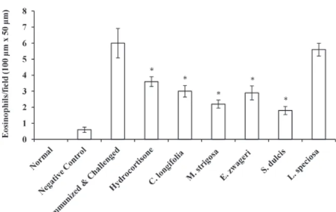
We measured the number of eosinophils in the ear section at 48 h after challenge and compared the number of them among groups of experiments, including non-treated, negative control, positive control groups, and those administered with hydrocortisone and five plants extracts used by Dayak people. We measured the number of eosinophils in 10 fields (100μm x 50μm) of ear sections from each group and the average number of eosinophils was determined (Fig. 4). The number of eosinophils in ear sections from a negative control group was slightly larger than those from non-treated group. This is probably caused by the immunization with PCl. Number of eosinophils of a positive control group was more than ten-fold compared with that of a negative control group. The lowest number of eosinophils was found in a group treated with S. dulcis extract (1. 8) followed by groups treated with M. strigosa, E. zwageri, and C. longifolia extract (2.2, 2.9 and 3, respectively). Interestingly, the group of mice administered with hydrocortisone had slightly higher of level of eosinophils (3.6) than those treated with plant extracts from S. dulcis, M. strigosa, E. zwageri, and C. longifolia except the mice treated with L. speciosa (5. 6) (Fig. 5). Higher the level of ear thickness, more eosinophils were found, suggesting the importance of the migration of eosinophils to the site of inflammation in this DTH response (Tominaga et al. 2011).
While eosinophils migrate from bone marrow to blood, differentiation and proliferation of eosinophil precursors are regulated by cytokines such as IL-3, granulocyte-monocyte colony-stimulating factor (GM-CSF), and IL-5. Among these cytokines, IL-5 is a most effective one to proliferate eosinophils selectively (Rogerio et al. 2010, Satoh et al.1997). In an allergic inflammatory response, several eotaxins such as CCL11 (eotaxin), CCL24 (eotaxin-2) and CCL26 (eotaxin-3) are also involved in the migration of eosinophils. The reduction of IL-4, IL-5, CCL11 and leukotriene B4 (LTB4)
may lead to the reduction of eosinophils at the site of inflammation (Fortunato et al. 2012, Menzies-Gow et al. 2002). Interrupting cell adherence or chemotaxis by the administration of hydrocortisone is also able to down-regulate the proliferation and the migration of eosinophils (Altman et al. 1981). Teixeira et al. (2001) stated that the expression of adhesion molecules (CAMs) on endothelial cells and leucocytes also have a role in eosinophil migration while the activation of eosinophils are induced by the presence of chemokines and (LTB4).
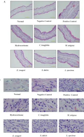
Fig. 4. Effects of plant extracts on the DTH response were shown as the ear swelling and the suppression of eosinophils migrated to the site of inflammation. DTH response was elicited against PCl as described in Materials and Methods. Forty-eight hours after challenge, ears were removed and fixed with 8% paraformaldehyde in a 0.2 M Phosphate buffer with pH 7.2, and embedded in paraffin, then stained with hematoxylin and eosion. Panel A. Mice ear sections with 100 x magnification Bars, 200μm. Panel B. Mice ear sections with 400 x magnification. Bars, 50μm. Leukocytes with red cytoplasm are eosinophils.
Most of the groups administered with medicinal plant extracts had lower number of eosinophils at the site of inflammation except that with L. speciosa, which had the number of eosinophils equivalent to that of a positive control. At 48 h after challenge, L. speciosa, however, still suppressed ear thickness. It is clear that extracts from S. dulcis, M. strigosa, E. zwageri and C. longifolia have anti-eosinophilia activity in their methanol extracts of leaves and those plant extracts may have the ability to down-regulate the inflammatory cytokines such as IL-5, or eotaxin resulting in the inhibition of the proliferation of eosinophils in bone marrow and spleens and the inhibition of the migration of eosinophils to the site of inflammation. Or, these plant extracts may contain molecules that suppress the migration of eosinophils.
Although the extract from L. speciosa suppressed the ear thickness moderately, it did not inhibit the migration of eosinophils, suggesting that L. speciosa extract may have an anti-inflammatory compounds which does not inhibit the migration of eosinophils to the site of inflammation. In other words, L. speciosa plant extract may have suppressed the inflammation by inhibiting the process other than the one involved in eosinophilia. It may be a good idea to administer L. speciosa plant extract together with that of M. strigosa or S. dulcis which has a potent activity to suppress the migration of eosinophils to the site of inflammation.
Various secondary metabolites of plants extracted from barks, stems, fruits, leaves and roots such as flavonoids, alkaloids, polyphenols, quionones, sesquiterpenes, terpenoids
and diterpenoids are scientifically proved to reduce the eosinophil migration to the site of inflammation (Rogerio et al. 2010). It is highly suggested that methanol extracts of Dayak Uud Danum medicinal plants especially those from S. dulcis, M. strigosa, E. zwageri and C. longifolia have the active compounds which inhibit the migration of eosinophils to the site of inflammation from bone marrow or spleen.
Prevention of colon epithelial cell damage by
plant extracts
In order to analyze the anti-inflammatory activity of methanol extracts of medicinal plants, an in vitro damage-prevention model of colon epithelial cells was employed. TER of FPCK-1-1 monolayer cells was measured during the co-culture with PMA-stimulated THP-1 cells.
On the third day of the co-culture, there was 13. 4% decrease in TER when FPCK-1-1 monolayer cells were co-cultured with PMA-stimulated THP-1 cells (Fig. 6). M. strigosa extract showed highest TER restoration and there is no significant difference between FPCK-1-1/THP-1 group and Fig. 5. Number of eosinophils migrated at the site of
inflammation. Eosinophil numbers of each group were counted at the magnification level of 400x. Results are shown as the mean of eosinophils in 10 squares of 100μm x 50μm of ear sections ± SE (n = 10). * Asterisks indicate that there are significant differences between positive control and other groups (Hydrocortisone, C. longifolia, M. strigosa, E. zwageri, and S. dulcis) (P < 0.05, Tukey HSD).
Fig. 6. Effects of plant extracts on the damage of FPCK-1-1 monolayer cells induced by PMA-stimulated THP-FPCK-1-1 cells. TER was measured during the co-culture of FPCK-1-1 cells with PMA-stimulated THP-1 cells. Results are shown as the mean ± SE (n = 4). *An asterisk indicates that there is a significant difference between FPCK-1-1/PMA-THP-1 group and M. strigosa group. There is no significant difference between control FPCK-1-1/THP-1 group and M. strigosa group (P < 0.01, Tukey-HSD).
M. strigosa group on the third day of the co-culture. Other plant extracts did not show any significant activity in this damage-prevention model of human colon epithelial cells.
One of the criteria to prevent the damage of FPCK-1-1 colon epithelial cells is the mucus production of epithelial monolayer cells. In order to analyze the level of mucopolysaccharide on the surface of FPCK-1-1 monolayer cells, we stained the FPCK-1-1 monolayer cells in the upper chamber three days after starting the co-culture. We compared the FPCK-1-1 monolayer cells after being treated with methanol extract of leaves with those co-cultured with PMA-stimulated THP-cells in the absence of plant extracts. The presence of mucopolysaccharides was detected by using the Alcian blue staining (Fig. 7). Alcian blue is a cationic dye technique that widely used for staining macromolecules like mucopolysaccharides and acidic mucin in tissues and cells (Dong et al. 2012).
As shown in Fig. 7, FPCK-1-1 monolayer cells treated with methanol extract from leaves of M. strigosa showed higher level of staining with Alcian blue compared with other groups, suggesting that mucopolysaccharides are produced by the monolayer cells in response to M. strigosa extract. It is relevant to the higher level of TER value suggesting that M. strigosa extract has the strongest protection against the damage signal from PMA-stimulated THP-1 cells. The
presence of mucopolysaccharides that covers the monolayer cells influences the TER value. The mucopolysaccharides becomes a shield that covers the surface of FPCK-1-1 epithelial monolayer cells and inhibits the movement of ions (Tominaga et al. 2013). Through TER measurement, the paracellular barrier function and tight junction integrity can be determined (Ren et al. 2012). The decreased TER value represents, in part, the increased level of the permeability of the tight junction (Balda et al. 1992). From our results it is suggested that M. strigosa extract has ameliorating effect on the barrier function of intestinal epithelial cells by reducing the paracellular permeability.
The increasing of paracellular permeability followed by the breakdown of tight junction may induce materials to penetrate from lumen to the basolateral side of the epithelium inducing inflammation that finally causes diarrhea (Guttman et al. 2006, Ren et al. 2012). Epithelial barrier disruption may cause the secretion of fluid and electrolytes inducing the loss of mucosal proteins and mal-absorption (Strauman et al. 2010). To preserve the function of intestinal barrier by preventing paracellular permeation induced by harmful agents to lumina, it is important to keep the epithelial tight junction intact (Al-Sadi and Ma 2007).
Next, we measured IL-22 levels released in the apical side of FPCK-1-1 monolayer cells treated with various plants extracts (Table 1). We measured levels of IL-22 in the upper chamber where FPCK-1-1 monolayer cells were cultured, while PMA-stimulated THP-1 cells were cultured in the lower chamber. FPCK-1-1 monolayer cells were cultured in the upper chamber in the presence of plants extracts for three days. Most plant extracts did not induce FPCK-1-1 colon epithelial cells to produce IL-22 except the extract from C. longifolia. Although we expected that M. strigosa extract would induce FPCK-1-1 cells to produce IL-22 because this extract prevented the decrease of TER by inducing FPCK-1-1 cells to produce mucopolysaccharides, it only induced the marginal level of IL-22 (Table 1).
It is reported that IL-22 produced by FPCK-1-1 cells has a role to recover the intestinal mucosal layer (Tominaga et al. 2012, 2013). IL-22 is one of a member of IL-10 cytokine family secreted mainly from CD4 positive T cells, NK cells and dendritic cells. In intestinal inflammation, main role of IL-22 is to induce the epithelial cells to produce proteins and strengthen the mucus barrier by promoting mucin secretion and increase the epithelial cells through the restitution of goblet cells that result in the intestinal recovery (Mizoguchi 2012). IL-22 regulates the tight junction between intestinal epithelial cells through promoting the cell proliferation and the wound healing (Dudakov et al. 2015).
TNF-α is a pro-inflammatory cytokine primarily produced by activated macrophages and T-lymphocytes. TNF-α has Fig. 7. Alcian-blue staining of FPCK-1-1 monolayer cells
treated with plant extracts. FPCK-1-1 monolayer cells were stained with Alcian-blue as described in Materials and Methods three days after starting the co-culture with PMA-stimulated THP-1 cells. Plant extracts were added at the beginning of the co-culture. Bars, 50μm.
role a role to protect hosts against infections by bacteria, parasites and viruses (Bradley 2008). In the inflammation of intestinal epithelial cells, however, TNF-α causes the intestinal damages. It induces the apoptosis of cells and the disruption of tight junctions through disturbing the structure and function of the tight junction resulting in the increase of paracellular permeability (Bruewer et al. 2003, Li et al. 2010). It is reported that the TER value of epithelial cells was recovered by adding the antibodies against TNF-α (Biasi et al. 2013, Tominaga et al. 2013).
Furthermore, Vezza et al (2016) reported that following flavonoid family compounds from several plants have potent anti-inflammatory activities of intestine: glycosides and aglycones, quercetins, flavonols, flavonones, flavones, catechins, isoflavons, anthocyanidins and chalones. These plant secondary metabolites are scientifically proved to have curative effects on colonic damage. These results suggest that methanol extract from M. strigosa leaves may have secondary metabolites that are able to block the signaling pathway of TNF-α, in the absence of IL-22.
Our results suggest that the extract from M. strigosa leaves has anti-inflammatory activity in both DTH response and colon epithelial inflammation. On the contrary, the extracts from S. dulcis, E. zwageri and C. longifolia leaves reduced the ear thickness and inhibited the migration of eosinophils to the site of inflammation in DTH response, but lacked the ability to prevent the damage of human intestinal cells, suggesting that the extracts from these plant leaves regulate different pathways from which M. strigosa manipulate in inflammation.
Inflammation has an important role as one of natural protection mechanism and it has a dual nature like a double-edged sword. Overproduction of several inflammatory mediators
leads to a chronic disease. Therefore, in order to explore anti-inflammatory reagents from natural products, various criteria must be applied to screen medicine.
It is well known that many indigenous people around the world use medicinal plants as anti-inflammatory medicine to treat several diseases. In West Kalimantan Indonesia, although government has been providing adequate health facilities and modern medicines, some people who live in poverty or in remote areas from government health facilities still rely on traditional medicinal plants to treat diseases such as fever, allergy, and diarrhea.
Dayak Uud Danum, indigenous people of West Kalimantan, Indonesia is one of the communities who still use and preserve knowledge of medicinal plants as their local wisdom. Through an ethnopharmacological approach, activities of many bioactive compounds from plants will be determined (Filho 2006). In this study, we examined anti-inflammatory activities of five medicinal plants used by Dayak Uud Danum. The results showed that all of these medicinal plants have anti-inflammatory activities in term of suppressing delayed-type hypersensitivity and one of these medicinal plants, M. strigosa has preventive effect on the damage of human colon epithelial cells.
In this study, the mechanisms of plant extracts that regulate the inflammatory responses are not yet examined. Further experiments are necessary to determine the mechanisms of potential plant extracts to regulate the inflammation.
Conclusions
We examined the anti-inflammatory activity of methanol extracts from leaves of C. longifolia, M. strigosa, E. zwageri, S. dulcis, and L. speciosa in terms of suppressing the delayed-Table 1. Effects of plant extracts on the production of IL-22 by FPCK-1-1 monolayer cells.
type hypersensitivity and the damage of human colon epithelial cells. As a result, we found that all of these medicinal plants have anti-inflammatory activities to ameliorate the DTH response and four plant extracts from C. longifolia, M. strigosa, E. zwageri, S. dulcis inhibit the migration of eosinophils to the site of inflammation. Only the extract from M. strigosa leaves has the preventive effect in the model of inflammation of colon epithelial cells. Further studies are necessary to clarify the function of compounds in plant extracts to regulate the inflammation.
Acknowledgements
We are grateful to Dr. Hiroshi Wakiguchi, President of Kochi University for his support, and we also would like to thank you to Dr. Sota Tanaka, Dr. Yoshiaki Iiguni, Dr. Satoshi Kubota and Dr. Farah Diba for the support and suggestion, and Dayak Uud Danum community in Ambalau district especially Mr. Telabang and his family for providing facilities during field research. Thank you for Syf. Astria, Aloysius Kahariayadi and Arief to help us collect the field information and samples. Methanol extracts from plant leaves were prepared in Dr. Ohtani’s laboratory with the support by Ms. Yui Hashimoto and Ms. Filzah Wahyudin. We appreciate Dr. Ohtani, Ms. Yui Hashimoto and Ms. Filzah Wahyudin for their help.
Conflict of Interest
The authors declare that there is no conflict of interest.
References
Abere, T.A., Okoye, C.J., Agoreyo, F.O., Eze, G.I., Jesuorobo, R.I., Egharevba, C.O. and Aimator, P.O. 2015. Antisickling and toxicology evaluation of the leaves of Scoparia dulcis Linn (Scrophulariaceae). BMC Complement. Altern. Med., 15: 2-7.
Amirghofran, Z. 2012. Herbal medicines for immunosuppresion. Iran J Allergy Asthma Immunol., 11: 111-119.
Alloy, S., Albertus. and Istiyani, C.P., Bamba, J. (ed.) 2008. “Mozaik dayak, keberagaman subsuku dan bahasa dayak di Kalimantan Barat”, Institut Dayakologi. Pontianak. Al-Sadi, R.M. and Ma, T.Y. 2007. IL-1β causes an increase in
intestinal epithelial tight junction permeability. J. Immunol., 178: 4461-4649.
Altman, L.C., Hills, J.S., Hairfield, W.M. and Mullarkey, M.F. 1981. Effects of corticosteroid on eosinophil chemotaxis and adherence. J. Clin. Invest., 67: 28-36.
Arung, E.T., Kusuma, I.W., Christy, E.O., Shimizu, K. and Kondo, R. 2009. Evaluation medicinal plants from Central Kalimantan for antimelanogenesis. J. Nat. Med.,
63: 473-480.
Balda, M.S., Fallon, M.B., Van Itallie, C.M. and Anderson, J. M. 1992. Structure, regulation and pathophysiology of tight junction in gastrointestinal tract. Yale J. Biol. Med., 65: 725-735.
Biasi, F., Leonarduzzi, G., Oteiza, P. I. and Poli, G. 2013. Inflammatory bowel disease: Mechanism, redox considerations, and therapeutic targets. Antioxid. Redox. Signal., 19: 1711-1746.
Binder, H.J. 2009. Mechanisms of diarrhea in inflammatory bowel disease: Molecular structure and function. Ann. N. Y Acad. Sci., 1165: 285-293.
Bradley, J.R. TNF- mediated inflammatory disease. J.Pathol., 214: 149-160.
Brijesh, S., Tetali, P. and Birdi, T.J. 2011. Study on effect of anti diarrheal medicinal plants on enteropathogenic Escherichia coli induced interleukin 8 secretion by intestinal epithelial cells. Altern. Med. Stud., 1: e16: 64-69.
Bruewer, M., Luegering, A., Kucharzik, T., Parkos, C.A., Madara, J.L., Hopkins, A. M., Nusrat, A. 2003. Proinflammatory cytokines disrupt epithelial barrier function by apoptosis-independent mechanisms. J. Immunol. 171: 6164-6172. Chan, E.W.C.C., Tan, L.N. and Wong, S.K. 2014. Phytochemistry
and pharmacology of Lagerstroemia speciosa: A natural remedy for diabetes. Int. J. Herb. Med., 2:100-105. Dawid, R. 2013. Medicinal plants used in treatment of
inflammatory skin disease. Postep. Dem. Alergol., XXX: 170-177.
Departemen Pertanian (ed.) 1976. “Vademikum Kehutanan Indonesia”. Direktorat Jendral Kehutanan, Jakarta. Diba, F., Yusro, F., Mariani, Y. and Ohtani, K. 2013. Inventory
and biodiversity of medicinal plants from tropical rain forest based on traditional knowledge by ethnic dayaknese communities in West Kalimantan Indonesia. Kuroshio Science, 7: 75-80.
Dinarello, C.A. 2000. Impact of basic research on tomorrow`s medicine: Proinflammatory cytokines. Chest, 118: 503-508.
Dong, W., Matsuno, Y.K. and Kameyama, A. 2012. A Procedure for alcian blue staining of mucins on polyvinylidene difluoride membranes. Anal.Chem., 16: 8461-8466. Dudakov, J.A., Hanash, A.M. and Van den Brink, M.R. 2015.
Interleukin-22: Immunobiology and pathology. Annu. Rev. Immunol, 33: 747-785.
Erwin, Nisa, R. A. and Daniel. 2015. Phytochemical test, toxicity leaves Kereahau (Callicarpa longifolia Lam.) with DPHH method. Indonesian Chim. Acta., 8: 55-59. Fortunato, L.R., Alves, C.F., Teixeira, M.M. and Rogerio, A.
P. 2012. Quercetion: A flavonoid with the potential to treat asthma. Braz. J. Pharm. Sci., 48: 489-599. Filho, R. R. M. 2006. Bioactive phytocompounds: New
approaches in the phytosciences. In Iqbal A et al., (eds.) “Modern Phytomedicine”. WILEY-VCH Verlag GmbH & Co. KGaA. Weinheim, pp. 1-24.
Guttman, J.A., Li, Y., Wickham, M.E., Deng, W., Vogl, A.W. and Finlay, B.B. 2006. Attaching and effacing pathogen-induced tight junction disruption in vivo. Cell. Microbiol., 8: 634-645.
Harborne, J. B. 1996. “Metode Fitokimia, Penuntun Cara Modern Menganalisis Tumbuhan”. Translated by: Padmawinata, K., Soediro, I., Niksolihin, S (ed.). Institut Teknologi Bandung. Bandung.
Hodges, K., Gill, R. 2010. Infectious diarrhea; Cellular and molecular mechanisms. Gut Microbes, 1: 4-21.
Hou, L.F., Dai, Y., Xia, Y.F. and Gong, Z.N. 2006. Alleviation of picryl chloride- induced delayed type hypersensitivity reaction by saponin fraction of Gleditsia sinensis. Biol. Pharm. Bull, 29: 1056-1059.
Houghton, P. J. and Raman, A. and Peter, J. (ed.) 1998. “Laboratory handbook for the fractionation of natural extracts”. Chapman & Hall, London.
Ishii, A., Oboki, K., Nambu, A., Morita, H., Ohno, T., Kajiwara, N., Arae, K., Sudo, H., Okumura, K., Saito, H. and Nakae, S. 2010. Development of IL-17 mediated delayed-type hypersensitivity is tot affected by down regulation of IL-25 expression. Allergol. Int., 59: 399-408.
Kaser, A., Zeissig, S. and Blumberg RS. 2010. Inflammatory bowel disease. Annu. Rev. Immunol., 28: 573-621. Kawaguchi, T., Miyaki, M., Masui, T., Watanabe, M., Ohta,
H., Maruyama, M., Utakoji, T. and Kitagawa, T. 1991. Establishment and characterization of an epithelial cell line with quasi-normal chromosomes from a tubular adenoma of a familial polyposis coli patient. Jpn. J. Cancer Res., 82: 138-141.
Kim, H. K., Namgoong, S. Y. and Kim, H. P. 1993. Antiinflammatory activity of flavonoid: Mouse ear edema inhibition. Arch. Pharm. Res., 16: 18-24. Kumar, S., Bajwa, B.S, Kuldeep, S. and Kalia, A.N. 2013.
Anti-inflammatory activity of herbal plants: A Review. IJAPBC, 2: 272-281.
Li, N., Gu, L., Qu, L., Gong, J., Li, Q., Zhu W and Li J. 2010. Berberine attenuates pro-inflammatory cytokine-induced tight junction disruption in an in vitro model of intestinal epithelial cells. Eur. J. of Pharm. Sci., 40: 1-8.
Menzies-Gow, A., Ying, S., Sabroe, I., Stubbs, V.L., Soler, D., William, T.J. and Kay, A.B. 2002. Eotaxin (CCL11) and Eotaxin-2 (CCL24) induce recruitment of eosinophils, basophils, neutrophils and macrophages as well as features of early-and late phase allergic reactions following cutaneous injection in human atopic and non-atopic volunteers. J. Immunol., 169: 2712-2718.
Mizoguchi, A. 2012. Healing of intestinal inflammation. Inflamm. Bowel Dis., 18: 1777-1784.
Mohammed, M. S., Osman, W. J. A., Garelnabe, E. A. E., Osman, Z., Osman, B., Khalid, H.S. and Mohamed, M.A. 2014. Secondary metabolites as anti-Inflammatory agents. J. Phyto. Pharm., 3: 275-285.
Mubashir, K., Ganai, B.A., Ghazanfar, K., Akbar, S., Malik, A. H. and Masood, A. 2013. Evaluation of Artemisia amygdalina D. for anti-inflammatory and immunomodulatory potential. ISRN Inflammation, 2013: 1-5.
Murti, K., Panchal, M., Taya, P. and Sigh, R. 2012. Pharmacological properties of Scoparia dulcis: A Review. Pharmacologia, 3: 344-347.
Nasrin, F., Ahmad, S. and Kamrunnahar. 2012. Evaluation of anti microbial, anti-oxidant and cytotoxic activities of methanolic extract of Lagerstroemia speciosa leaves and barks. JAPS., 2: 142-147.
Oskoueian, A., Haghighi, R.S., Ebrahimi, M. and Oskoueian, E. 2012. Bioactive compounds, antioxidant, tyrosinase inhibition, xantine oxidase inhibition, anticholinesterase and anti-Inflammatory activities of Prunus mahaleb L. seed. J. Med. Plants. Res., 62: 225-233.
Othman, A. R., Abdullah, N., Ahmad, S., Ismail, I. S. and Zakaria, M. P. 2015. Elucidation on in vivo anti-inflammatory bioactive compounds isolated from Jatropa curcas L. Plant root. BMC Complement. Altern. Med., 15: 2-10.
Pari, L. and Latha, M. 2004. Protective role of Scoparia dulcis plant extract on brain status and lipidperoxidation in STZ diabetic male wistar Rats. BMC Complement. Altern. Med., 4: 1-8.
Perumal, P.S., Anaswara, P.V., Muthuraman, A. and Krishan, S. 2014. Therapeutic potency of saponin rich aqueous extract of Scoparia dulcis L. in alloxan induced diabetes in rats. AYU., 35: 211-216.
Priya, T.T., Sabu, M.C. and Jolly, C.I. 2008. Free radical scavenging and anti-inflammatory properties of Lagerstroemia speciosa (L). Inflammopharmacology, 16: 182-187.
Rankin, S.M., Conroy, D.M. and William, T.J. 2000. Eotaxin and eosinophils recruitment: Implication for human disease. Mol. Med. Today, 6: 20-27.
Ren, A., Zhang, W., Thomas, H.G., Barish, A., Berry, S., Kiel, J.S. and Naren, A.P. 2012. A Tannic acid-based medical food, Cesinex®, exhibits broad-spectrum antidiarrheal properties: A mechanistic and clinical study. Dig. Dis. Sci., 57: 99-108.
Rogerio, A.P., Sa-Nunes, A. and Faccioli, L.H. 2010. The activity of medicinal plants and secondary metabolites on eosinophilic inflammation. Pharmacol. Res., 62: 298-307. Sardana, A., Hernawati, J., Dharma, N.G.Y., Nugroho, A.E. and Aliyah, N. 2011. “Potret hutan provinsi Kalimantan
Barat”. Kementrian Kehutanan. Direktorat Jenderal Planologi Kehutanan. Balai Pemantapan Kawasan Hutan Wilayah III Pontianak.
Satoh, T., Chen, Q.J., Sasaki, G., Yokozeki, H., Katayama, I. and Nishioka, K. 1997. Cyclophosphamide-induced blood and tissue eosinophilia in contact sensitivity: mechanism of hapten induced eosinophil recruitment into the skin. Eur. J. Immunol., 27: 85-91.
Soni, R. K., Dixit, V., Irchhaiya, R. and Alox, S. 2014. Callicarpa macrophylla: A review update on its botany, ethnobotany, phytochemistry and pharmacology. Int. J. Pharmacogn., 1: 87-94.
Silva, G. L., Lee, I. K. and Kinghorn, A. D. 1998. Special problems with the extraction of plants. In: Cannell, RJP. (eds.) “Natural products isolation”, Humana Press Inc. New Jersey, pp. 343-364.
Sjostrom, E. 1981. “Wood chemistry fundamental and application”, Academic Press. New York.
Strauman, M.C., Harper, J.M., Harrington, S.M., Boll, E.J. and Nataro, J.P. 2010. Entreroaggregative Escherichia coli distrupt epithelial tight junctions. Infec. Immun., 1-34. doi:10.1128/IAI.00580-10.
Texeira, M.M., Talvani, A., Tafuri, W.L., Lukacs, N.W. and Hellewell, P.G. 2001. Eosinophil recruitment into sites of delayed-type hypersensitivity reactions in Mice. J. Leukoc. Biol., 69: 353-360.
Tsuchiya, S., Kobayashi, Y., Goto, Y., Okumura, H., Nakae, S., Konno, T. and Tada, K. 1982. Induction of maturation in cultured human monocytic leukemia cells by a phorbol diester. Cancer Res., 42: 1530-1536.
Tominaga, A., Okuyama, H., Fukuoka, S., Taguchi, T., Kusumoto, Y., Shimizu, K., and Ono, S. 2010. Effects of edible algae polysaccharides on allergic, inflammatory, and anti-tumor responses through toll-like receptor 4. Anti-Inflammatory & Anti-Allergy Agents in Medicinal Chemistry, 9: 238-150.
Tominaga, A., Fujii, T., Okuyama, H., Taguchi, T., Kusumoto, Y., and Ono, S. 2011. Effects of edible algae on immune responses: Algae polysaccharides regulate delayed-type hypersensitivity and tumor growth. Kuroshio Science, 5: 59-65.
Tominaga, A., Konishi, Y. and Taguchi, T. 2012. Establishment of a new culture model of intestinal inflammation autonomous cure of damaged human colon epithelial FPCK-1-1 cells. Kuroshio Science, 6: 145-154.
Tominaga, A., Konishi, Y., Taguchi, T., Fukuoka, S., Kawaguchi, T., Noda, T. and Shimizu, K. 2013. Autonomus cure of
damaged human intestinal epithelial cells by TLR2 and TLR4-dependent production of IL-22 in response to Spirulina polysaccharides. Int. Immunopharmacol., 17: 1009-1019.
Tsoumis G. 1991. “Science and technology of wood”, Van Nostrand Reinhold, New York.
Tu, Y., Sun, L., Guo, M. and Chen, W. 2013. The medicinal uses of Callicarpa L. in traditional chinese medicine: An ethnopharmacological, phytochemical and pharmacological review. J. Ethnopharmacol., 146: 465-481.
Unno, T., Sugimoto, A. and Kakuda, T. 2004. Xanthine oxidase inhibitors from the leaves of Lagerstroemia speciosa (L.) Pers. J. Ethnopharmacol, 93: 391-395.
Unno, T., Sakane, I., Masumizu, T., Kohno, M. and Kakuda, T. 1997. Antioxidative activity of water extracts of Lagerstroemia speciosa leaves. Biosci. Biotech. Biochem., 61: 1772-1774.
Vezza, T., Rodríguez-Nogales, A., Algieri, F., Utrilla, M.P., Rodríguez-Cabezas, M. E. and Galvez, J. 2016. Flavonoids in inflammatory bowel disease: A Review. Nutrients, 8: 1-22. Pii: E211. Doi: 10.3390/nu8040211. Wankhar, W., Srinivasan, S., Rajan, R. and Rathinasarmy, S.
2015. Phytochemicals screening and anti-microbial efficacy of Scoparia dulcis Linn (Scrophulariaceae) againts clinical isolates. J. Pharmacogn. Phytochem., 3: 17-21. Yadav, V., Jayalakshmi, S., Singla, R.K. and Patra, A. 2012a.
Ex vivo screening of stem extracts of Callicarpa macrophylla Vahl. for anti fungal activity. IGPS., 2: 103-107. Yadav, V., Jayalakshmi, S., Singla, R.K. and Patra, A. 2012b.
Investigation of analgesic & anti-pyretic potential of Callicarpa macrophylla Vahl. leaves extracts. J. Med. Mol. Med., Article WMC. 003447: 1-7.
Yusro, F., Mariani, Y., Diba, F. and Ohtani, K. 2014. Inventory of medicinal plants for fever used by four dayak ethnic in West Kalimantan, Indonesia. Kuroshio Science, 8: 33-38.
Yoshino, K., Kawaguchi, T., Yamazaki, K. and Sano, M. 2010. Preventive effects of (-) -epigallocatechin-3-O-gallate on mouse type IV allergy induced by Oxazolone and its anti-inflammatory activities. J. Tech. Ed., 17: 57-65.
Yoshimoto, T., Wang, C. R., Yoneto, T., Matsuzawa, A., Cruikshank. W.W. and Nariuchi, H. 2000. Role of IL-16 in delayed type hypersensitivity reaction. Blood, 95: 2869-2874.
Zhang, J. M. and An, J. 2007. Cytokines, Inflammation and Pain. Int. Anesthesiol. Clin., 45: 23-37.