Japan Advanced Institute of Science and Technology
JAIST Repository
https://dspace.jaist.ac.jp/
Title
Amino-Carrageenan@Polydopamine Microcomposites as Initiators for the Degradation of Hydrogel by near-Infrared Irradiation for Controlled Drug Release
Author(s) Nonsuwan, Punnida; Matsumura, Kazuaki
Citation ACS Applied Polymer Materials, 1(2): 289-297 Issue Date 2019-01-09
Type Journal Article
Text version author
URL http://hdl.handle.net/10119/16674
Rights
Punnida Nonsuwan and Kazuaki Matsumura, ACS Applied Polymer Materials, 1(2), pp.289-297. This document is the Accepted Manuscript version of a Published Work that appeared in final form in ACS Applied Polymer Materials, copyright (c) American Chemical Society after peer review and technical editing by the publisher. To access the final edited and published work see
https://doi.org/10.1021/acsapm.8b00209. Description
Amino-Carrageenan@ Polydopamine Microcomposites as
Initiator for the Degradation of Hydrogel by Near Infrared
Irradiation for Controlled Drug Release
Punnida Nonsuwan1,2, Kazuaki Matsumura1*
1School of Materials Science, Japan Advanced Institute of Science and Technology, 1-1 Asahidai, Nomi, Ishikawa 923-1292, Japan
2Department of Chemistry, Faculty of Science, Chulalongkorn University, 254 Phayathai Road,
Pathumwan, Bangkok 10330, Thailand
KEYWORDS: carrageenan derivative, temperature-sensitive material, NIR irradiation, hydrogel, degradation
2
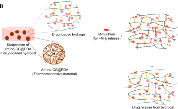
ABSTRACT: A hydrogel immobilized with light-responsive amino group was developed by using kappa carrageenan (CG) and polydopamine (PDA). In the hybrid amino-CG@PDA microcomposite system, amino-CG was used to encapsulate PDA microspheres that act as a photothermal agent. Near infrared (NIR) irradiation is absorbed by the PDA to generate heat, releasing the amino-CG via gel-to-sol phase transition. By precisely controlling the transition temperature of amino-CG gel at 40 °C by adding a KCl hardening solution at the appropriate concentration, we successfully developed a composite hydrogel that dissolves at 40 °C when heated with NIR irradiation. The amino-CG@PDA was further composited into an aldehyde-introduced dextran hydrogel, which was reported to degrade by reaction with amino compounds. Utilizing the amino-CG release via NIR irradiation, we can successfully control the release of drug incorporated in the hydrogel, in which the NIR irradiation helps to diffuse amino-CG into the dextran hydrogel to initiate the hydrogel degradation by the reaction with aldehyde.
3 1. INTRODUCTION
The human health and quality of life have greatly benefited from the design, development, fabrication, and use of polymeric biomaterials for biomedical applications, such as tissue
engineering, artificial organs, regenerative medicine, and controlled drug release systems. Some of these polymers have been incorporated into smart, intelligent, or stimuli-responsive
materials.1-3 Various stimuli such as temperature, pH, enzyme, and light can affect the properties
of smart polymers.4-7 In particular, thermoresponsive polymer hydrogels are widely employed as
smart materials with the important property of tunable phase transition in response to
temperature change. 8, 9 This unique property allows the smart polymer hydrogels to function as
on-off controls that could be switched by temperature change.
We have previously developed a degradable polysaccharide hydrogel for bioadhesion.10 An
aqueous solution of aldehyde with introduced oxidized dextran was mixed with a polyamine solution to form the hydrogel via Schiff base formation as crosslinking points.10 Interestingly, the
hydrogel was degraded by main chain scission of the polysaccharide part via Maillard reaction. To control the degradability of this hydrogel, we introduced methacrylate moiety into the oxidized dextran to form crosslinking points with dithiothreitol via thiol-ene Michael addition reaction with the remaining aldehyde groups.11 In that system, we found that a posteriori addition
of amino compounds could initiate the hydrogel degradation through the reaction of aldehyde with amine, and such degradation could be controlled independently from the mechanical properties of the hydrogel11. In this report, we further create a posteriori delivery system of
4
a drug delivery system could be especially useful for the temperature-responsive release of amino compounds.
A few natural polymers and their modification products have been considered as temperature-sensitive materials. Cellulose derivatives such as methyl cellulose show reverse thermogelation. When an optimum balance of hydrophilic and hydrophobic moieties is established, this
methylcellulose solution gels upon heating and returns to the liquid state upon cooling.12 Ethyl
(hydroxyethyl) cellulose also showed thermoresponsive gelation in the presence of lysine-based surfactants. An increase of temperature triggers the formation of mixed micelles and causes the network to swell due to charge repulsion between polymer chains.13 Another
temperature-sensitive polysaccharide is chitosan (CS). The CS/β-glycerol phosphate (GP) system provides themoresponsive gelling induced by heat, due to the hydrophobic interaction to form a colloidal network. When the temperature decreases, it slowly turns into a flexible network due to the existence of additional hydrogen bonds.14 This unique thermosensitive hydrogel can be
potentially applied in drug delivery15-17 and regenerative medicine (tissue regeneration).18, 19
Kappa-carrageenan (κ-CG) is a temperature-sensitive polysaccharide that is receiving attention for pharmaceutical applications. The sol-gel transition temperature of κ-CG could be tuned by a variety of physical crosslinking conditions near the physiological temperature. Thus, this work focused on using themoresponsive carrageenan for the controlled release of active compounds by using heating as external stimuli.
κ-CG is a linear sulfated polysaccharide extracted from marine red algae, and it is composed of D-galactose and 3,6 anhydrogalactose units. The gelation of κ-CG involves a coil-to-helix conformational transition followed by helix aggregation.20, 21 The gel formation process is
5
thermoreversible, and it can be induced by cooling or crosslinking by monovalent cations such as potassium ions.22-24 The cross-linking density and the charge of the gel affect the phase transition
temperature and control the release profile of entrapped active molecules.22, 24 The
thermo-sensitive nature of κ-CG hydrogels makes them an interesting candidate for temperature-activated drug delivery applications.24-26 In addition, the structure of κ-CG contains abundant
hydroxyl groups, providing the possibility for further derivatization and bioconjugation. The chemical modification of polysaccharides is considered to enhance their properties. In this paper, κ-CG was functionalized to produce the amino-CG derivative. The gelation of beads was
promoted by K+, and the gel-to-sol transition of amino-CG was controlled by increasing the
temperature from 37 to 40 °C.
To create a smart platform for controlled molecule release, near-infrared (NIR) irradiation is an especially attractive candidate external stimuli, as it can be remotely applied for a short period of time with on-off switching and temporal precision. To enhance the potential of NIR,
photothermal agents have been intensively explored. These photothermal agents can readily absorb NIR light and convert the photon energy into thermal energy, and the heating triggers the polymer matrices to release entrapped molecules.27 Polydopamine (PDA) particle is a widely
used photothermal agent in photothermal therapy and controlled release system by NIR.27-31 This
paper reports a new platform of entrapment material, by trapping PDA microspheres into a thermo-sensitive polysaccharide (amino-CG derivative). Upon exposure to an NIR laser, the generated heat triggers the gel-to-sol phase transition, leading to the diffusion of amino carrageenan.
6
We intend to utilize the triggered release of amino compounds to further control the release of a drug by hydrogel degradation. From our previous report, we can control the degradation of oxidized glycidyl methacrylate immobilized dextran (oxidized Dex-GMA)-based hydrogel by amino group addition via the Maillard reaction, triggered by the Schiff base formation between aldehyde and amino groups11. In this work, a dispersion of amino-CG@PDA in oxidized
Dex-GMA based hydrogel was prepared. Irradiated of NIR light caused the diffusion of amino-CG into the hydrogel, which reacts with aldehyde remaining in the hydrogel. As a result of hydrogel degradation, drugs loaded in it were released. The above concept is illustrated in Figure 1A and B.
2. Experimental section 2.1 Materials
Kappa-Carrageenan (κ-CG, molecular weight 4.3×10⁵) and dopamine hydrochloride were
obtained from TCI (Tokyo, Japan). 3-Bromopropylamine hydrobromide was purchased from Sigma Aldrich (St. Louis, MO, USA). Ammonium hydroxide (NH4OH, 28–30%) was acquired
from Kanto Chemical Co. Inc. (Tokyo, Japan). Ethanol and other chemicals were purchased from Nacalai Tesque, Inc. (Kyoto, Japan). All chemicals were used without purification.
2.2 Preparation of degradable hydrogel via Schiff base formation reaction with amino compound Oxidized glycidyl methacrylate derivatized dextran (Ox-GMA-Dex) was synthesized as
described in the previous report.11 First, 5 g of dextran was dissolved in 20 mL of dimethyl
sulfoxide (DMSO), and N2 gas was flowed through the transparent solution for 30 min. Then, 0.8
7
nitrogen gas for 30 min, and the reaction mixture was allowed to stand at 50 °C for 12 h. An equimolar amount of hydrochloric acid (HCl) was added to neutralize the DMAP, before dialysis against distilled water for one week. After drying, the pale yellow-brown flake product of Dex-GMA was obtained.
Sodium periodate (NaIO4) was used as oxidizing agent to prepare oxidized Dex-GMA. In brief,
2.5 g of Dex-GMA was dissolved in 10 mL of distilled water, and NaIO4 (0.375 g in 5 mL DI
water) was added into the solution. The reaction was allowed to proceed at 50 °C for 1 h. The oxidized Dex-GMA product was obtained after dialysis and drying.
The synthesized products were characterized by 1H nuclear magnetic resonance (NMR) (400
MHz, Bruker). The 1H-NMR results were used to investigate the degree of substitution (%DS) of
GMA, by comparing the ratio of the areas under the proton peaks at 1.9 ppm (methyl protons in GMA) to the peak at 3.2–4.1 ppm (dextran sugar unit protons). In addition, The aldehyde content or the degree of oxidation (%oxidation) in Ox-GMA-Dex was determined using the fluorometric method, based on the reaction between acetoacetanilide (AAA) and aldehyde in the presence of ammonia.32
The hydrogel was prepared by the crosslinking of Ox-GMA-Dex with dithiothreitol (DTT). Equal volumes of Ox-GMA-Dex (10% w/v) and DTT in phosphase buffer solution (PBS) (1.36% w/v) were mixed. The hydrogel was easily formed via thiol Michael addition reaction between methyl acrylate (from Ox-GMA-Dex) and thiol (from DTT) in 1:1 mole ratio at 37 °C. Removal of DTT was not required because we previously confirmed that most of thiol had reacted with the GMA and aldehyde parts.11
8 2.3 Synthesis and characterization of amino-CG
Amino-CG was synthesized by applying the method reported by Tranquilan-Aranilla et al. in 2012.33 κ-CG (1 g) was suspended in 10 mL of 80% (w/v) solution of 2-propanol in a 100-mL
round bottom flask equipped with a reflux cooler, and stirred vigorously at room temperature. NaOH (40%, 1.2 mL) was added dropwise to the mixture, and the stirring was continued for another 1 h at 40 °C. After alkaline activation, 3-bromopropylamine hydrobromide (0.55 g) was added. The reaction was allowed to proceed for 24 h at 40 °C. After the reaction was finished, the mixture was filtered, suspended in 80% (w/v) of 2-propanol (aq), and neutralized with 1 M HCl. The products were collected after filtration, washed several times with 80% (w/v) 2-propanol (aq) followed by pure 2-2-propanol, freeze-dried for a day, and kept in the refrigerator until use. The synthesized amino-CG was characterized by 1H and 13C NMR, differential
scanning calorimetry (DSC), and zeta potential analysis.
To confirm the structure of products, 1H NMR and 13C NMR spectra were collected using a
Bruker 400 MHz NMR spectrophotometer after dissolving the sample in D2O.
The zeta potential at pH 7.0 was determined using a Zetasizer Nana-ZS (Malvern Instruments, UK) instrument. The samples were diluted to 0.1% (w/v) with DI water at pH 7. Three zeta potential measurements were taken for each sample.
DSC (Seiko SII Exstar6000) was used to determine the thermal transition of modified samples. The experiment was done under N2 gas atmosphere from 35 to 150 °C at a heating rate of
10 °C/min.
9
Cell viability was determined by measuring the ability of cells to convert 3-(4,5-dimethyl thial-2-yl)-2, 5-diphenyltetrazalium bromide (MTT) to a purple formazan dye. L929 cells suspended in 0.1 mL medium at a concentration of 1.0 × 10⁴/mL were placed in 96-well culture plates. After
incubation for 72 h at 37 °C, 0.1 mL of the medium containing different concentrations of amino-CG or non-treated CG as a control was added to the cells, followed by incubation for 48 h. To evaluate cell viability, 0.1 mL MTT solution (300 mg/mL in medium) was added to the cultured cells, which were further incubated for 4 h at 37 °C. After removing the remaining medium, 0.1 mL DMSO was added to each well to dissolve the precipitate. The resulting color intensity, which was proportional to the number of viable cells, was measured by a microplate reader (versa max, Molecular Devices Co., CA, USA) at 540 nm.
2.5 Determination of amino content and of amino-CG and its ability to function as amino source 2,4,6-Trinitrobenzene sulfonic acid (TNBS) is a rapid and sensitive assay reagent for quantitative determination of amino groups.34 In brief, 1 mL of standard glycine (0–25 µg/mL) or analysis
sample was added to 1 mL of 4% (w/v) NaHCO3 (pH 8.5) and 1 mL of 0.1% (w/v) TNBS in
water. The solution was then incubated at 40 °C in the dark for 2 h. After that, 1 mL of 10% (w/v) of SDS and 0.5 mL of 1 M HCl were added to each sample solution. The absorbance of the solution at 335 nm was read. A curve was generated using the measurement from standard samples, and used to determine the concentration in the analysis sample.
To confirm that the amino-CG could function as an amino source, the reduction in the molecular weight (Mw) of Ox-GMA-Dex after adding amino-CG was investigated. Ox-GMA-Dex (2%
10
w/v, 24% oxidation, 23% DS of GMA) was mixed with the same volume of 5% (w/v) amino-CG solution or glycine and then incubated at 37 °C. To determine the Mw of oxidized Dex-GMA at a given time after reaction, gel permeation chromatography (GPC) (Shimadzu, Japan, BioSep-s2000 column, Phenomenex, Inc., CA, USA) was utilized. PBS was used as the mobile phase (flow rate = 0.50 mL/min) and pullulan was used as the standard.
2.6 Synthesis and characterization of polydopamine (PDA) microspheres
PDA microspheres were synthesized by using a previously reported method.27 Dopamine
hydrochloride (0.3 g) was dissolved in 5 mL of DI water. The prepared solution was slowly added to a mixture of 28−30% NH4OH (1 mL), ethanol (20 mL), and DI water (45 mL). The
reaction was allowed to proceed for 12 h at room temperature under mild magnetic stirring. Subsequently, the prepared PDA microspheres were collected by centrifugation and washed with DI water several times. The synthesized products were dried and characterized.
The functional groups in the products were confirmed using a FT/IR-4200 spectrophotometer in 4000–600 cm-1. The surface morphologies were observed by a Hitachi S-4100 SEM and a
Hitachi H-7100 TEM systems. UV-vis spectra were obtained using a UV-1800 spectophotometer. 2.7 Preparation of amino-CG@PDA microcomposite beads
An amino-CG aqueous solution (2–4% w/v) was prepared by heating at 60 °C with constant stirring. After that, 0.02% (w/v) of PDA microspheres were suspended into each of solution. The beads were prepared using an ionotropic gelation method. Briefly, 5 mL of the heated amino-CG solution was extruded dropwise through a syringe needle (0.838 mm of inner diameter) into 50 mL of KCl solution (5% w/v). The beads were allowed to harden for 30 min in the KCl
cross-11
linking solution. The gel beads were filtered, washed with DI water 3 times, and freeze-dried until constant weight was achieved. The amino-CG@PDA was thus obtained.
2.8 Swelling study for determination of so-gel transition of amino-CG
Water uptake of the prepared beads is defined as the weight percent with respect to that of the dried beads. To measure the swelling ratio of amino-CG@PDA, dried samples were weighted (Wdry) and immersed in PBS at 37 °C (or 40 °C) for different periods of time. The wet samples
were carefully blotted with filter paper to absorb the excess water, and then weighted again (Wwet). The water uptake is defined by the following equation (1):
Water uptake (%) = [(Wwet-Wdry)/Wdry] × 100 (1)
In this study, we used two temperatures (37 and 40 °C) to find the suitable condition to prepare amino-CG@PDA microcomposite that is stable at 37 °C but undergoes gel-to-sol phase
transition at 40 °C. Beads prepared under this condition will be further used to prepare the NIR-induced temperature-responsive microcomposite.
2.9 Temperature and light-responsive sol-gel transition test of amino-CG@PDA microcomposites
To evaluate the dissolution of the thermoresponsive gel, amino-CG@PDA microcomposites were suspended in PBS and incubated at 37 or 40 °C for 24 h. At different time intervals, the
suspension was collected for the amino quantification. For NIR-triggered dissolution, the
suspension of amino-CG@PDA composite was exposed to 808-nm NIR light (at a power density of 1 W/cm2) for 30 min at different time points (0.5, 1.5, 2.5, 3.5, and 4.5 h). One milliliter of the
12
supernatant was collected at each predetermined time to analyze the amino group amount by TNBS assay. Then, the sample was re-dispersed for further irradiation. A suspension incubated in dark condition was used as a negative control. The cumulative amount of amino-CG released from the composite beads was calculated according to the following equation (2):
Amino release efficiency (%) = (Wt/Wi) × 100 (2)
Where Wi and Wt are the amino content in the obtained microcomposites at equilibrium state and
that released from the loaded nanocomposites at time t during the release process, respectively. 2.10 Drug loading into Ox-GMA-Dex based hydrogel
Doxorubicin hydrochloride (DOX) was loaded into Ox-GMA-Dex. DOX was dissolved at the concentration of 0.05 mg/mL in a solution of 10 wt% Ox-GMA-Dex (10% oxidation, 23% DS of GMA) in PBS. The DOX-loaded hydrogel was prepared as follows. Briefly, 0.5 mL of the mixed solution of DOX and Ox-GMA-Dex, 0.5 mL of 1.36 wt% DTT, and 0.01 g of amino-CG@PDA were mixed in a test tube and then incubated at 37 °C for 30 min to allow the gelation, and the product was denoted as DOX@hydrogel. Here, we assumed that 100% of the drug was loaded into hydrogel.
2.11 Light-responsive drug releasing test
For NIR-triggered drug release from hydrogel, the prepared DOX@hydrogel was immersed in 5 mL of PBS, and exposed to NIR light (808 nm, 1.0 W/cm2) for 30 min. One milliliter of the
solution was collected at different time points (0.5, 1.5, 2.5, 3.5, and 4.5 h) to analyze the DOX release by fluorescence spectroscopy (λex 480 nm and λem 590 nm). Then, 1 mL of PBS was
re-13
added the rest of the mix for further irradiation. The experimental setup is shown in Figure 1A. The cumulative amount of DOX released from the hydrogel was calculated according to the following equation (3):
DOX release efficiency (%) = (Wt/Wi) × 100 (3)
Where Wi and Wt are the DOX content in the oxidized Dex-GMA based hydrogel, and that
released from the loaded hydrogel at time t during the release process, respectively.
14
Figure 1 (A) Experimental setup for light-responsive drug release test. (B) Schematic presentation of drug release via hydrogel degradation under NIR irradiation.
3. Results and discussion
3.1 Synthesis and characterization of amino-CG
The amino-CG was synthesized in a simple reaction as shown in Figure 2A. The κ-CG was activated by NaOH to increase the nucleophilicity by the formation of alkoxides,35 and
3-bromopropylamine hydrobromide was then added to react with carrageenan alkoxides to form amino-derivatized carrageenan. The 1H and 13C NMR spectra of κ-CG and modified CG are
shown in Figures 2B and 2C, respectively. The disaccharide polymer κ-CG contains 3-linked β-D-galactopyranose (G unit) and 4-linked 3,6-anhydro-α-β-D-galactopyranose (DA unit) as a
15
repeating unit which showed the 1H chemical shifts in Figure 2B-b. In the spectrum for
amino-CG (Figure 2B-c), new proton peaks appeared at the chemical shift of 2.25–3.20 ppm, possibly due to the mixing of shifted chemical shift of the methylene protons in the propylamine and that of amino proton of amino-CG. The proton decoupled 13C NMR analyses gave more information
on the structural properties of amino-CG derivatives. The spectrum of untreated κ-CG (Figure 2C-a) shows strong signals of the twelve carbon atoms in the disaccharide repeating unit.33, 35
New signals were present in the amino-CG spectrum (Figure 2C-b), namely additional
resonances at the chemical shifts of 31.05 and 30.08 ppm, which were assigned to the methylene carbon from propylamine substitution. The intensities of the new signals are very weak due to the low amount of DS.
17
Figure 2 (A) Schematic presentation of the synthesis of amino-CG. (B) 1H NMR spectra of
3-bromopropylamine hydrobromide (a), original κ-CG (b) and amino-CG (c). (C) 13C NMR spectra
of original κ-CG (a) and amino-CG (b).
The charge of polysaccharide is reflected by the zeta potential (ζ-potential) of the solution, determined as the electrical potential produced around the surface of particles dispersed in solvent.36, 37 The ζ-potential resultsat pH 7.0 can help to confirm the synthesized product (Table
1 and Figure S1). The negative zeta potential of -58.6 ± 0.8 mV in the original κ-CG can be explained by the anionic structure which contains –SO3- groups. However, the functionalized
amino-CG showed a relatively higher zeta potential (-37.3 ± 1.4 mV), because the functionalized amino groups carry positive charge to partially neutralize the high negative value of unmodified κ-CG. Consequently, a higher ζ-potential was observed.
The cytotoxicity of intact CG and amino-CG to L929 cells was evaluated through MTT assay, and the results are provided in Figure S2. As amino-CG and intact CG formed hydrogel-like viscous fluid at higher
concentration, we evaluated their cytotoxicity at ≤ 5000 and 2000 µg/mL in the medium, respectively. Even at the maximum tested concentrations, we did not observe any viability decrease due to their low cytotoxicity. Generally, cationic compounds show cytotoxicity. But as the DS of amino group was low in amino-CG, the cytotoxicity might be almost the same as that of intact CG. As we previously reported,11 the
cytotoxicity of Ox-GMA-Dex was 3000 times lower than that of glutaraldehyde, due to the stability of the aldehyde groups in the polymer backbone. From these results, our composite hydrogel could be a cytocompatible system.
18 Table 1 The Zeta Potential of κ-CG and Amino-CG
polysaccharide solution zeta potential (mV)
κ-CG -58.6 ± 0.8
amino-CG (0.87% DS) -37.3 ± 1.4
* Concentration of the polysaccharides was 1 mg/ml; **Average of three measurements at 25°C
The thermal analysis results also supported the successful synthesis of amino-CG. Figure 3 showed DSC thermograms of pure CG and amino-CG for detecting the glass transition temperature (Tg). The Tg of amino-CG was 102.2 °C whereas that of CG was lower (70.7 °C),
because in amino-CG the intra- and/or intermolecular interaction is enhanced by the methylene groups.38, 39 Consequently, restricted segments of polymer chains were formed, and hence it takes
more energy to break the bonds. All these results confirmed the successful modification of κ-CG with amino groups.
19
3.2 Amino content in amino-CG and the reactivity with Ox-GMA-Dex
The degree of substitution (%DS) was calculated from the amount of amines contained within the amino-CG sample, by using a rapid and sensitive assay reagent (TNBS).34 The calculated DS
value of amino-CG was 0.87 %. In our previous report, Ox-GMA-Dex reacted with thiol groups in the GMA part to form hydrogel, and reacted with amine in the aldehyde part to start the degradation.10, 11, 40, 41 To confirm the reactivity of amino-CG with aldehyde group in
Dex and the degradability of the polysaccharide, we checked the molecular weight of Ox-GMA-Dex by using GPC. The %DS of GMA and %oxidation in Ox-GMA-Ox-GMA-Dex were determined by previously reported methods.11 The proton chemical shifts of Ox-GMA-Dex are: 1.9 ppm
(methyl proton of GMA), 3.2–4.1 ppm (dextran sugar unit proton), 4.9 ppm (dextran anomeric proton), and 5.7–6.2 ppm (vinyl proton of GMA), as shown in Figure S3. The DS value was determined to be 23% by comparing the ratio of the areas under the proton peaks at 1.9 ppm (methyl protons in GMA) to the peak at 3.2–4.1 ppm (dextran sugar unit protons, H2-H6). The aldehyde content was evaluated from the fluorometric method by the reaction of AAA and aldehyde. From the fluorescence intensity, %oxidation was controlled to be 10%. In the
remainder of this paper, we denote this oxidized Dex-GMA as Ox(10%)-GMA(23%)-Dex, where 10% is the degree of oxidation and 23% is the DS of the GMA.
In this study, the decrease of Mw in the oxidized Dex-GMA (containing aldehyde groups) after the reaction with amino-CG or glycine solution (model of primary amine) was determined by mixing oxidized Dex-GMA and amino-CG (or glycine) at 37 °C. The Mw was measured by GPC every 20 min for 3 h. As shown in Figure 4, the Mw of oxidized Dex-GMA (1% w/v) did not
20
change after addition of intact CG. However, it dramatically decreased in glycine solution (2.5% w/v) after 30 min, then decreased more slowly and became stable after 3 h (red line). This agreed well with our previous study that showed the amino group reacts with aldehyde to cause the degradation.11,40 The same trend of Mw was observed after adding 2.5% w/v amino-CG, but the
Mw value after degradation was higher than that in glycine solution (blue line). The reason behind this result is that the amino content in amino-CG was lower than that of free amino groups from the glycine solution, leading to a low capacity for degradation. However, Mw still decreased at 120 min in the amino-CG added group, suggesting the lower diffusion of amino-CG due to its high molecular weight (4.3 ×10⁵). Since the amino-CG was confirmed to trigger the
degradation, it was further used in the degradation of hydrogel.
21
3.3 Synthesis and characterization of PDA@amino-CG microcomposite
This study reports a delivery system based on amino groups using NIR light and temperature for remote triggering, using PDA as a photothermal agent. The PDA microspheres were
encapsulated in amino-CG gel beads, which undergo conformation transition when the
temperature is increased,23 leading to the release of compounds containing the amino group. The
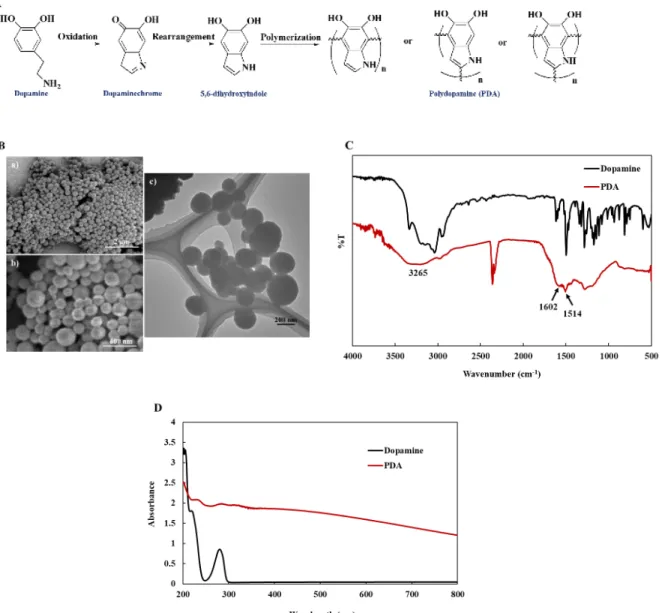
synthesis of PDA is presented in Figure 5A, where the dopamine monomer was oxidized and then cyclized to form dopaminechrome in a mixture of water, ethanol, and ammonia. After that, the molecular structure of dopaminechrome was rearranged to produce an 5,6-dihydroxyindole intermediate, which further formed PDA microspheres by intermolecular polymerization (which is clearly seen by the solution color change from pale to dark brown).
SEM and TEM images were used to confirm the successful synthesis of PDA microspheres. The obtained PDA microspheres had regular spherical structure with an average size of 150–300 nm (Figure 5B) and showed good dispersibility.
The chemical composition of free dopamine and PDA were characterized by FTIR spectrometry. Unlike pure dopamine, the FTIR spectrum of PDA showed absorption bands at 1520 and 1615 cm-1, corresponding to the vibration of N−H in the aromatic rings. The signal at 3265 cm-1
revealed the existence of –OH groups in 1,2-dihydroxybenzene or catechol (Figure 5C).42 In the
UV-Vis spectra (Figure 5D), characteristic absorption peak of free dopamine was observed at 280 nm.27 The curves of dopamine and PDA were dissimilar. PDA showed an absorption ranging
22
dihydroxyindole, which subsequently self-polymerized.43 A change in the solution color from
colorless to deep brown was detected, suggesting the successful synthesis of polydopamine, which can function as a photothermal agent in the NIR range.
Figure 5 (A) Schematic presentation of the production of PDA. (B) SEM (a: 15k magnification, b: 50 k magnification) and TEM (c) images of the PDA microspheres. (C) FTIR spectra of dopamine and PDA. (D) UV−vis spectra of dopamine and PDA.
23
The PDA@amino-CG microcomposite can be easily prepared by using an ionotropic gelation method. κ-CG is an anionic polyelectrolyte hydrogel due to its sulfate groups. In aqueous solutions and in the presence of cations such as K+, Na+, or Ca2+, κ-CG easily forms
thermoreversible gels. In this process, there is an initial coiling to produce the helical conformation, and then the helices aggregate to form infinite networks.44, 45 The cations can
induce conformational changes in the polymer.46 Due to the instantaneous nature of such ionic
gelation, drugs, enzymes, and other substances can be encapsulated in carrageenan. In this study, a κ-CG derivative, amino-CG, was used for encapsulation of PDA microspheres in the presence of K+ to form thermoreversible gel. SEM images in Figure 6A show the morphology of dried
bead samples. Samples produced by amino-CG (Figure 6Aa-c) display spherical form with a smoother surface compared to the amino-CG@PDA (Figure 6Ad-f), which showed a rough surface morphology since the PDA microspheres prevented the carrageenan chains from approaching each other.
Water uptake is an important property in release studies. The swelling of hydrogel promotes the release of substances, because water molecules penetrate the structure of dry hydrogel and boost the relaxation of chains. This increases the pore size of porous hydrogel for the release of drugs or other target substances.24, 47 The hydrogel we developed here is supposed to be stable at 37 °C
and dissolves upon heating with external stimuli such as NIR. Thus, the swelling behavior of amino-CG@PDA beads at 37 and 40 °C in PBS was studied (Figure 6B). The polysaccharide concentration (2, 3, or 4% w/v) affected the swelling ratio, due to the changing gel charges and network density. From the swelling ratio in Figure 6B, an initial burst of quick water penetration occurred within the first 3 h at both temperatures. The more crosslinked beads (prepared with 4% amino-CG at 37 °C, red solid line) provides a lower swelling ratio compared to those prepared
24
with 2% and 3 % amino-CG (blue and yellow solid lines). The reason is that, at a higher
carrageenan concentration, the more abundant SO3- on the carrageenan backbone can form more
crosslink with K+ in KCl, resulting in a high degree of crosslinking. The thermoreversible gel
beads prepared with 4% amino-CG were stable at 37 °C after 10 h with a constant swelling ratio. In contrast, the 2% and 3% of amino-CG@PDA beads dissolved within 5 h, due to their lower– SO3- contents and therefore reduced ionic crosslinking (low crosslink density). The water uptake
by all types of bead samples at 40 °C are shown by the dashed lines. Besides the 2% and 3% of amino-CG@PDA beads, the 4% amino-CG@PDA thermoreversible gel beads were dissolved after 5 h under incubation at 40 °C. Thus, the thermoreversible 4% amino-CG@PDA preparation was selected for further experiments, since it formed stable swelling beads at body temperature and became a liquid at 40 °C.
25
Figure 6 (A) SEM images of 4% amino-CG beads (a-c) and 4% amino-CG@PDA beads (d-f) prepared with 5% KCl. (B) Water uptake for different bead formulations at 37 °C and 40 °C.
3.4 Temperature and light-responsive amino-CG release
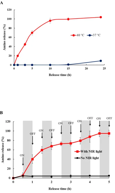
In this study, the hydrogel beads of 4% amino-CG@PDA crosslinked with 5% KCl were chosen for use in thermo-sensitive sol-gel transition. Since the driving force for dissolving the amino-CG should be controlled precisely by small temperature changes, the supernatant amino group concentration is plotted in Figure 7A after immersing amino-CG@PDA in PBS at 37 or 40 °C. As shown in Figure 7A, the amount of released amino group was negligible at 37 °C, since at this temperature the beads just swelled without dissolution. In comparison, the amino release was much faster at 40 °C, up to 70% after 5 h and 100% at around 10 h. Under this higher
temperature, the gel dissolved into a sol after reaching swelling equilibrium. These results agree with the swelling behavior studies. It exhibits that the 4% amino-CG@PDA thermoreversible gel
26
beads could dissolve at 40 °C incubation within 5 h, while it can remain stable at 37 °C with a constant swelling ratio after 10 h. To sum up, the beads prepared by 4% amino-CG@PDA crosslinked with 5% KCl indeed showed a thermosensitive releasing capability, and the release behavior could be easily managed via controlling the environment temperature.
In the temperature-responsive release test, the amino-CG@PDA is sensitive to high temperature above the body temperature. To further confirm the ability of external stimulus (NIR light) to trigger the amino-CG release, amino-CG@PDA microcomposites were exposed to NIR light irradiation, and the cumulative amino release was evaluated by using a UV−vis
spectrophotometer. As plotted in Figure 7B, the amino-CG release was negligible in the absence of NIR light. When the NIR light was turned on, the temperature of the suspension increased to 42 °C as shown in Figure S4, and thus the amino release was greatly enhanced. When the external trigger (NIR light) was turned off, a slower release was observed. For example, the measured cumulative release of amino groups was 40.14% after 30 min of NIR irradiation. After the light was turned off, the release increased by another ~20% 30 min later (to 59.68%). Either with or without NIR light, the release of amino groups gradually increased after 2 h. The reason is the prominent gel-to-sol phase transition of amino-CG@PDA gel beads when the PDA converted NIR light to heat, leading to the re-dissolution of amino-CG chain. As a result, the encapsulated PDA microspheres were liberated and dispersed in the solution. Now, with less encapsulated PDA in the amino-CG@PDA microcomposites during the subsequent irradiation, the temperature of the suspension clearly decreased in the third cycle of irradiation to 38 °C, leading to less amino compounds being released. This amino release pattern occurred
photothermal-27
responsive ability, and that the release rate of amino compounds can be controlled by switching NIR-light irradiation.
Figure 7 (A) Release profiles of 4% amino-CG@PDA microcomposites prepared with 5% KCl under direct heating to 37 or 40 °C for different periods of time. (B) Cumulative release profiles
28
of amino compound from amino-CG@PDA microcomposites with and without NIR-light irradiation.
3.5 NIR light-responsive DOX release from hydrogel degradation
In this work, the Ox-GMA-Dex was used for hydrogel preparation. Equal volumes of Ox(10%)-GMA(23%)-Dex (10% w/v) and DTT solution (1.36% w/v) were mixed to form hydrogel including amino-CG@PDA beads and used for drug-loaded template. The process of drug release via hydrogel degradation under NIR irradiation was shown in Figure 1B.
Hydrogels provide a platform for achieving controlled therapeutic delivery to specific sites. The stimulus responsive hydrogels undergo changes (such as degradation) in response to
environmental triggers. This property suggests the possible use of these materials for specific functions, especially drug release.48, 49 Doxorubicin is one of the most widely used commercial
anticancer drugs in clinical application. Its derivative DOX·HCl is soluble in water, and hence it was used as a model drug to investigate the release from Ox-GMA-Dex based hydrogel. Figure 8A, B show the cumulative drug release from hydrogels loaded with 10 μg/ml drug over 5 h and 10 days in PBS, respectively. Only a very small amount of DOX was released in the absence of NIR light (black line), and it may come from free DOX liberated from the outer surface of hydrogel. In the presence of NIR light, we proposed a model of drug release as shown in Figure 8C. The NIR light is irradiated on the DOX@hydrogel (step A), generating heat in the PDA of amino-CG@PDA microcomposites. This heating triggers the gel-to-sol phase transition of amino-CG to produce a flowing sol (step B).50, 51 Then, the released amino-CG reacts with the
remaining aldehyde on the hydrogel to form Schiff base in step C, initiating the degradation process which facilitates drug release (step D). In this experiment, the amino-CG@PDA gel
29
beads started to transform to sol when irradiated under NIR light. After 4 h of incubation the beads dissolved completely, suggesting that the Schiff base reaction occurred. The cumulative DOX release is significantly increased under NIR irradiation compared that with without NIR light. Thus, the release of drug from hydrogel can be controlled by NIR trigger, and drug release continues to some extent even after the NIR light was turned off. However, the cumulative DOX %release was only 5.5% over 5 h, and the hydrogel was not completely degraded. The release of DOX was likely to come from two factors: degradation and hydrogel swelling. The small amount of release may be explained as follows. First, the DOX molecules are positively chargedand can be easily adsorbed on the negatively charged Ox-GMA-Dex chain by
electrostatic interaction.52 Hence, the release of DOX by diffusion from the Ox-GMA-Dex
hydrogel network was slow. The second reason was the low speed of hydrogel degradation, which is the critical step for drug release from the hydrogel. Because the amount of amino source used (5.4×10⁻⁶ mole –NH2) was 11.5 times lower than the aldehyde content in oxidized
Dex-GMA (6.2×10⁻⁵ mole), the degradation points (Shift base formation) was subsequently fewer,
hence the slow release of the drug. Figure 8B shows the cumulative DOX release profile over 10 days without NIR light irradiation after the first 5 h. We estimated that as much time as 10 days was required for main chain scission of DOX@hydrogel to enhance the DOX release. The main chain degradation of oxidized Dex-GMA hydrogel proceeded gently for the first 2 days to reach a DOX release of almost 10%. After that, the main chain scission was significantly accelerated, reaching a cumulative drug release of up to 83% over 7 days, and 100% release can be obtained for 10 days of incubation with complete degradation of hydrogel. These findings indicate that we can control the release of drug by hydrogel degradation triggered from NIR irradiation.
30
Figure 8 (A) Cumulative release profiles of DOX from hydrogel with and without NIR-light irradiation over 5 h. (B) Cumulative release profiles of DOX from hydrogel over 10 days. (c) Schematic presentation of drug release under NIR irradiation.
4. Conclusion
Amino-CG was composited with PDA to form a hydrogel. This hydrogel showed gel-sol transition at 40 °C, therefore it is stable at physiological temperatures. NIR irradiation could dissolve the amino-CG@PDA gel beads due to the photothermal property of PDA. When the
31
amino-CG@PDA gel beads were incorporated into the Ox-GMA-Dex hydrogel, the aldehyde group in the latter can react with the external amino group in the former, degrading the main chain through the Schiff base formation reaction. Under NIR irradiation, hydrogel degradation was initiated to release DOX incorporated in it. This kind of controlled drug release model may be a promising candidate for therapeutic hydrogels, for use in external stimuli-responsive drug delivery, although the rate of degradation should be considered.
ASSOCIATED CONTENT Supporting Information.
The Supporting Information is available free of charge on the ACS Publications website at DOI: Schematic illustration of the concept of NIR induced drug release, Zeta potential distribution of κ-CG and amino-κ-CG, cytotoxicity assay of amino-κ-CG, 1H NMR spectrum of Ox-GMA-Dex and
temperature profile of amino-CG@PDA microcomposites by NIR irradiation. AUTHOR INFORMATION
Corresponding Author * [email protected] Author Contributions
Both authors have contributed to the study and given approval to the final version of the manuscript.
Notes
32 REFERENCES
(1) Hardy, J. G.; Palma, M.; Wind, S. J.; Biggs, M. J. Responsive Biomaterials: Advances in Materials Based on Shape-Memory Polymers. Adv. Mater. 2016, 28, 5717-5724.
(2) Liu, J.; Detrembleur, C.; Mornet, S.; Jérôme, C.; Duguet, E. Design of Hybrid Nanovehicles for Remotely Triggered Drug Release: An Overview. J. Mater. Chem. B 2015, 3, 6117-6147.
(3) Roy, D.; Brooks, W. L. A.; Sumerlin, B. S. New Directions in Thermoresponsive Polymers. Chem. Soc. Rev. 2013, 42, 7214-7243. (4) Jochum, F. D.; Theato, P. Temperature- and Light-Responsive Smart Polymer Materials. Chem. Soc. Rev. 2013, 42, 7468-7483. (5) Patel, M.; Kaneko, T.; Matsumura, K. Switchable Release Nano-Reservoirs for Co-Delivery of Drugs via a Facile Micelle–
Hydrogel Composite. J. Mater. Chem. B 2017, 5, 3488-3497.
(6) Schmaljohann, D. Thermo- and pH-Responsive Polymers in Drug Delivery. Adv. Drug Deliv. Rev. 2006, 58, 1655-1670. (7) Thornton, P. D.; Mart, R. J.; Webb, S. J.; Ulijn, R. V. Enzyme-Responsive Hydrogel Particles for the Controlled Release of
Proteins: Designing Peptide Actuators to Match Payload. Soft Matter 2008, 4, 821-827.
(8) Hoffman, A. S. Stimuli-Responsive Polymers: Biomedical Applications and Challenges for Clinical Translation. Adv. Drug Deliv.
Rev. 2013, 65, 10-16.
(9) Das, E.; Matsumura, K. Tunable Phase-Separation Behavior of Thermoresponsive Polyampholytes Through Molecular Design.
J. Polym. Sci. A 2017, 55, 876-884.
(10) Hyon, S. H.; Nakajima, N.; Sugai, H.; Matsumura, K. Low Cytotoxic Tissue Adhesive Based on Oxidized Dextran and Epsilon-poly-L-lysine. J. Biomed. Mater. Res. A 2014, 102, 2511-2520.
(11) Nonsuwan, P.; Matsugami, A.; Hayashi, F.; Hyon, S. H.; Matsumura, K. Controlling the Degradation of an Oxidized Dextran-Based Hydrogel Independent of the Mechanical Properties. Carbohydr. Polym. 2019, 204, 131-141.
(12) Li, L.; Thangamathesvaran, P. M.; Yue, C. Y.; Tam, K. C.; Hu, X.; Lam, Y. C. Gel Network Structure of Methylcellulose in Water.
Langmuir 2001, 17, 8062-8068.
(13) Calejo, M. T.; Cardoso, A. M. S.; Marques, E. F.; Araújo, M. J.; Kjøniksen, A.-L.; Sande, S. A.; de Lima, M. C. P.; Jurado, A. S.; Nyström, B. In Vitro Cytotoxicity of a Thermoresponsive Gel System Combining Ethyl(Hydroxyethyl) Cellulose and Lysine-Based Surfactants. Colloids Surf. B 2013, 102, 682-686.
(14) Lu, S.; Yang, Y.; Yao, J.; Shao, Z.; Chen, X. Exploration of the Nature of a Unique Natural Polymer-Based Thermosensitive Hydrogel. Soft Matter 2016, 12, 492-499.
(15) Peng, Y.; Li, J.; Li, J.; Fei, Y.; Dong, J.; Pan, W. Optimization of Thermosensitive Chitosan Hydrogels for the Sustained Delivery of Venlafaxine Hydrochloride. Int. J. Pharm. 2013, 441, 482-490.
33
(16) Wu, Y.; Wei, W.; Zhou, M.; Wang, Y.; Wu, J.; Ma, G.; Su, Z. Thermal-Sensitive Hydrogel as Adjuvant-Free Vaccine Delivery System for H5N1 Intranasal Immunization. Biomaterials 2012, 33, 2351-2360.
(17) Zhang, D.; Sun, P.; Li, P.; Xue, A.; Zhang, X.; Zhang, H.; Jin, X. A Magnetic Chitosan Hydrogel for Sustained and Prolonged Delivery of Bacillus Calmette–Guérin in the Treatment of Bladder Cancer. Biomaterials 2013, 34, 10258-10266.
(18) Kwon, J. S.; Kim, G. H.; Kim, D. Y.; Yoon, S. M.; Seo, H. W.; Kim, J. H.; Min, B. H.; Kim, M. S. Chitosan-Based Hydrogels to Induce Neuronal Differentiation of Rat Muscle-Derived Stem Cells. Int. J. Biol. Macromol. 2012, 51, 974-979.
(19) Stanish, W. D.; McCormack, R.; Forriol, F.; Mohtadi, N.; Pelet, S.; Desnoyers, J.; Restrepo, A.; Shive, M. S. Novel Scaffold-Based Bst-Cargel Treatment Results in Superior Cartilage Repair Compared with Microfracture in a Randomized Controlled Trial. J.
Bone Joint Surg. Am. 2013, 95, 1640-1650.
(20) Daniel-da-Silva, A. L.; Ferreira, L.; Gil, A. M.; Trindade, T. Synthesis and Swelling Behavior of Temperature Responsive Κ-Carrageenan Nanogels. J. Colloid Interface Sci. 2011, 355, 512-517.
(21) Mangione, M. R.; Giacomazza, D.; Bulone, D.; Martorana, V.; San Biagio, P. L. Thermoreversible Gelation of κ-Carrageenan: Relation between Conformational Transition and Aggregation. Biophys. Chem. 2003, 104, 95-105.
(22) Bono, A.; Anisuzzaman, S. M.; Ding, O. W. Effect of Process Conditions on the Gel Viscosity and Gel Strength of Semi-Refined Carrageenan (SRC) Produced from Seaweed (Kappaphycus alvarezii). JKSUES 2014, 26, 3-9.
(23) Daniel-da-Silva, A. L.; Fateixa, S.; Guiomar, A. J.; Costa, B. F.; Silva, N. J.; Trindade, T.; Goodfellow, B. J.; Gil, A. M.
Biofunctionalized Magnetic Hydrogel Nanospheres of Magnetite and Kappa-Carrageenan. Nanotechnology 2009, 20, 355602. (24) Santo, V. E.; Frias, A. M.; Carida, M.; Cancedda, R.; Gomes, M. E.; Mano, J. F.; Reis, R. L. Carrageenan-Based Hydrogels for the
Controlled Delivery of PDGF-BB in Bone Tissue Engineering Applications. Biomacromolecules 2009, 10, 1392-1401. (25) Azizi, S.; Mohamad, R.; Abdul Rahim, R.; Mohammadinejad, R.; Bin Ariff, A. Hydrogel Beads Bio-Nanocomposite based on
Kappa-Carrageenan and Green Synthesized Silver Nanoparticles for Biomedical Applications. International Journal of Biological Macromolecules 2017, 104, 423-431.
(26) Leong, K. H.; Chung, L. Y.; Noordin, M. I.; Onuki, Y.; Morishita, M.; Takayama, K. Lectin-Functionalized Carboxymethylated Kappa-Carrageenan Microparticles for Oral Insulin Delivery. Carbohydr. Polym. 2011, 86, 555-565.
(27) Xu, X.; Bai, B.; Wang, H.; Suo, Y. A Near-Infrared and Temperature-Responsive Pesticide Release Platform through Core–Shell Polydopamine@PNIPAm Nanocomposites. ACS Appl. Mater. Interfaces 2017, 9 (7), 6424-6432.
(28) Ge, R.; Li, X.; Lin, M.; Wang, D.; Li, S.; Liu, S.; Tang, Q.; Liu, Y.; Jiang, J.; Liu, L.; Sun, H.; Zhang, H.; Yang, B. Fe3O4@polydopamine
Composite Theranostic Superparticles Employing Preassembled Fe3O4 Nanoparticles as the Core. ACS Appl. Mater. Interfaces
2016, 8, 22942-22952.
(29) Han, J.; Park, W.; Park, S.-j.; Na, K. Photosensitizer-Conjugated Hyaluronic Acid-Shielded Polydopamine Nanoparticles for Targeted Photomediated Tumor Therapy. ACS Appl. Mater. Interfaces 2016, 8, 7739-7747.
34
(30) Han, L.; Zhang, Y.; Lu, X.; Wang, K.; Wang, Z.; Zhang, H. Polydopamine Nanoparticles Modulating Stimuli-Responsive PNIPAM Hydrogels with Cell/Tissue Adhesiveness. ACS Appl. Mater. Interfaces 2016, 8, 29088-29100.
(31) Li, Y.; Jiang, C.; Zhang, D.; Wang, Y.; Ren, X.; Ai, K.; Chen, X.; Lu, L. Targeted Polydopamine Nanoparticles Enable Photoacoustic Imaging Guided Chemo-Photothermal Synergistic Therapy of Tumor. Acta Biomater. 2017, 47, 124-134. (32) Li, Q.; Sritharathikhun, P.; Motomizu, S. Development of Novel Reagent for Hantzsch Reaction for the Determination of
Formaldehyde by Spectrophotometry and Fluorometry. Anal. Sci. 2007, 23, 413-417.
(33) Tranquilan-Aranilla, C.; Nagasawa, N.; Bayquen, A.; Dela Rosa, A. Synthesis and Characterization of Carboxymethyl Derivatives of Kappa-Carrageenan. Carbohydr. Polym. 2012, 87, 1810-1816.
(34) Kale, R.; Bajaj, A. Ultraviolet Spectrophotometric Method for Determination of Gelatin Crosslinking in the Presence of Amino Groups. J. Young Pharm. 2010, 2, 90-94.
(35) Leong, K. H.; Chung, L. Y.; Noordin, M. I.; Mohamad, K.; Nishikawa, M.; Onuki, Y.; Morishita, M.; Takayama, K.
Carboxymethylation of Kappa-Carrageenan for Intestinal-Targeted Delivery of Bioactive Macromolecules. Carbohydr. Polym. 2011, 83, 1507-1515.
(36) Carneiro-da-Cunha, M. G.; Cerqueira, M. A.; Souza, B. W. S.; Teixeira, J. A.; Vicente, A. A. Influence of Concentration, Ionic Strength and pH on Zeta Potential and Mean Hydrodynamic Diameter of Edible Polysaccharide Solutions Envisaged for Multinanolayered Films Production. Carbohydr. Polym. 2011, 85, 522-528.
(37) Rungtiwa, W.; Sujin, S.; Bovornlak, O.; Saiyavit, V. Zeta Potential (ζ) and Pasting Properties of Phosphorylated or Crosslinked Rice Starches. Starch - Stärke 2005, 57, 32-37.
(38) Fujishima, M.; Matsuo, Y.; Takatori, H.; Uchida, K. Proton-Conductive Acid–Base Complex Consisting of κ-Carrageenan and 2-Mercaptoimidazole. Electrochem. commun. 2008, 10, 1482-1485.
(39) Liew, J. W. Y.; Loh, K. S.; Ahmad, A.; Lim, K. L.; Wan Daud, W. R. Synthesis and Characterization of Modified κ-Carrageenan for Enhanced Proton Conductivity as Polymer Electrolyte Membrane. PloS one 2017, 12, 1-15.
(40) Chimpibul, W.; Nagashima, T.; Hayashi, F.; Nakajima, N.; Hyon, S. H.; Matsumura, K. Dextran Oxidized by a Malaprade Reaction Shows Main Chain Scission through a Maillard Reaction Triggered by Schiff base Formation Between Aldehydes and Amines. J. Polym. Sci. A 2016, 54, 2254-2260.
(41) Matsumura, K.; Nakajima, N.; Sugai, H.; Hyon, S.-H. Self-Degradation of Tissue Adhesive based on Oxidized Dextran and Poly-l-lysine. Carbohydr. Polym. 2014, 113, 32-38.
(42) Liu, X.; Cao, J.; Li, H.; Li, J.; Jin, Q.; Ren, K.; Ji, J. Mussel-Inspired Polydopamine: A Biocompatible and Ultrastable Coating for Nanoparticles in Vivo. ACS Nano 2013, 7, 9384-9395.
(43) Xu, H.; Liu, X.; Wang, D. Interfacial Basicity-Guided Formation of Polydopamine Hollow Capsules in Pristine O/W Emulsions – Toward Understanding of Emulsion Template Roles. Chem. Mater. 2011, 23, 5105-5110.
35
(44) Naim, S.; Samuel, B.; Chauhan, B.; Paradkar, A. Effect of Potassium Chloride and Cationic Drug on Swelling, Erosion and Release from Kappa-Carrageenan Matrices. AAPS PharmSciTech 2004, 5, e25.
(45) Rochas, C.; Rinaudo, M. Mechanism of Gel Formation in κ-Carrageenan. Biopolymers 1984, 23, 735-745.
(46) MacArtain, P.; Jacquier, J. C.; Dawson, K. A. Physical Characteristics of Calcium Induced κ- Carrageenan Networks. Carbohydr.
Polym. 2003, 53, 395-400.
(47) Weng, L.; Gouldstone, A.; Wu, Y.; Chen, W. Mechanically Strong Double Network Photocrosslinked Hydrogels from N,N-Dimethylacrylamide and Glycidyl Methacrylated Hyaluronan. Biomaterials 2008, 29, 2153-2163.
(48) Buwalda, S. J.; Vermonden, T.; Hennink, W. E. Hydrogels for Therapeutic Delivery: Current Developments and Future Directions. Biomacromolecules 2017, 18, 316-330.
(49) Gao, L.; Sun, Q.; Wang, Y.; Zhu, W.; Li, X.; Luo, Q.; Li, X.; Shen, Z. Injectable Poly(ethylene glycol) Hydrogels for Sustained Doxorubicin Release. Polym. Adv. Technol. 2017, 28, 35-40.
(50) Kara, S.; Tamerler, C.; Bermek, H.; Pekcan, Ö. Cation Effects on Sol–Gel and Gel–Sol Phase Transitions of κ-Carrageenan– Water System. Int. J. Biol. Macromol. 2003, 31, 177-185.
(51) Pekcan, Ö.; Tari, Ö. A Fluorescence Study on The Gel-to-Sol Transition of κ-Carrageenan. Int. J. Biol. Macromol. 2004, 34, 223-231.
(52) Wu, S.; Zhao, X.; Li, Y.; Du, Q.; Sun, J.; Wang, Y.; Wang, X.; Xia, Y.; Wang, Z.; Xia, L. Adsorption Properties of Doxorubicin Hydrochloride onto Graphene Oxide: Equilibrium, Kinetic and Thermodynamic Studies. Materials 2013, 6, 2026-2042.
36
S-1 Supporting Information
Amino-Carrageenan@ Polydopamine Microcomposites as
Initiator for the Degradation of Hydrogel by Near Infrared
Irradiation for Controlled Drug Release
Punnida Nonsuwan1,2, Kazuaki Matsumura1*1School of Materials Science, Japan Advanced Institute of Science and Technology, 1-1 Asahidai,
Nomi, Ishikawa 923-1292, Japan
2Department of Chemistry, Faculty of Science, Chulalongkorn University, 254 Phayathai Road,
Pathumwan, Bangkok 10330, Thailand
S-2
S-3
Figure S2 Cytotoxicity of dextran, intact CG, and amino-CG.
Intact CG
S-4
S-5
Figure S4 Time course of temperature profile of the suspension containing amino-CG@PDA
microcomposites in the photothermal conversion experiment under intermittent NIR irradiation (gray shaded areas).