Constant extraction of DNA from
paraffin-embedded gastric surgical and biopsy
specimens for polymerase chain reaction (PCR)
analysis
著者
HASUI Kazuhisa, HIGASHI Michiyo, LOU Hong,
YASHIKI Shinji, NAKAMURA Takao, SUEYOSHI
Kazunobu, TASHIRO Yukie, SHIRAHAMA Hiroshi,
SATO Eiichi
journal or
publication title
鹿児島大学医学雑誌=Medical journal of
Kagoshima University
volume
51
number
Suppl.
page range
32-37
URL
http://hdl.handle.net/10232/18354
Constant extraction of DNA
from paraffin-embedded gastric surgical and biopsy specimens
for polymerase chain reaction (PCR) analysis
Hasui Kazuhisa1, Higashi Michiyo1, Lou Hong2, Yashiki Shinji2, Nakamura Takao3,
Sueyoshi Kazunobu4, Tashiro Yukie5, Shirahama Hiroshi5 and Sato Eiichi6
'Second Department of Pathology, department of Virology, Kagoshima University Faculty of Medicine,
'Department of Pathology, Kagoshima Institute of Preventive Medicine,
"Department of Pathology, Kagoshima Municipal Hospital, department of Pathology, Imakiire General Hospital
and 6Kagoshima University
Summary
In order to see how large paraffin-embedded tissue is enough for extraction of DNA, of which the solution is employed as a template DNA solution of polymerase chain
reaction (PCR), this study compare in the PCR for human
/? -globin (HBG) gene the DNAsolutions gotten from par
affin-embedded tissue sections of various largeness by means of TaKaRa DEXPAT™ and the DNA solutions about 10 times concentrated by means of Amicon Microcon Microconcentrator. The about 10 times concen trated DNA solution from a section of 2 mm x 2 mm x 100/J.m largeness could be the template DNA for the PCR,
whereas the DNA solution extracted from a section of 2 cm x 1 cm x 20 jUm largeness by means of DEXPAT™ was
enough for the DNA template solution for the PCR. The
DNA solution gotten by means of TaKaRa DEXPAT™ included a large amount of protein so that the phenol ex tracted and ethanol sedimentation are suggested to be good
procedure to concentrate DNA and diminish the back
ground in agar-gel electrophoresis of PCR product. The DNA extracted from thin sections included larger amount of short stranded DNA so that the positive control PCR was important to see whether the extracted DNA is enough for PCR analysis. The DNA in the paraffin-embedded tissue stored long time included an amount of the shorter than 250 bp and longer than 110 bp long stranded DNA.
Figure 1. The largeness of the specimens, from which DNA was extracted
SP01) One section of gastric wall with mod erate to poorly differentiated adenocarcinoma.
SP03) One section of gastric wall without
carcinoma.
SP02) One tiny piece of the gastric mucosa, which is of the same largeness as an usual
endoscopic biopsy specimen. The mucosa includes
adenocarcinoma.
SP04) One tiny piece of gastric mucosa, which is of the same largness as an usual endoscopic
gastric mucosa. Carcinoma is not recognized in this specimen.
SPOT
SP02
Introduction
Since polymerase chain reaction (PCR) analysis was introduced in surgical pathology, it became easy to detect specific DNA sequences of micro-organisms, vi ruses and several kind of human genes by means of PCR employing DNA extracted from paraffin sections of biopsy or surgical material. Method of extracting DNA from par affin-embedded tissue section has been developing. TaKaRa DEXPAT™ that is a fluid product to extract DNA
from paraffin-embedded tissue section gives an easy per formance of DNA extraction in comparison with the rou
tine method that comprises dewax in xylene, proteinase K
digestion, phenol extraction and ethanol sedimentation.
On the other hand, Amicon Microcon microconcentrator
gives an easy way to concentrate DNA solution or to
change its buffer, although the routine DNA extraction method can concentrrate DNA by adding an adequate vol ume of buffer after ethanol sedimentation of DNA.
But it is difficult sometimes to see the amount of
paraffin-embedded tissue section enough for a template
DNA solution of the PCR analysis, especially when the
paraffin-embedded tissue is small such as that of
endoscopic mucosal biopsy material. Then, this study aimed to see how large the paraffin-embedded tissue sec
tion is necessary to extract constantly an enough DNA
amount for the PCR analysis.
SP03 1.0 cm 2.5 cm SP04 1.2 mm 2.0 mm ,:',' .'..'.;,: •'• . fa •^1 2.3 ram 2 0 mm
Constant extraction of DNA [33]
Figure 2. The nature of DNA extracted from
paraffin-embedded tissue by means of DEXPAT™
The DNA was extracted from one 20jUm thick section of SP01 and SP03 by means of TaKaRa
DEXPAT. A trace band of amplified DNA is seen in
the product of the PCR of HGB GH20-21 primers in
the both SP01 and SP03. The product of the PCR
employing the four primers shows bands at 110, 204, 250, 268 bp length, suggesting that the most extracted
DNA strands are less than 300bp long.
SPut, DNA extracted from one ascoonof SP03, DNA extracted horn one section of 20 n m thickness by means of DEXPAT 20 f*m thickness by means of DEXPAT
Marker HBG HBG Marker HBG HBG
9 PC03-04 PC03.04 9 PC03-04 PC03.04
HBG GH20.21 HBG GH20.21
GH20-21 GH20-21
Material and method
Paraffin-embedded gastric wall sections with carci noma (SP01) and without carcinoma (SP03) and tiny
pieces of mucosal tissue with carcinoma (SP02) and with
out carcinoma (SP04) were prepared. Paraffin sections were 2.3x1.5 cm in SP01, 2.5x1.0 cm in SP03, 2.3x2.0 mm
in SP02, and 2.0x1.2 mm in SP04, as shown in Fig. 1.
Each one section of 3, 10 and 20 /Zrn thickness of
the paraffin-embedded tissue (SP01 and SP03) was cut into
a 1.5 ml microtube. Each one section of 3, 10, 20, 50, 100
and 200 jUm thickness of the paraffin-embedded tissue (SP02 and SP04) was cut into a 1.5 ml microtube.
From two 20 /Zm thick sections of paraffin-embed
ded gastric mucosa with and without adenocarcinoma
resected one years before (1998), 6 years before (1993), 11 years before (1988), 16 years before (1983), and 21 years before (1978), DNA was extracted in order to see the na ture of the DNA in the paraffin-embedded tissue that was
stored long time.
DNA extraction by means of TaKaRa DEXPAT
According to the operating mannual of TaKaRa
DEXPAT™', the microtubes with a paraffin section and
0.5 ml TaKaRa DEXPAT™ solution were heated by means
of heat block at 100°C for 10 min. Before the heating, a
small hole was made on the cap of the microtubes by means of a needle, to aviod the open of the cap by a high pressure that yields in boiling. After the heating, the solutions were sedimentated at 12,000 rpm for 10 min at room tempera
ture. Three hundred to 400 Id1of supernatant under paraf fin layer was removed by using a micropipette from the microtube to a new microtube (Fig. 3). The supernatant is
the template DNA solution for the PCR (Fig. 4).
DNA concentration by means of microconcentrator
Three hundred jUl of the supernatant DNA solution was concentrated about 10 times by means of
microconcentrator (Microcon, Amicon INC.). According
to the mannual of the microconcentrator2, a trace amount of glycerin that maintains moist ultrafiltration membrane of
device was removed by spin-rinse with 0.1N NaOH and
H20. The device was immersed by Tris-EDTA buffer un
til its usage. Before usage, after spin-rinse of TE buffer, the device was spin-rinsed once by H,0. Three hundred fl 1of the supernatant DNA solution was applied to the de vice, was dry-uped, and was diluted in 20 to 30 jU\ of TE buffer (Fig. 3).
The concentrated DNA solution was employed for the PCR (Fig. 5).
Phenol extraction, ethanol sedimentation and about 10
times concentration of the DNA extracted
Three hundred JU\ of DNA solution extracted from
two 20 p.\ thick sections of paraffin-embedded tissue of
stomach with and without adenocarcinoma was processed
though phenol extraction and ethanol sedimentation and
was concentrated about 10 times.
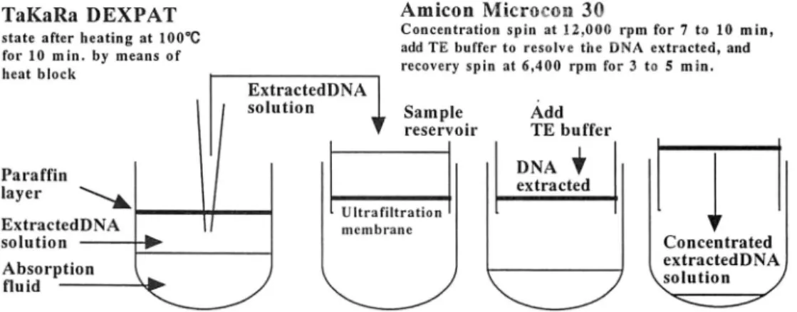
TaKaRa DEXPAT state after heating at 100°C for 10 min. by means of
heat block
Amicon Microcon 30
Concentration spin at 12.000 rpm for 7 to 10 min, add TE buffer to resolve the DNA extracted, and recovery spin at 6,400 rpm for 3 to 5 min.
Paraffin layer ExtractedDNA solution Absorption fluid ExtractedDNA solution Sample reservoir Ultrafiltration membrane Add TE buffer Concentrated extractedDNA solution /
Figure 4. PCR of HBG PC03-04 primes, employing
the DNA extracted solution by means of TaKaRa
DEXPAT™
In each of SP01, SP02, SP03 and SP04, the tem
plate DNA solution was5 jU\ of thesolution
1; DNA extracted from a 3 Aim thick section
2: DNA extracted from a 10 jUm thick section 3: DNA extracted from a 20 Aim thick section 4: DNA extracted from a 50 Aim thick section 5: DNA extracted from a 100 Aim thick section 6: DNA extracted from a 200 Aim thick section
PCR of human /3-globin (HBG) gene
Primers employed were PC03 and 04, and GH20
and 21. The amplified DNA of the PCR employing PC03 and 04 is 110 bp long and that of the PCR employingGH20
and 21 is 408 bp or 204 bp long. PCR employing the 4 primers yields 110,204,250,268 and 408 bp long bands of amplified DNA.
One hundred Ail of PCR solution includes 10 Ail of
lOx Buffer, 8 Ail of dNTP mixed solution, 0.5 Ail of TaKaRa Ex Taq, 2 Ail of each primers and autoclaved H,0. The PCR was performed according to the following proto
col, predenature for 5 min at 94°C, 30 cyclesof denature for 30 seconds at 94°C, annealing for 30 seconds at 55°C, and extension for 30 seconds at 72°C, and post-extension for 5 min at 72°C. Twenty Ail of the PCR product was charged
on 4% agar gel electrophoresis stained by ethidium bro mide.
Measurement ofDNA in the extracted DNA solution The amount of DNA extracted from the sections of SP01 and SP02 by means of TaKaRa DEXPAT™ and con
centrated by means of Amicon Microcon microcon
centrator was measured by a spectrophotometer before and
after phenol etraction and ethanol sedimentation of DNA.
The DNA amount exctracted the paraffin sections of gas
tric wall tissue stored long time was also measured. The
ratio (A260/A280) of the absorption at 260 nm (A260) ver sus the absorption at 280 nm (A280) was calculated for an index of protein contamination. The absorption at 260 nm (A260) represented the amount of DNA.
Result
The length of DNA strands extracted was examined
by means of PCR employing HBG primers and the DNA solution extracted from one 20 Aim thick section of the
SP01 and of the SP03 (Fig. 1). Five p. 1of the supernatant
DNA solution was applied to the PCR of HBG PC03 and 04 primers, of HBG GH20 and 21 primers, and of HBG PC03, 04, GH20 and 21 primers. The PCR employing the
SP03
extracted DNA solutions from the sections of the SP01 and of the SP03 indicated the same results, as shown in Fig. 2. In the PCR of HBG PC03 and 04, the amplified DNA was
of 110 bp long DNA. In the PCR of HBG GH20 and 21, the amplified DNA showed an obvious band at 204 bp
length and a trace band at 408 bp length. In the PCR of the four primers, the amplified DNA showed bands at 110, 204, 250 and 268 bp length in a smear. The DNA strands extracted from the paraffin sections were of less than 268 bp length, although a trace amounts of DNA of longer than 408 bp was recognized.
It was examined by means of the PCR employing HBG PC03 and 04 primers whether each extracted DNA
solution included an enough amount of DNA for the PCR
analysis. The products of PCR, which employed the ex tracted DNA solutions extracted from one 3, 10 and 20 Ai
m thick sections of the SP01 and SP03 paraffin-blocks as the template DNA solution, revealed a band of amplified
DNA at 110 bp length on the agar gel electrophoresis, sug gesting that these extracted DNA solutions included an amount of DNA enough for the PCR, as shown in Fig. 4 SP01 and SP03. But the extracted DNA solutions extracted from one of 3, 10, 20, 50, 100 and 200 Aim thick sections
of the SP02 and SP04 paraffin-blocks did not include an
amount of DNA enough for the PCR, as shown in Fig. 4
SP02 and SP04, although the PCR of that from one 200 Ai m thick section of the SP02 paraffin-block yielded a band
of the amplified DNA at 110 bp length.
The about 10 times concentrated DNA solutions of the extracted DNA solutions from the sections of the SP01 and SP03 paraffin-blocks included an amount of DNA enough for the PCR, revealing a band of amplified DNA at 110 bp length (Fig. 5, SP01 and SP03). Concatamer for mation was not seen (Fig. 5).
Employing the about 10 times concentrated DNA solutions of the extracted DNA solutions from the sections of the SP02 paraffin-block, the PCR amplified DNA enough to reveal a band at 110 bp length on the agar gel electrophoresis in the lanes of 20, 50, 100 or 200 micron meters thick sections in Fig. 5 SP02. From the lane of 20
Constant extraction of DNA [35]
SP01 SPU3
Figure 5. PCR of HBG PC03-04 primes, employ ing the about 10 times concentrated DNA solution In each of SP01, SP02, SP03 and SP04, the tem
plate DNA solution was 5 Ail of the solution
1; DNA extracted from a 3 Aim thick section 2: DNA extracted from a 10 Aim thick section 3: DNA extracted from a 20 Aim thick section 4: DNA extracted from a 50 jUm thick section 5: DNA extracted from a 100 Aim thick section 6: DNA extracted from a 200 Aim thick section
1 2 3
SP02
p m thick section to that of 200 Ai m thick section, the band
of the amplified DNA became stronger, probably reflecting
the DNA amont of the concentrated DNA solutions. The Fig. 5 SP04 showed the same tendency as the Fig. 5 SP02. But the band of the amplified DNA at the 110 bp length was seen in the lanes of the 50, 100 and 200 Aim thick sections.
The A260/A280 of the SP01 and SP02 concentrated DNA solutions was from 0.465 to 0.610, as indicated in the table 1. Contamination of a large amount of protein in the solutions was indicated. The A260/A280 of the DNA so lutions after phenol extraction and ethanol sedimentation
was from 1.493 to 1.740, indicating a quite low degree of the protein contamination. The DNA in the solutions was
from 50 ng/Ail x 50 to 1218 ng/jUl x 50. From the 10 to 20 Aim thick sections (SP01 and SP02) the largest amount of DNA was extracted. The volume of DNA extracted from one 10 Aim thick section indicated the lowest amount of DNA.
The nature of DNA in the SP01 and SP02 solutions
after phenol extraction and ethanol sedimentation was ex
amined by means of the PCR for HBG (Fig. 6). As shown in Fig. 4, three bands of amplified DNA at 110, 250 and
SP(M
268 bp were recognized in SP01. In the SP02, a band of
amplified DNA was recognized at 110 bp in the lane 4, 5 and 6.
The DNA solutions extracted from paraffin-embed ded tissue of stomach with and without adenocarcinoma in cluded shorter than 250 bp and longer than 110 bp strands
of DNA, as shown in Fig. 7.
The DNA extraction fluid extracted from two 20 p m thick sections of the paraffin-embedded gastric tissue
stored long time included a large amount of DNA. But the
amplified DNA bands of the PCR of HBG (Fig. 7) did not
run parallel with the amount of the DNA in table 2.
Discussion
It is quite important to see the nature of DNA ex
tracted from paraffin section, because fixation of tissue,
procedurs to make paraffin block, and storage duration of the paraffin-block effect on the nature of the DNA, espe cially in the length of the DNA strands. As shown in Fig. 2, the DNA extracted DNA comprised dominantly strands of less 268 bp length, although the product of the PCR em ploying HBG GH20-21 primers suggested a small amount
of DNA strands longer than 408 bp length. Because most
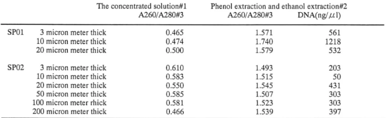
Table 1. The content of DNA in 300 micron liters extracted DNA solution of DEXPAT treatment The concentrated solution#l
A260/A280#3 SP01 3 micron meter thick
10 micron meter thick 20 micron meter thick SP02 3 micron meter thick 10 micron meter thick 20 micron meter thick 50 micron meter thick 100 micron meter rhick 200 micron meter thick
0.465 0.474 0.500 0.610 0.583 0.550 0.585 0.581 0.466
Phenol extraction and ethanol extraction#2 A260/A280#3 DNA(ng/Ail) 1.571 561 1.740 1218 1.579 532 1.493 203 1.515 50 1.545 431 1.507 303 1.523 303 1.539 397
#1: The about 10 times concentrated DNA solution by means of the microconcentrater
#2: The DNA slution extracted from the concentrated solution (#1) by means of phenol extraction and ethanol extraction #3; Ratio of the absorption at 260 nm versus the absorption at 280 nm by spectrophotometry
Figure 6. PCR od HBG gene, employing primers, PC03 and 04, and GH20 and 21, and DNA solu
tions of SP01 and SP01 after phenol extraction and
ethanol extraction in table 1
In SP 01, bands of amplified DNA are seen at 110, 250 and 260 bp are recognized in teh clear
background. In SP02 faint but gradual increaseof bands are recognized in the lane 4, 5 and 6. 1; DNA extracted from a 3 Aim thick section 2: DNA extracted from a 10 Aim thick section 3: DNA extracted from a 20 Aim thick section 4: DNA extracted from a 50 Aim thick section 5: DNA extracted from a 100 Aim thick section 6: DNA extracted from a 200 Aim thick section
DNA sequences that can be tragets of PCR analysis are shorter than 200 bp length, the DNA extracted in this study was proved to be enough for PCR analysis. The PCR em ploying HBG PC03, PC04, GH20 and GH21 is useful to
see the length of the extracted DNA.
Usually 0.1 Aig of template DNA is applied to PCR. The amount of DNA extracted could be measured. And the template DNA amount could be adjusted to 0.1 Aig
DNA per 5 Ail- But in a surgical pathology laboratory
there would not be an apparatus to measure a quite small
amount of DNA. Then, it is useful to see the enough
amount of tissue for PCR by estimating the areas where
target cells exist under microscope.
It was shown in Fig. 4 SP03 that the concentration of DNA extracted from a section of 2.5 cm xl.O cm x 3 Ai m largeness is enough for the PCR. But the concentration of DNA extracted from a section of 2.0 mm x 1.2 mm x 200
Aim largeness was not enough for the PCR. The concen
tration of DNA extracted from a SP02 section of 2.3 mm x 2.0 mm x 200 Aim largeness, about two times larger than
the SP04 section, was enough for the PCR. The minimum
largeness of the tissue section, from which the concentra
tion of DNA extracted by means of TaKaRa DEXPAT™ is
enough for the PCR was shown to be between 2.0 mm xl.2 mm x 200 Aim and 2.3 mm x 2.0 mm x 200 Aim.
An extremely small amount of DNA can be the tem
plate DNA for 2 times PCR employing the same primer set or the inner primers. Such an extremely small amount DNA can be gotten by means of micro-resection method. Depending on the areas where the target cells exist, an ad equate sensivity of the PCR must be employed. On the
other hand, this study indicated that the about 10 times con
centrated solution of the extracted DNA solution from one 100 Aim thick section of gastric mucosa biopsy material
more than 2 mm x 2 mm largeness is enough for PCR analysis. Therefore, the section of 100 Aim thickness with target cells areas larger than 2 mm x 2 mm is needed in the
PCR. Recently, Taq DNA polymerase that can amplify more the target sequence of DNA than TaKaRa Ex Taq DNA polymerase employed in this study has been intro duced so that thinner section would be enough for such Taq DNA polymerase.
Figure 7. The nature of DNA in the paraffin-em
bedded tissue stored long time
One band of amplified DNA at 110 bp and two bands of comcatamer of primers at about 20 and 40 bp were recognized in each lane. In the lanes of 1988 Ca and 1988 Non-Ca, additonal weal bands at the longer length were seen.
Ca: The DNA solution extracted from the section
with adenocaricnoma was employed as a template
DNA solution. Non-Ca: The DNA solution ex tracted from the section without adenocarcinoma
was employed as a template DNA solution.
Mtrk<r 1978 1978 1983
9 Ca NoDrCa Ca
1983 1988 1988 1993 1993 1998 Non-Ca Ca Non-Ca Ca Non-Ca Ca
1998 Non-Ca
Table 2. The content of DNA in the extraction fluid from two 20Aim thick sections of gastric wall sections with and without adenocarcinoma, which were stored for 1 year (1998), 6 years (1993), 11 years (1988), 16 years (1983) and 21 years (1978).
A260/A280"3 DNA(ng///l)
1978 1983 1988 1993 1998
Ca"1 Non-Ca*2 Ca Non-Ca Ca Non-Ca Ca Non-Ca Ca Non-Ca
1.559 2232 1.552 2247 1.564 2378 1.477 2547 1.400 2575 1.494 2482 1.588 1959 1.564 2282 1.569 2061 1.577 2187
#1: The DNA extraction fluid from the sections with adenocarcinoma. #2: The DNA extraction fluid from the sections without adenocarcinoma.
Constant extraction of DNA [37]
Concatamer formation was not seen in the PCR em ploying the about 10 times concentrated DNA solution,
suggesting that the about 10 times concentration is not the overconcentration to yield concatamer in the PCR.
The complete spine-rinse of a trace of glycerin in
ultrafiltration membrane of the microconcentrator2 is quite
important in the concentration of the template DNA solu tion for PCR, because an amount of DNA enough for the template DNA solution is absorbed even by the trace amount of glycerin. Although the routine method can con
centrate DNA solution and remove substances other than
DNA, the easy and rapid procedure of DNA concentration by means of the Amicon Microcon microconcentrator is important in practise.
The amount of DNA can be estimated by photoab-sorption at 260 nm in spectrophotometry. Because of the
contamination of a large amount of protein as quite low ra tio of A260/A280 in Table 1, the exact amount of DNA in
the DNA extraction fluid gotten by TaKaRa DEXPAT™ could not be measured, although the bands of the amplified DNA in the PCR employing the DNA extraction fluid sug gested its quality enough for PCR, as shown in Figures 2, 3 and 4.
The DNA in the DNA extraction fluid processed thorugh phenol extraction and ethanol sedimentation re vealed high values of A260/A280 ratios and indicated its
enough amount. The amount of DNA extracted (Table 1)
suggested that the largest amount of DNA can be extracted
from the 10 to 20 JUL m thick sections. Gradual increase of
DNA extraced accroding to the thcikness of the section was proved by the PCR in Fig. 5 and 6, although the amount of
DNA measured were of the almost same amount in SP02.
Probably a large amount of short-stranded DNA was ex
tracted from thin sections in SP02. Then, the DNA ex
tracted from paraffin-embedded sections must be exam
ined by means of PCR for HBG in order to see whether the DNA includes long stranded DNA enough for the PCR
analysis.
The DNA extracted from paraffin sections of stom
ach with and without adenocarcinoma included shorter
than 250 bp and longer than 110 bp strands of DNA enough for usual PCR analysis, as shown in Fig. 7. In the not-saturated PCR amplification of the target DNA, the amount of amplified DNA runs parallel with that of the target DNA in the template DNA solution. The stain of the bands of the amplified DNA by ethidium bromide has an quality to suggest the amount of the amplified DNA. Therefore, It suggested a large amount of the short-stranded DNA in the DNA extraction fluid that the bands of the amplified DNA in Fig. 7 did not run parallel with the amount of the DNA in the corresponding the extraction fluid in Table 2. On the other hand, it was suggested that there was an amount of the shorter than 250 bp and longer than llObp long-stranded DNA in the paraffin-embedded tissue. The stor age duration of the paraffin-blocks did not effect on the amount of the such long-stranded DNA.
References
1, Operation manual of TaKaRa DEXPATTM, Code No. 9091, TaKaRa Biocheical (in Japanese)
2, Operating manual of MICROCON, Microcincentrators, Publication 1-394 H, AMICON, Inc, Beverly, USA