Organogenesis of the Midgut Gland in the Prawn
Penaeus japonicus
著者
NAKAMURA Kaworu, TSURU Hiroyuki
journal or
publication title
鹿児島大学水産学部紀要=Memoirs of Faculty of
Fisheries Kagoshima University
volume
36
number
1
page range
221-225
別言語のタイトル
クルマエビ中腸腺の器官形成
Vol. 36,No.l, pp. 221-225 (1987)
Organogenesis of the Midgut Gland
in the Prawn Penaeus japonicus
Kaworu Nakamura and Hiroyuki Tsuru
Abstract
Development of the midgut gland was observed histologically during larval stages of the prawn Penaeus japonicus. The diverticula of the organ showed drastic increases of the volume and number according to the larval growth. Especially passing through the stage of postlarva 10, they increased rapidly more than at previous stages as from the zoea 3 to the postlarva 9. It suggested that the early larvae so far as of the postlarva 10 would suffer diseases more easily than at the later period, if the midgut gland functioned as a member of the defence mechanism during their developmental process.
Seed production of the prawn Penaeusjaponicus has developed its culture system under a
stable technicalmanagement and improved feeding of pellets1*. However, it suffers recently
a mass mortality during its early period of development. The elaboration upon its countermeasure is difficult because of deficient informations on the larval growth under an intact condition. Differentiations of internal organs, especially concerning to the defensemechanism, have been out of objects of the scientific study hitherto treated2).
In this report on the larval growth, the investigation of the midgut gland development has been carried out histologically at each stage from the zoea 3 to the postlarva 30. And its diverticular number has been computed as an index of the differentiation degree of the midgut gland which would be one of the most essential sites to the vital protection against the cause of diseases.
Materials and Methods
Larvae of the prawn Penaeus japonicus spawned in 500 / tank prepared at the laboratory were used. Rearing of the larvae was done by the routine of method, feeding with
Chaetoceros, Brachionus and yeast. During about 1 month of rearing, water temperature was
22. 5-27. 0°C (Apr. 10-Mar. 10). The larvae of each stage were sampled and fixed in 10%
formalin or Bouin solution. Under a dissecting microscope, their external characters were observed for the samples in formalin to certify their normal development. For the specimen in Bouin, dehydration through ethanol series and embedding in paraffin were treated. After
Laboratory of Propagation Physiology, Faculty of Fisheries, Kagoshima University, 50-20 Shimoarata 4, Kagoshima, 890 Japan.
222 Mem. Fac. Fish., Kagoshima Univ. Vol.36, No.l (1987)
sectioning into thin slices of 5 fxm, they were stained with PAS-hematoxylin. Sampled numbers were different as 2-7 among stages, and stages subjected to this experiment were each larval stage from the zoea 3 to the postlarva 12 (P12), P15, P20, P25 and P30. The
histological preparations sectioned transversally were applied to the following investiga
tions. 1) Observation of the diverticular development and calculation of the diverticular
number presenting in the slice which showed its maximum. The number was counted as half the total diverticula, because of their symmetrial arrangement and its relativity to the
developmental degree. 2) Transcription of the successive slices of the midgut gland at the
chosen stages of larvae. For it, the camera lucida was used.
Results and Discussion
Larvae of the prawn reached the stage of postlarva passing through periods of 6 stages of nauplius, 3 stages of zoea and 3 stages of mysis after hatching. Their development was
normal, like as the reference of the published record3).
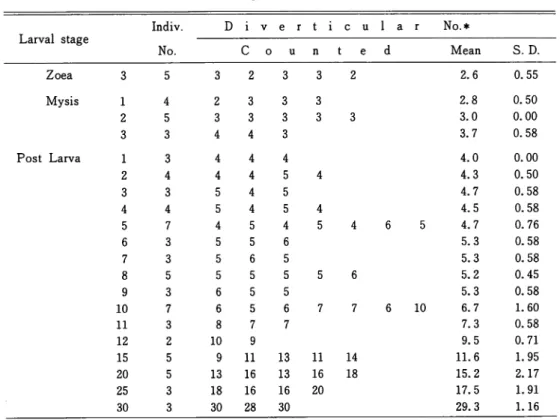
Relation between the diverticular number and each stage of larvae was indicated in Table
1. The average of 5 individuals at the zoea 3 showed 2. 6. At the mysis 1, the value became Table 1 . Increase of the diverticular number of the midgut gland according to
each of the larval stages from the zoea 3 to the postlarva 30.
Larval stage Indiv. D i v e r t i c u l a r No.* No. C o u n t e d Mean S. D. Zoea 3 5 3 2 3 3 2 2.6 0.55 Mysis 1 4 2 3 3 3 2 5 3 3 3 3 3 3 3 4 4 3 Post Larva 1 3 4 4 4 2 4 4 4 5 4 3 3 5 4 5 4 4 5 4 5 4 5 7 4 5 4 5 4 6 3 5 5 6 7 3 5 6 5 8 5 5 5 5 5 6 9 3 6 5 5 10 7 6 5 6 7 7 11 3 8 7 7 12 2 10 9 15 5 9 11 13 11 14 20 5 13 16 13 16 18 25 3 18 16 16 20 30 3 30 28 30 10 2.8 0.50 3.0 0.00 3.7 0.58 4.0 0.00 4.3 0.50 4.7 0.58 4.5 0.58 4.7 0.76 5.3 0.58 5.3 0.58 5.2 0.45 5.3 0.58 6.7 1.60 7.3 0.58 9.5 0.71 11.6 1.95 15.2 2.17 17.5 1.91 29.3 1.16 number as half the total diverticula counted in the section which showed the
NAKAMURA • TSURU ! Organogenesis of Prawn Midgut Gland
m i n i I I 1 2 3 * 5 6 7 8 9 10 12 15
Zoea Mysis Post larva
LARVAL STAGE
Fig. 1. Graphic representation of the increase of the
diverticular number calculated in Table 1. The value means a half the total diverticula counted in the section which showed the maximum number.
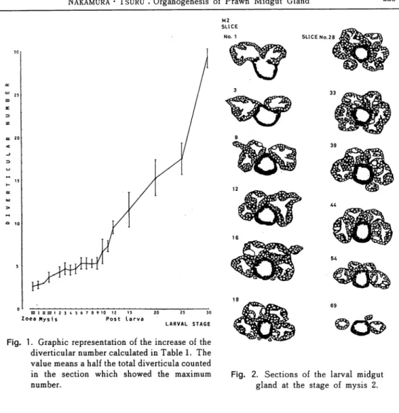
M2
SLICE
Fig. 2. Sections of the larval midgut gland at the stage of mysis 2.
2. 8. And at the successive stages of mysis, it increased to 3. 0 or 3. 7. The rate of increase
was large especially at the mysis 2-3, as well as at the later periods as P5-6 and P9-10. After passing the P10, the diverticular number showed a steep line of increase as represented in Fig. 1. For example, each average of P2, P4, P6, P8, P10, P12, P15, P20 and P30 was 4. 3, 4.5, 5.3, 5.2, 6.7, 9.5, 11.6, 15. 2 and 29. 3, respectively. It seemed that the organogenesis of the midgut gland would progress rapidly after the P10.
Sections transcribed from the histological preparation of the midgut gland in the mysis 2 larva were shown in Fig. 2. In the figure, the diverticula were represented as the black circle
provided with white spots which meant the nuclei of the diverticular cells. The black ring
situated at the central area was the midgut. Anterior midgut, in the slice number 1, corresponded to the stomach. From these drawings, it would be easy to understand that the diverticula were symmetrically arranged and the maximum number in the slice was 6 (in the slice number 28 and 33). For the specimen of the P2, the wall of the stomach indicated its beginning of morphological complexity and the shape of the diverticula showed a weak224
Mem. Fac. Fish., Kagoshima Univ. Vol.36, No.l (1987) P2
SLICE No. 1
Fig. 3. Sections of the larval midgut gland at the stage of postlarva 2. mg, midgut; st,
stomach.
SLICE No. 26
Fig. 4. Sections of the larval midgut gland at the stage of postlarva 10. mg, midgut; st,
stomach.
transformation (Fig. 3). The diverticular number was 8 (maximum in the slice number 19).
For the specimen of the P5, the wall of the stomach showed the complexity, possessing an intrusion into the lumen. The diverticula were affected by some artifact, and their maximum
number became 10. For the specimen of the P10, the diverticula developed extremely their
volume extending around the midgut as shown in Fig. 4. Especially their ventral and lateral areas spread, because the upper portion corresponded to the ventral of the midgut gland, in these all figures. Transformation of the diverticula was also remarkable. Their sections
showed irregular folds. And the maximum number was 12 (in the slice number 26).
From the results, it would be deduced that the organogenesis of the midgut gland of the prawn has a specific period of development which shows a rapid increase of the volume and number of the diverticla. That corresponds to the later period than the postlarva 9. It would be considered that early larvae until the postlarva 10 suffer diseases easily than at the later
Nakamura • Tsuru : Organogenesis of Prawn Midgut Gland 225
stages, if the midgut gland functioned as a member of the defence mechanism in the vital protection against the cause of diseases.
Acknowledgements
This work was supported in part by a grant from the Japan Ministry of Agriculture, Forestry and Fishery.
References
1 ) SHIGUENO, K. (1975) : "Shrimp Culture in Japan", pp. 3-110, Assoc. Internat. Techn. Prom., Tokyo.
2 ) McLaughlin, P. A. (1983) : Digestive system. "The Biology of Crustacea" (ed. by L. H. Man tel), Vol.5, pp. 26-52, Academic Press, New York.
3) HuDINAGA, M. (1942) '. Reproduction, development and rearing of Penaeus japonicus BATE.