ORIGINAL
Effect of controlled ventilation during assist-control ventilation
on diaphragm thickness : a post hoc analysis of
an observational study
Taiga Itagaki1, Nobuto Nakanishi1, Takuya Takashima2, Yoshitoyo Ueno2, Natsuki Tane3, Yumiko Tsunano1, Toshiyuki Nunomura4, and Jun Oto3
1Department of Emergency and Critical Care Medicine, Tokushima University Graduate School, Tokushima, Japan, 2Department of Emer-gency and Critical Care Medicine, Tokushima University Hospital, Tokushima, Japan, 3Department of Emergency and Disaster Medicine, Tokushima University Hospital, Tokushima, Japan, 4Division of Critical Care Center, Kochi Red Cross Hospital, Kochi, Japan
Abstract : Background : Since diaphragm passivity induces oxidative stress that leads to rapid atrophy of di-aphragm, we investigated the effect of controlled ventilation on diaphragm thickness during assist-control ventilation (ACV). Methods : Previously, we measured end-expiratory diaphragm thickness (Tdiee) of patients
mechanically ventilated for more than 48 hours on days 1, 3, 5 and 7 after the start of ventilation. We retrospec-tively investigated the proportion of controlled ventilation during the initial 48-hour ACV (CV48%). Patients were
classified according to CV48% : Low group, less than 25% ; High group, higher than 25%. Results : Of 56 patients
under pressure-control ACV, Tdiee increased more than 10% in 6 patients (11%), unchanged in 8 patients (14%)
and decreased more than 10% in 42 patients (75%). During the first week of ventilation, Tdiee decreased in both
groups : Low (difference, -7.4% ; 95% confidence interval [CI], -10.1% to -4.6% ; p < 0.001) and High group (differ-ence, -5.2% ; 95% CI, -8.5% to -2.0% ; p = 0.049). Maximum Tdiee variation from baseline did not differ between Low (-15.8% ; interquartile range [IQR], -22.3 to -1.5) and High group (-16.7% ; IQR, -22.6 to -11.1, p = 0.676). Conclu-sions : During ACV, maximum variation in Tdiee was not associated with proportion of controlled ventilation higher than 25%. J. Med. Invest. 67 : 332-337, August, 2020
Keywords : Mechanical Ventilation, assist-controlled ventilation, diaphragm, ventilator-induced diaphragm disfunction, atrophy
INTRODUCTION
It has been established that mechanical ventilation can cause diaphragm dysfunction. (ventilator-induced diaphragm dysfunc-tion : VIDD) (1) which is associated with higher mortality in in-tensive care units (ICU) and hospitals (2, 3). The most important cause of VIDD and diaphragm atrophy is disuse owing to the suppression of inspiratory effort (4, 5). In animal experiments, short periods of passive controlled mechanical ventilation (CMV) has induced oxidative stress that leads to protein degradation, resulting in diaphragm-muscle atrophy and weakness (6-8). In one of the few human investigations, Levine et al. (9) reported that prolonged diaphragmatic inactivity in brain-dead organ donors was associated with marked atrophy of diaphragm myofi-bers. Furthermore, these detrimental effects of CMV on the dia-phragm could be largely ameliorated by assist-control ventilation (ACV) (10), intermittent spontaneous breathing during CMV (11), pressure support ventilation (PSV) (12, 13), and adaptive support ventilation (14). Consequently, to preserve spontaneous effort, most critically ill patients who require mechanical ventila-tion receive assisted and spontaneous modalities (1, 15).
ACV is the most frequently applied mode during critical care, with 60% of mechanically ventilated patients receiving ACV (16, 17). In ACV, mechanical ventilation can be triggered by the ventilator (controlled ventilation) or the patient (assisted ventilation) (18). During controlled ventilation, triggering occurs
after detection of a certain period without inspiratory effort ; when this happens, diaphragm contraction does not precede the mechanical ventilation. Theoretically, the more the proportion of controlled ventilation increases during ACV, the more the diaphragm is disused. Marin-Corral et al. (19) recently observed Maastricht III organ donors who can stimulate their diaphragm spent 41% of ventilation time in assisted and spontaneous modalities. This resulted in less diaphragm myofiber damage than in brain-dead organ donors relying on full support. In most studies cited above (6-15), diaphragm atrophy from disuse, however, has been evaluated with traditional CMV, which ren-ders the diaphragm completely inactive, or by comparing CMV with partially assisted mechanical ventilation. To the best of our knowledge, our clinical study is the first to investigate the effects on diaphragm atrophy, diagnosed by ultrasonography, of controlled ventilation through the course of a single ventilation mode, ACV.
We aimed to test the hypothesis that a higher proportion of controlled ventilation during the initial 48 hours of ACV would correlate with greater diaphragm atrophy among general criti-cally ill patients.
METHODS
We conducted this study in a university hospital. Ethical approval for this study was provided by the ethics committee of Tokushima University Hospital (protocol number 3220). Written informed consent from each patient was waived as approved by the committee. This study was registered on a clinical trial (UMIN clinical trial registry : 000032944).
The Journal of Medical Investigation Vol. 67 2020
Received for publication May 25, 2020 ; accepted July 27, 2020. Address correspondence and reprint requests to Taiga Itagaki M.D., Ph.D., Department of Emergency and Critical Care Medicine, Tokus-hima University Graduate School, 3-18-15 Kuramoto-cho, Tokushi-ma, 770-8503, Japan and Fax : +81-88-633-9339.
Study design and patients
This study is a post-hoc analysis of our prospective observa-tional study (20) which evaluated changes in diaphragm and in-tercostal muscle thickness in mechanically ventilated patients. In that study, we recruited adult patients who were consecutively admitted to the ICU and expected to require mechanical ventila-tion for more than 48 hours. Exclusion criteria were : age under 18 years ; trauma or chest tube at the measurement point ; and diagnosis of primary neuromuscular disease. Among this pop-ulation of the previous study, we selected patients who were on pressure-control ACV throughout at least the first 54 hours of mechanical ventilation, and also excluded patients who were continuously paralyzed with neuromuscular blocking agents.
Measurement of diaphragm thickness
In the initial study (20), on days 1, 3, 5 and 7 after the start of mechanical ventilation, we used ultrasonography to measure diaphragm thickness at peak inspiration and end-expiration (Tdiee). Diaphragm thickening was calculated for each measure-ment as follows : Thickening fraction (%) = [(thickness at peak inspiration – thickness at end-expiration) / thickness at end-ex-piration] × 100. Data collection was discontinued, whichever occurs first, at extubation, at patient discharge from the ICU, or at death. Measurement procedures are fully detailed in our previous study. In brief, referring to previously reported meth-ods, measurement was performed using B mode ultrasound with the liner transducer perpendicularly placed on the right chest wall at the zone of apposition (21). Of note, each recording was performed by the same investigator and actual measurement of diaphragm thickness was retrospectively done by the same investigator with the stored images in order to blind the data analysis from each patient’s status.
Proportion of controlled ventilation
Reviewing patient charts, we retrospectively calculated the proportions of controlled ventilation and assisted ventilation during the initial 48 hours of ACV. To eliminate the possible effects of neuromuscular blocking agents that might have been used during tracheal intubation or in the operating theater, we focused on the 48-hour-period starting 6 hours after the start of mechanical ventilation in the ICU. That is why inclusion criteria required ICU patients who were on ACV for more than 54 hours. Actual respiratory frequency is automatically recorded by the minute in our electrical system (PrimeGaia, Nihon Kohden, Tokyo, Japan). Set values for respiratory rate were manually entered into the same system by bedside nurses. While actual respiratory frequency was equal to the set value for respiratory rate, we assumed that mechanical breaths delivered in the min-ute were all controlled ventilation triggered by the time. While actual respiratory frequency was greater than the set value for respiratory rate, mechanical breaths delivered in the minute were all considered as assisted ventilation triggered by the patient. Then, we calculated the proportion of controlled ventila-tion during the initial 48 hours of ACV (CV48%) according to the formula : CV48% (%) = [total minutes of controlled ventilation / 48 × 60] × 100.
Endpoints of the study
The primary endpoints of this study were the direction and maximum variation in Tdiee during mechanical ventilation.
According to median CV48%, we assigned patients to either of two groups and performed intergroup comparison of direction and maximum variation in Tdiee. Secondary endpoints were
duration of mechanical ventilation, incidence of reintubation and tracheostomy, and ICU mortality.
Clinical data collection
Demographic data, APACHE (acute physiology and chronic health evaluation) II score on admission to the ICU, reason for mechanical ventilation and clinical outcomes were gathered from medical charts. Then, for the initial 48-hour period of ACV, we collected the following physiological and ventilator variables : set values for respiratory rate ; actual respiratory frequen-cy ; plateau pressure ; positive end-expiratory pressure (PEEP) ; tidal volume ; and arterial blood gas analysis.
Statistical analysis
Continuous data are presented as medians with interquartile range (IQR), whereas categorical variables are expressed as numbers and percentages. Direction of changes in Tdiee over time were analyzed in each group by repeated measures of analysis of variance with multiple comparisons to reveal effects over time. Differences between continuous variables, that is maximum variation in Tdiee, thickening fraction, ventilatory and physi-ologic variables, and duration of mechanical ventilation were assessed using t tests or Mann-Whitney U tests. Categorical variables, that is, incidence of reintubation, tracheostomy, and death in the ICU, were assessed as appropriate with chi-squared tests or Fisher exact tests. Statistical calculations were carried out with statistics software (SPSS, version 26, SPSS Inc., Chica-go, IL). P < 0.05 was considered statistically significant.
RESULTS
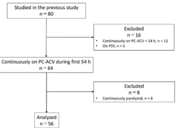
From the 80 patients in the preliminary study, we selected the 64 patients who were on pressure-control ACV throughout at least the first 54 hours of mechanical ventilation. We then excluded 8 patients who had been continuously paralyzed with neuromuscular blocking agents, and finally analyzed valid data from 56 patients (Fig. 1). Assessing median CV48% as 24.9%, we assigned patients to either of two groups : Low group, CV48% less than 25% ; High group, CV48% more-than-or-equal-to 25%. Table 1 shows the characteristics of the Low and High groups. The most frequent reason for mechanical ventilation was acute respiratory failure (34%) for Low group and post-operative care (30%) for High group.
Figure 1. Flow chart of study participants.
PC-ACV, pressure-control assist-control ventilation ; PSV, pressure support ventilation.
Diaphragm thickness was measured in 100%, 100%, 73% and 43% of patients on days 1, 3, 5 and 7, respectively. Tdiee was 2.1 mm [IQR, 1.6 to 2.5], 1.9 mm [IQR, 1.5 to 2.5], 1.8 mm [IQR, 1.6 to 2.5] and 2.0 mm [IQR, 1.4 to 2.7], and thickening fraction was 8.3% [IQR, 4.0 to 17.1], 8.3% [IQR, 4.9 to 12.4], 6.9% [IQR, 3.2 to 16.2] and 5.6% [IQR, 0.0 to 10.6] on days 1, 3, 5 and 7, re-spectively. Tdiee increased by more than 10% from the baseline in 6 patients (11%), unchanged without a 10% increase or decrease in 8 patients (14%) and decreased by more than 10% in 42 pa-tients (75%). Primary and secondary outcomes are summarized in Fig. 2 and Table 2. Over the first week of mechanical ventila-tion, Tdiee decreased in both Low group (difference, -7.4% ; 95% confidence interval [CI], -10.1% to -4.6% ; p < 0.001) and High group (difference, -5.2% ; 95% CI, -8.5% to -2.0% ; p = 0.049) (Fig. 2). Maximum variation in Tdiee from the baseline showed no
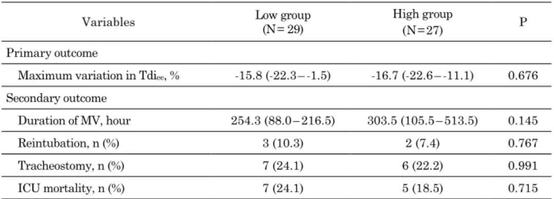
intergroup difference (Low, -15.8% [IQR, -22.3 to -1.5] vs. High, -16.7% [IQR, -22.6 to -11.1], p = 0.676). There was no statistically significant intergroup difference in duration of mechanical ven-tilation, incidence of reintubation and tracheostomy, and ICU mortality (Table. 2).
Table 3 shows ventilation and physiological variables during the initial 48 hours of ACV. Set value for respiratory rate was significantly lower in Low group than in High group (13.6 per min [IQR, 12.2 to 15.1] vs. 16.4 per min [IQR, 14.4 to 18.9], p = 0.003). CV48% was 5.1% [IQR, 1.6 to 13.7] in Low group and 53.8% [IQR, 36.4 to 61.6] in High group (p < 0.001). Tidal volume was significantly greater in Low group than in High group (9.1 mL / kg predicted body weight (PBW) [IQR, 7.7 to 10.1] vs. 7.8 mL / kg PBW [IQR, 7.1 to 8.8], p = 0.029) ; this was despite lower plateau pressure (18.9 cmH2O [IQR, 16.7 to 21.4] vs. 21.1 cmH2O [IQR, 18.9 to 22.7], p = 0.017) and PEEP (7.8 cmH2O [IQR, 6.0 to 8.4] vs. 8.0 cmH2O [IQR, 7.2 to 11.0], p = 0.030). PaO2 / FIO2 ra-tio were similar in both groups. In Low group, pH was slightly greater than in High group, although variables in both groups were within the normal range (7.44 [IQR, 7.41 to 7.47] vs. 7.41 [IQR, 7.34 to 7.43], p = 0.003). Thickening fraction did not differ between Low and High group (12.5% [IQR, 8.3 to 22.5] vs. 17.4% [IQR, 11.1 to 22.2], p = 0.367).
Table 1. Patient characteristics
Low group (N = 29) High group (N = 27) Age, yr 74 (63 – 80) 66 (5 – 73) Male, n (%) 18 (62) 17 (63) Height, cm 158 (155 – 163) 160 (152 – 170) Weight, kg 58 (48 – 69) 61 (51 – 69) APACHE II score 27 (21 – 32) 22 (15 – 28)
Reason for mechanical ventilation, n (%)
Acute respiratory failure 10 (34) 4 (15)
Stroke 5 (17) 2 (7)
Post CPR 4 (14) 3 (11)
Post-operative care 4 (14) 8 (30)
Sepsis / septic shock 3 (10) 4 (15)
Acute heart failure 2 (7) 4 (15)
Others 1 (4) 2 (7)
Data are expressed as median (interquartile range). APACHE, acute physiology and chronic health evaluation ; CPR, cardio-pul-monary resuscitation.
Figure 2. Changes in mean end-expiratory diaphragm thickness over time during assist-control ventilation in Low and High group. Error bars indicate 95% confidence intervals. *P < 0.05 vs. day 1.
Table 2. Primary and secondary outcomes
Variables Low group(N = 29) High group
(N = 27) P
Primary outcome
Maximum variation in Tdiee, % -15.8 (-22.3 – -1.5) -16.7 (-22.6 – -11.1) 0.676
Secondary outcome
Duration of MV, hour 254.3 (88.0 – 216.5) 303.5 (105.5 – 513.5) 0.145
Reintubation, n (%) 3 (10.3) 2 (7.4) 0.767
Tracheostomy, n (%) 7 (24.1) 6 (22.2) 0.991
ICU mortality, n (%) 7 (24.1) 5 (18.5) 0.715
Data are expressed as median (interquartile range) unless otherwise noted. Tdiee, end-expiratory
DISCUSSION
This study revealed four important findings. (1) Contrary to our hypothesis, Tdiee decreased over time in both groups
and maximum variation in Tdiee was not associated with high
proportion of controlled ventilation during early-phase of ACV. (2) Higher proportion of controlled ventilation during ACV was associated with higher set value for respiratory rate. (3) Despite higher plateau pressure and PEEP, tidal volume was significant-ly less in the High group than in the Low group. (4) There was no significant intergroup difference in clinical outcomes.
The best established causal evidence for diaphragm myotrau-ma is disuse atrophy (4, 5). Consequently, we focused on dia-phragm inactivity during ACV, which is sometimes overlooked because respiratory muscle monitoring is not standard in most ICUs (22), and because every delivered breath during ACV looks the same (18). Marin-Corral et al, (19) recently compared the structural characteristics of diaphragms in brain-dead organ donors and Maastricht III organ donors who showed diaphrag-matic stimulation. They found that Maastricht III organ donors presented less VIDD than brain-dead organ donors. Despite this histological evidence, we were unable to detect such adverse ef-fects of controlled ventilation on patient diaphragms during ACV. In our study, we arbitrarily assigned patients to two groups with median CV48% : High, 53.8% ; and Low, 5.1%. We found that the proportion of assisted ventilation was 46.2% in High and 94.9% in Low. On the other hand, in the study by Marin-Corral et al., the median percentage of time in assisted and spontaneous modalities was 41% [IQR, 12 to 76] in Maastricht III donors and 1% [IQR, 0 to 23] in brain-dead donors. Our different results are probably accounted for by greater diaphragm activity in our pa-tients. Future studies may clarify the risk of diaphragm atrophy by revealing a clear cut-off value for CV48%.
Our findings are essentially consistent with two studies by Goligher et al. (3, 15) that show that diaphragm thickness de-creases both during controlled modes of ventilation and during
partially assisted ventilation. In one of these studies (15), con-tractile activity decreased according to the number of days of controlled modes of ventilation. Moreover, in patients receiving controlled modes of ventilation, contractile activity was slightly but significantly greater if respiratory frequency was higher than the set rate. Even in PSV, high pressure levels of prolonged PSV developed diaphragm atrophy and contractile dysfunction (23). Such results suggested that presence of triggering, or oc-currence of diaphragm activity in itself may not be sufficient to prevent diaphragm atrophy. Moreover, the protective effect of diaphragm contraction seems to depend on the amount of inspi-ratory effort. Actually, we did not observe significant intergroup differences in inspiratory efforts represented by the thickening fraction. While we remain unsure what level of inspiratory effort is optimal, results from other investigations (3, 14, 24) suggest that the most effective approach may be to keep inspiratory ef-fort similar to quiet breathing at rest. To do this, it is necessary to optimize ventilation settings and sedation level (25) and thus control neural respiratory drive.
In this study, Low group showed higher tidal volume in spite of lower driving pressure (plateau pressure minus PEEP). Be-cause there was no intergroup difference in thickening fraction, Low group might have more of compliant lungs than increased inspiratory efforts. This was seen as one reason for lower tidal volume and higher PEEP in High group. In a recent animal study (26), lung protective ventilation with low tidal volume and high PEEP worsened VIDD by inducing oxidative stress and succeeding downregulation of peroxisome proliferator-activated receptor γ coactivator 1alpha (PGC-1α), reactive oxygen species inhibitor in diaphragm. This result is consistent with another study (27) showing that mechanical ventilation with a very large tidal volume of 35 mL / kg results in less oxidative stress in the diaphragm than ventilation with a moderate tidal volume of 9 mL / kg. Although the relationship between high PEEP and oxidative stress is unknown, excessive PEEP is associated with a rapid reduction in the number of sarcomeres and the Table 3. Ventilation and physiological variables during the initial 48-hour period of assist-controlled ventilation
Variables Low group(N = 29) High group
(N = 27) P
Set respiratory rate, per min 13.6 (12.2 – 15.1) 16.4 (14.4 – 18.9) 0.003
Actual respiratory frequency, per min 18.7 (18.1 – 21.4) 17.7 (16.3 – 20.5) 0.245
CV48%, % 5.1 (1.6 – 13.7) 53.8 (36.4 – 61.6) <0.001
Peak pressure, cm H2O 19.5 (17.4 – 21.9) 21.3 (19.0 – 23.2) 0.026
Plateau pressure, cm H2O 18.9 (16.7 – 21.4) 21.1 (18.9 – 22.7) 0.017
Positive end-expiratory pressure, cm H2O 7.8 (6.0 – 8.4) 8.0 (7.2 – 11.0) 0.030
Tidal volume, mL per kg PBW 9.1 (7.7 – 10.1) 7.8 (7.1 – 8.8) 0.029
FIO2, % 32 (25 – 39) 39 (29 – 51) 0.184
Arterial blood gas analysis ;
pH 7.44 (7.41 – 7.47) 7.41 (7.34 – 7.43) 0.003
PaCO2, mm Hg 35.3 (31.2 – 39.3) 40.4 (35.7 – 46.2) 0.352
PaO2, mm Hg 88.1 (75.5 – 92.9) 91.5 (81.7 – 101.6) 0.220
PaO2 /FIO2, mm Hg 269 (206 – 366) 269 (153 – 340) 0.533
Thickening fraction, % 12.5 (8.3 – 22.5) 17.4 (11.1 – 22.2) 0.367
Data are expressed as median (interquartile range). CV48% represents the proportion of controlled venti-lation for the initial 48 hours of assist-control ventiventi-lation. CV, controlled ventiventi-lation ; PBW, predicted body weight.
longitudinal atrophy of diaphragm fibers (28). Observing lower tidal volume and higher PEEP in the High group, we speculated that the VIDD developed in the High group might be worsened by incidental lung protective ventilation ; we did not detect, however, any detrimental effects. To prevent development of VIDD in clinical settings, further study may reveal what levels of tidal volume and PEEP are optimal during lung protective ventilation.
This study has several limitations. First, we employed previ-ously used definition for proportion of controlled ventilation (15, 19) assuming that all breaths were controlled breaths if actual respiratory frequency was equal to the set value for respiratory rate, and assisted breaths if actual frequency was greater than the set value. However, without detailed monitoring, this as-sumption may not be always accurate. For example, there is the potential that patients are breathing spontaneously even if re-spiratory frequency is equal to the set value. In this case, CV48% may be overestimated, especially when the set rate is high. Second, we focused on an initial 48-hour period of ACV, a dura-tion that might be inadequate to detect the significant effects of controlled ventilation on the diaphragm. Even so, the 28 hours of controlled ventilation in the High group was within the range (18-69 hours) of the period of diaphragm inactivity in an autopsy study demonstrating rapid atrophy owing to disuse (9). Third, as mentioned above, according to the CV48% median value, we arbi-trarily assigned patients to either of two groups and carried out intergroup comparison. As a result, we did not precisely find the discriminating cut-off value for CV48% ; we did not conduct this analysis because the sample population was small, especially the number of patients with very high CV48%. Fourth, since we stud-ied only patients on pressure-control ACV, our findings cannot be applied to patients on volume-control ACV. Finally, since we studied adult mechanically ventilated patients with and with-out respiratory failure, inspiratory efforts affecting diaphragm thickness were highly variable in our population. Future studies focused on patients with respiratory failure, who are at greater risk of VIDD, may have more useful clinical implications.
CONCLUSIONS
In a mixed population, the proportion of controlled ventilation more-than-or-equal-to 25% during early-phase ACV was not associated with maximum variation in diaphragm thickness detected by ultrasonography. More studies are required to un-derstand how the degree and duration of diaphragm inactivity during ACV affects diaphragm atrophy.
CONFLICTS OF INTEREST
All authors disclose no conflict of interest.
ACKNOWLEDGEMENTS
None
FUNDING
This research did not receive any specific grant from funding agencies in the public, commercial, or not-for-profit sectors.
REFERENCES
1. Dres M, Goligher EC, Heunks LMA, Brochard LJ : Critical illness-associated diaphragm weakness. Intensive Care Med 43(10) : 1441-52, 2017
2. Dres M, Dube BP, Mayaux J, Delemazure J, Reuter D, Brochard L, Similowski T, Demoule A : Coexistence and impact of limb muscle and diaphragm weakness at time of liberation from mechanical ventilation in medical intensive care unit patients. Am J Respir Crit Care Med 195(1) : 57-66, 2017
3. Goligher EC, Dres M, Fan E, Rubenfeld GD, Scales DC, Herridge MS, Vorona S, Sklar MC, Rittayamai N, Lanys A, Murray A, Brace D, Urrea C, Reid WD, Tomlinson G, Slutsky AS, Kavanagh BP, Brochard LJ, Ferguson ND : Mechanical ventilation-induced diaphragm atrophy strongly impacts clinical outcomes. Am J Respir Crit Care Med 197(2) : 204-13, 2018
4. Goligher EC, Brochard LJ, Reid WD, Fan E, Saarela O, Slutsky AS, Kavanagh BP, Rubenfeld GD, Ferguson ND : Diaphragmatic myotrauma : a mediator of prolonged ventilation and poor patient outcomes in acute respiratory failure. Lancet Respir Med 7(1) : 90-8, 2019
5. Schepens T, Dres M, Heunks L, Goligher EC : Dia-phragm-protective mechanical ventilation. Curr Opin Crit Care 25(1) : 77-85, 2019
6. Sassoon CS, Caiozzo VJ, Manka A, Sieck GC : Altered di-aphragm contractile properties with controlled mechanical ventilation. J Appl Physiol 92(6) : 2585-95, 2002
7. Shanely RA, Zergeroglu MA, Lennon SL, Sugiura T, Yimlamai T, Enns D, Belcastro A, Powers SK : Mechanical ventilation-induced diaphragmatic atrophy is associated with oxidative injury and increased proteolytic activity. Am J Respir Crit Care Med 166(10) : 1369-74, 2002
8. Powers SK, Shanely RA, Coombes JS, Koesterer TJ, McKenzie M, Van Gammeren D, Cicale M, Dodd SL : Me-chanical ventilation results in progressive contractile dys-function in the diaphragm. J Appl Physiol 92(5) : 1851-8, 2002
9. Levine S, Nguyen T, Taylor N, Friscia ME, Budak MT, Rothenberg P, Zhu J, Sachdeva R, Sonnad S, Kaiser LR, Rubinstein NA, Powers SK, Shrager JB : Rapid disuse atrophy of diaphragm fibers in mechanically ventilated humans. N Engl J Med 358(13) : 1327-35, 2008
10. Sassoon CS, Zhu E, Caiozzo VJ : Assist-control mechanical ventilation attenuates ventilator-induced diaphragmatic dysfunction. Am J Respir Crit Care Med 170(6) : 626-32, 2004
11. Gayan-Ramirez G, Testelmans D, Maes K, Rácz GZ, Cadot P, Zádor E, Wuytack F, Decramer M : Intermittent sponta-neous breathing protects the rat diaphragm from mechani-cal ventilation effects. Crit Care Med 33(12) : 2804-9, 2005 12. Hudson MB, Smuder AJ, Nelson WB, Wiggs MP, Shimkus
KL, Fluckey JD, Szeto HH, Powers SK : Partial support ventilation and mitochondrial-targeted antioxidants protect against ventilator-induced decreases in diaphragm muscle protein synthesis. PLoS One 10(9) : e0137693, 2015
13. Futier E, Constantin JM, Combaret L, Mosoni L, Roszyk L, Sapin V, Attaix D, Jung B, Jaber S, Bazin JE : Pressure support ventilation attenuates ventilator-induced protein modifications in the diaphragm. Crit Care 12(5) : R116, 2008
14. Jung B, Constantin JM, Rossel N, Le Goff C, Sebbane M, Coisel Y, Chanques G, Futier E, Hugon G, Capdevilla X, Petrof B, Matecki S, Jaber S : Adaptive support ventilation prevents ventilator-induced diaphragmatic dysfunction
in piglet : an in vivo and in vitro study. Anesthesiology 112(6) : 1435-43, 2010
15. Goligher EC, Fan E, Herridge MS, Murray A, Vorona S, Brace D, Rittayamai N, Lanys A, Tomlinson G, Singh JM, Bolz SS, Rubenfeld GD, Kavanagh BP, Brochard LJ, Ferguson ND : Evolution of diaphragm thickness during mechanical ventilation. Impact of inspiratory effort. Am J Respir Crit Care Med 192(9) : 1080-8, 2015
16. Esteban A, Anzueto A, Alía I, Gordo F, Apezteguía C, Pálizas F, Cide D, Goldwaser R, Soto L, Bugedo G, Rodrigo C, Pimentel J, Raimondi G, Tobin MJ : How is mechanical ventilation employed in the intensive care unit? Am J Re-spir Crit Care Med 161(5) : 1450-8, 2000
17. Esteban A, Anzueto A, Frutos F, Alía I, Brochard L, Stewart TE, Benito S, Epstein SK, Apezteguía C, Nightingale P, Arroliga AC, Tobin MJ ; Mechanical Ventilation Interna-tional Study Group : Characteristics and outcomes in adult patients receiving mechanical ventilation : a 28-day inter-national study. JAMA 287(3) : 345-55, 2002
18. Mancedo J. Assist-control ventilation. In : Tobin MJ, editor. Principles and practice of mechanical ventilation, third edition. New York : The McGraw-Hill Companies, Inc p. 159-74, 2013
19. Marin-Corral J, Dot I, Boguña M, Cecchini L, Zapatero A, Gracia MP, Pascual-Guardia S, Vilà C, Castellví A, Pérez-Terán P, Gea J, Masclans JR : Structural differences in the diaphragm of patients following controlled vs assisted and spontaneous mechanical ventilation. Intensive Care Med 45(4) : 488-500, 2019
20. Nakanishi N, Oto J, Ueno Y, Nakataki E, Itagaki T, Nishimura M : Change in diaphragm and intercostal mus-cle thickness in mechanically ventilated patients : A pro-spective observational ultrasonography study. J Intensive Care 7 : 56, 2019
21. Bunnell A, Ney J, Gellhorn A, Hough CL : Quantitative
neuromuscular ultrasound in intensive care unit-acquired weakness : a systematic review. Muscle Nerve 52(5) : 701-8, 2015
22. Heunks LM, Doorduin J, van der Hoeven JG : Monitoring and preventing diaphragm injury. Curr Opin Crit Care 21(1) : 34-41, 2015
23. Hudson MB, Smuder AJ, Nelson WB, Bruells CS, Levine S, Powers SK : Both high level pressure support ventilation and controlled mechanical ventilation induce diaphragm dysfunction and atrophy. Crit Care Med 40(4) : 1254-60, 2012
24. Jubran A, Grant BJ, Laghi F, Parthasarathy S, Tobin MJ : Weaning prediction esophageal pressure monitoring complements readiness testing. Am J Respir Crit Care Med 171(11) : 1252-9, 2005
25. Spinelli E, Mauri T, Beitler JR, Pesenti A, Brodie D : Re-spiratory drive in the acute reRe-spiratory distress syn-drome : pathophysiology, monitoring, and therapeutic interventions. Intensive Care Med 46(4) : 606-18, 2020 26. Zhou XL, Wei XJ, Li SP, Ma HL, Zhao Y : Lung-protective
ventilation worsens ventilator-induced diaphragm atrophy and weakness. Respir Res 21(1) : 16, 2020
27. Marín-Corral J, Martínez-Caro L, Lorente JA, de Paula M, Pijuan L, Nin N, Gea J, Andrés E, Barreiro E : Redox balance and cellular inflammation in the diaphragm, limb muscles, and lungs of mechanically ventilated rats. Anes-thesiology 112(2) : 384-94, 2010
28. Lindqvist J, van den Berg M, van der Pijl R, Hooijman PE, Beishuizen A, Elshof J, de Waard M, Girbes A, Spoelstra-de Man A, Shi ZH, van den Brom C, Bogaards S, Shen S, Strom J, Granzier H, Kole J, Musters RJP, Paul MA, Heunks LMA, Ottenheijm CAC : Positive end-expiratory pressure ventilation induces longitudinal atrophy in dia-phragm fibers. Am J Respir Crit Care Med 198(4) : 472-85, 2018