Secreted factors from cultured dental pulp
stem cells promoted neurite outgrowth of
dorsal root ganglion neurons and ameliorated
neural functions in streptozotocin-induced
diabetic mice
Emiri Miura-Yura
1, Shin Tsunekawa
1, Keiko Naruse
2, Nobuhisa Nakamura
2, Mikio Motegi
1, Hiromi Nakai-Shimoda
1,
Saeko Asano
1, Makoto Kato
1, Yuichiro Yamada
1, Takako Izumoto-Akita
3, Akihito Yamamoto
4, Tatsuhito Himeno
1,
Masaki Kondo
1, Yoshiro Kato
1, Jiro Nakamura
1, Hideki Kamiya
1*
1Division of Diabetes, Department of Internal Medicine, Aichi Medical University School of Medicine, Nagakute,2Department of Internal Medicine, School of Dentistry, Aichi Gakuin
University,3Department of Oral and Maxillofacial Surgery, Nagoya University Graduate School of Medicine, Nagoya, and4Department of Histology and Oral Histology, Institute of
Biomedical Sciences, Tokushima University Graduate School, Tokushima, Japan
Keywords
Conditioned medium, Diabetic polyneuropathy, Stem cells from human exfoliated deciduous teeth *Correspondence Hideki Kamiya Tel.: +81-561-62-1683 Fax: +81-561-62-1276 E-mail address: hkamiya@aichi-med-u.ac.jp J Diabetes Investig 2020; 11: 28–38 doi: 10.1111/jdi.13085 ABSTRACT
Aims/Introduction: Transplantation of stem cells promotes axonal regeneration and angiogenesis in a paracrine manner. In the present study, we examined whether the secreted factors in conditioned medium of stem cells from human exfoliated deciduous teeth (SHED-CM) had beneficial effects on diabetic polyneuropathy in mice.
Materials and Methods: Conditioned medium of stem cells from human exfoliated deciduous teeth was collected 48 h after culturing in serum-free Dulbecco’s modified Eagle’s medium (DMEM), and separated into four fractions according to molecular weight. Dorsal root ganglion neurons from C57BL/6J mice were cultured with SHED-CM or DMEM to evaluate the effect on neurite outgrowth. Streptozotocin-induced diabetic mice were injected with 100lL of SHED-CM or DMEM into the unilateral hindlimb muscles twice a week over a period of 4 weeks. Peripheral nerve functions were evaluated by the plantar test, and motor and sensory nerve conduction velocities. Intraepidermal nerve fiber densi-ties, capillary number-to-muscle fiber ratio, capillary blood flow and morphometry of sural nerves were also evaluated.
Results: Conditioned medium of stem cells from human exfoliated deciduous teeth sig-nificantly promoted neurite outgrowth of dorsal root ganglion neurons compared with DMEM. Among four fractions of SHED-CM, the only fraction of<6 kDa promoted the neu-rite outgrowth of dorsal root ganglion neurons. In addition, SHED-CM significantly pre-vented decline in sensory nerve conduction velocities compared with DMEM in diabetic mice. Although SHED-CM did not improve intraepidermal nerve fiber densities or mor-phometry of sural nerves, SHED-CM ameliorated the capillary number-to-muscle fiber ratio and capillary blood flow.
Conclusions: These results suggested that SHED-CM might have a therapeutic effect on diabetic polyneuropathy through promoting neurite outgrowth, and the increase in capillaries might contribute to the improvement of neural function.
Received 21 December 2018; revised 18 May 2019; accepted 21 May 2019
INTRODUCTION
The number of diabetes patients in the world is reported to be >400 million in the IDF DIABETES ATLAS 2017. The pur-pose of diabetes treatment is to prevent and suppress the pro-gress of diabetic complications that reduce the quality of life and life expectancy of diabetes patients. Diabetic polyneuropa-thy (DPN) is the most common complication of diabetes mel-litus, and affects approximately half of patients with diabetes1. The pathological alteration of DPN is degeneration of periph-eral nerve fibers and microvascular abnormalities feeding the nerves2,3. Existing anti-DPN drugs merely alleviate the symp-toms of pain derived from DPN, but there are no curative remedies effective for pathologically and fundamentally treating DPN4.
It has been reported that transplantation of various types of stem cells strengthened nerve regeneration in animal models of several diseases, including spinal cord injury and cerebral infarction5–7. Recent studies by the present authors and others have also shown that transplantation of various stem cells into limb skeletal muscles improves the reduction of nerve conduc-tion velocities, nerve blood flow, intraepidermal nerve fiber densities and capillary number to musclefiber ratio in diabetic animal models8–11. However, these studies showed poor differ-entiation and low survival rates of transplanted cells, suggesting that the improvement of nerve functions and nerve regenera-tion were attributable to the paracrine effects of transplanted cells, and that the soluble factors secreted from stem cells played a vital role. In addition, it has been reported that many kinds of cells, including Schwann cells and stem cells, secrete exosomes that contain several microribonucleic acids, and show beneficial effects on both the central and peripheral nervous system12–14. The administration of let-7i, which is one of the microribonucleic acids, improved DPN in diabetic mice15. Therefore, the use of secreted factors, which can be collected as a serum-free conditioned medium (CM) of stem cells, without the need for cell transplantation and immunosuppressive agents, has recently become a target of scientific research16,17.
Stem cells from human exfoliated deciduous teeth (SHED), which reside within the perivascular niche of the dental pulp, express markers for both neural stem cells and mesenchymal stem cells18. SHED have a high capacity of proliferation and differentiate into various types of cells, including neural cells, adipocytes, osteoblasts and chondrocytes18, and express many genes encoding extracellular and cell-surface proteins at levels that are at least twofold higher than those in human bone mar-row-derived mesenchymal stem cells5,19. Furthermore, SHED are reported to secrete trophic factors that promote axonal regeneration and angiogenesis20. These reports have led us to believe that SHED might have greater potential for the treat-ment of DPN.
In the present study, we aimed to elucidate the effect of SHED-CM on DPN in streptozotocin (STZ)-induced diabetic mice.
METHODS
Preparation of CM from SHED
Exfoliated human deciduous teeth were obtained from 6- to 12-year-old children and reserved for clinical purposes at Nagoya University Hospital in Nagoya, Japan using approved guidelines set by Nagoya University (H-73, 2003). SHED were collected from the dental pulp and cultured as previously described18. To prepare CM from SHED, SHED were cultured
up to 80% confluence in 10-cm dishes, which contained
approximately 69 106 cells, washed three times with phos-phate-buffered saline and then cultured in serum-free Dul-becco’s modified Eagle’s medium (DMEM) for 48 h at 37°C. The CM was collected and centrifuged for 5 min at 3,000 g at 4°C, and the supernatant was collected and used as SHED-CM, as previously described21.
Extraction of exosome from SHED-CM
SHED-CM was subjected to filtration on 0.22-lm pore filters (Millipore, Billerica, MA, USA), then the CM was ultracen-trifuged at 100,000 g for 110 min (L-70; Beckman Coulter, Indianapolis, IN, USA). The precipitate was resuspended with DMEM and used as exosome. The existence of exosome was confirmed by a transmission electronic microscope.
Separation of SHED-CM according to molecular weight SHED-CM was separated into four fractions (>100 kDa, 20– 100 kDa, 6–20 kDa and <6 kDa) according to molecular weight using Amicon ultra centrifugalfilter devices with 50,000, 10,000 or 3,000 molecular weight (Millipore).
Measurement of angiogenic and neurotrophic factors
The concentrations of angiogenic and neurotrophic factors in SHED-CM were measured with a human enzyme-linked immunosorbent assay kit (vascular endothelial growth factor [VEGF], basic fibroblast growth factor [FGF2; R&D Systems, Minneapolis, MN, USA], nerve growth factor, brain-derived neurotrophic factor, neurotrophin-3, neurotrophin 4/5 [Biosen-sis Pty Ltd., Adelaide, SA, Australia]), according to the manu-facturer’s instructions. Absorbance was measured at 450 and 570 nm with a microplate reader (SpectraMax M5; Molecular Devices, Sunnyvale, CA, USA).
Primary culture of dorsal root ganglion neurons and evaluation of neurite outgrowth
Dorsal root ganglion (DRG) neurons were prepared from 4- to
6-week-old normal male C57BL/6 mice, as previously
described22. After anesthesia and taking blood from the hearts, the spines were removed. Using a dissecting microscope, the spinal cord was exposed, and approximately 20 DRGs (Th8– L5) were immediately isolated from each mouse. All the proce-dures were finished within 30 min per mouse. DRGs were col-lected into DMEM/F12 (1:1) medium (Gibco; Invitrogen, Carlsbad, CA, USA) and incubated in Leibovitz’s L-15 medium
(Gibco; Invitrogen) containing 0.42% collagenase (Wako Pure Chemical Industries, Osaka, Japan) for 30 min at 37°C. After spinning at 230 g for 5 min and removing the supernatant, DRGs were dissociated in fetal bovine serum with aflame nar-rowed glass pipette. After spinning at 230 g for 5 min and removing the supernatant again, DRGs were diluted in DMEM or SHED-CM, which contained 30 nmol/L selenium. Further-more, either exosome or a fraction of SHED-CM (>100 kDa, 20–100 kDa, 6–20 kDa or <6 kDa) was added to DRG neu-rons in DMEM. The isolated DRG neuneu-rons were seeded on glass coverslips coated with poly-L-lysine in 24-well plates. Two or three mice were killed per experiment, and DRG neurons were seeded on from six to eight glass coverslips. After cultur-ing for 48 h, DRG neurons werefixed with 4% paraformalde-hyde for immunostaining. Neurons were permeabilizated with 0.3% Triton X–phosphate-buffered saline for 10 min at room temperature and incubated in 1% bovine serum albumin–phos-phate-buffered saline for 30 min for blocking. DRG neurons were stained with Anti-Neurofilament H Antibody (1:200, MAB5256X; MilliporeSigma, Burlington, MA, USA) for 1 h. This antibody only detects the medium-to-large population of DRG neurons. Neurofilament heavy chain belongs to the group of type IV intermediate filaments and has an important func-tion in mature axons. Therefore, we considered that anti-neuro-filament heavy chain antibody was enough to visualize neurons and neurite outgrowth regardless of the population of DRG neurons. For staining the nucleus, coverslips were also stained with 40,6-diamidino-20-phenylindole dihydrochloride (Merck, Tokyo, Japan) for 5 min. Images were captured by a charge-coupled device camera (DP70; Olympus Optical, Tokyo, Japan) using afluorescence microscope (BX51; Olympus Optical). We observed neurite outgrowth of at least 10 neurons per coverslip four times. All neurites were traced and measured by free-hand using ImageJ (Research Services Branch of the National Insti-tutes of Health, Bethesda, MD, USA). The average of total neu-rite length per neuron was analyzed.
Cell viability of human umbilical vein endothelial cells
The effect of SHED-CM on viability of human umbilical vein endothelial cells (HUVECs; Lonza, Tokyo, Japan) was examined by 3-(4,5-dimethylthiazol-2-yl)-2,5-diphenyltetrazolium bromide (MTT) assay (Dojindo Laboratories, Kumamoto, Japan). HUVECs were seeded at a density of 19 104 cells/100lL in 96-well plates. After 24 h, the medium was changed into M199 for serum starvation and HUVECs were incubated overnight. Then, the medium was changed into six different culture media (DMEM, DMEM with 0.5 nmol/L [19.1 ng/mL] VEGF, SHED-CM,>6 kDa of SHED-CM, <6 kDa of SHED-CM and DMEM with exosome). After 24 h, HUVECs were incubated with 100lL MTT solution (0.5 mg/mL) for 4 h at 37°C. Then 100lL of 0.04 mol/L HCL/isopropanol was added to dissolve the formed formazan for 10 min at room temperature. The optical density was measured at a wavelength of 570 nm with
a microplate reader (SpectraMax M5; Molecular Devices, Sun-nyvale, CA, USA).
In vivo study
Five-week-old male C57BL/6 mice (Japan SLC, Hamamatsu, Japan) were used for experiments according to a protocol approved by the Department of Animal Experiments in Aichi Medical University. The mice were housed in an aseptic animal room at a temperature of 20–24°C with a 12-h light cycle and 12 fresh air changes per hour, and allowed free access to food and water. Diabetes was induced by intraperitoneal injection of STZ (150 mg/kg; Sigma-Aldrich). One week after STZ adminis-tration, we checked blood glucose from 09.00 to 12.00 hours under ad libitum feeding, and the mice with plasma glucose concentrations >350 mg/dL were considered to be diabetic mice. Twelve weeks after the induction of diabetes, mice were injected with 100lL of SHED-CM or DMEM into their right soleus muscles twice a week over a period of 4 weeks (n= 7– 10). Before (12 weeks after the induction of diabetes) and after (16 weeks after the induction of diabetes) the injection, weight and blood glucose levels were measured.
Thermal plantar test
Hind paw withdrawal response against thermal stimuli of radi-ant heat was measured before and after the treatment in dia-betic mice and age-matched normal mice, using a plantar test 7370 device (Ugo Basile, Comerio, Italy) according to the method Himeno et al.10 reported. Mice were put on a glass pane and separated into a Perspex enclosure in which the ani-mal is unrestrained. Radiant heat was placed below a glass pane and beamed onto the plantar surface of the hind paw. The paw withdrawal latencies were recorded automatically by a sensor. The latencies were measured six times per session, separated by a minimum interval of 5 min. Paw withdrawals due to locomo-tion or weight shifting were not counted. Data are expressed as paw withdrawal latency in seconds.
Measurement of nerve conduction velocities
Mice anesthetized with isoflurane were placed on a heated pad at 37°C in a room maintained at 25°C to ensure a constant rectal temperature of 37°C. Motor nerve conduction velocities (MNCVs) were determined between the sciatic notch and ankle, and sensory nerve conduction velocities (SNCVs) were measured between the knee and ankle, with retrograde stimula-tion with a Neuropak NEM-3102 instrument (Nihon-Koden, Osaka, Japan), as previously described10,23.
Measurement of bloodflow in hind paw and sciatic nerve Hind paw and sciatic nerve bloodflow was measured by a laser Doppler Blood Flow Meter (Flo-N1; Omega Wave Inc., Tokyo, Japan). Mice anesthetized with isoflurane were placed on a heated pad at 37°C in a room maintained at 25°C to ensure a constant rectal temperature of 37°C. A laser probe was placed
1 mm above the plantar skin and the exposed sciatic nerve of mice, as previously reported10,24.
Measurement of intraepidermal nervefiber densities
Nerve fibers of plantar skin were stained with anti-PGP 9.5 antibody, as previously reported25. Each individual nerve fiber with branching inside the epidermis was counted as one, and nerve fibers with branching in the dermis were counted sepa-rately. Sixfields from each section were randomly selected for the intraepidermal nerve fiber densities (IENFDs). IENFDs were derived and expressed as epidermal nerve fiber numbers according to the length of the epidermal basement membrane (fibers per mm).
Capillary number-to-musclefiber ratio
The sections of soleus muscles were cut into 10lm and
used for immunostaining as previously reported, with minor modifications8,9. The vascular capillaries were immunostained with rat monoclonal anti-mouse CD31 (1:20; Dianova, Ham-burg, Germany), and visualized with Alexa Fluor 594 goat anti-rat immunoglobulin G (H+L) antibody (1:200; Abcam, Tokyo, Japan). Images were captured by a charge-coupled device camera (DP70; Olympus Optical) using a fluorescence microscope (BX51; Olympus Optical). The capillary endothe-lial cells and the muscle fiber were counted under light microscopy to determine the capillary density. Five fields from each section were randomly selected for the capillary counts. To avoid overestimating the capillary density due to muscle atrophy or underestimating it due to interstitial edema, the capillary density was expressed as the capillary-to-muscle fiber ratio.
Morphometry of sural nerves
Semithin cross-sections of the proximal sural nerves were cut into 2lm and stained with toluidine blue. The total comple-ment of sural nerve myelinated fibers was assessed, and the following parameters were obtained: fascicular area, occupancy rate, mean fiber area, mean axonal area, mean myelin area, fiber diameter, axonal diameter, myelin thickness, axonal cir-cularity and G-ratio. These parameters were obtained by using image processing and ImageJ (Research Services Branch of the National Institutes of Mental Health, Bethesda, MD, USA).
Statistical analysis
All of the group values were expressed as the mean– standard deviation. Statistical analyses were made by one-way ANOVA,
with the Bonferroni correction for multiple comparisons.
RESULTS
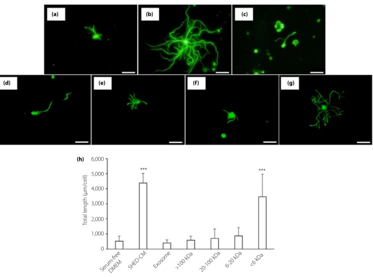
Effect of SHED-CM on neurite outgrowth of DRG neurons We initially examined the effect of SHED-CM on DPN using a primary culture of DRG neurons from normal mice. DRG neu-rons were cultured with serum-free DMEM or SHED-CM for
48 h. The neurite outgrowth in DRG neurons cultured with SHED-CM (Figure 1b) were significantly longer than that with
DMEM (Figure 1a; SHED-CM, 4380.3– 642.5 lm/neuron;
DMEM, 523.4– 337.1 lm/neuron, P < 0.001).
Effects of soluble factors in SHED-CM on neurite outgrowth of DRG neurons and on the cell viabilities of HUVECs
To evaluate the factors in SHED-CM that have a beneficial effect on neurons, we extracted exosome from SHED-CM and separated SHED-CM into four fractions (<6 kDa, 6–20 kDa, 20–100 kDa and >100 kDa). Incubation with exosome did not
improve neurite outgrowth of DRG neurons
(401.7– 198.7 lm/neuron; Figure 1c), indicating that exosomes purified from SHED-CM might be not be enough to contribute to the neurite outgrowth of DRG neurons. Among four frac-tions of SHED-CM, only the <6 kDa fraction of SHED-CM promoted the neurite outgrowth of DRG neurons (Figure 1g), and the other three fractions did not show any effect (Fig-ure 1d–f; >100 kDa; 590.4 – 271.0 lm/neuron, 20–100 kDa; 704.7– 619.8 lm/neuron, 6–20 kDa; 873.7 – 553.4 lm/neu-ron,<6 kDa; 3467.0 – 1504.1 lm/neuron, respectively).
We evaluated the concentrations of well-known neurotrophic or angiogenic factors in SHED-CM. Whole SHED-CM con-tained nerve growth factor, brain-derived neurotrophic factor,
FGF2 and VEGF at concentrations of 9.1– 0.3 pg/mL,
69.4– 2.4 pg/mL, 7.7 – 0.8 pg/mL and 1455.9 – 21.2 pg/mL, respectively. The concentrations of neurotrophin-3 and neu-rotrophin 4/5 were too small to detect. As expected, none of these factors, of which the defined molecular weight was >6 kDa, were detected in the fraction of <6 kDa (Figure S1a– d). These results suggested that unidentified soluble factors of <6 kDa in SHED-CM might have the desired effect on neurite outgrowth.
In MTT assay, SHED-CM significantly increased the cell via-bility of HUVECs compared with DMEM. Furthermore, SHED-CM was more effective compared with DMEM with 0.5 nmol/L (19,100 pg/mL) VEGF (Figure S1e). The concentra-tion of VEGF in SHED-CM was approximately 1,400 pg/mL, which was very low compared with VEGF added to DMEM. These results suggested that not only VEGF, but also other angiogenic factors in SHED-CM might have improved the via-bility of HUVECs.
Next, the effect of SHED-CM on DPN was investigated in a diabetic animal model. Whole SHED-CM was administered into the mice, because it contained both the neurotrophic and angiogenic factors in a fraction of >6 kDa and the unidentified factors promoting the neurite outgrowth of DRG in the fraction of<6 kDa, as described above.
Bodyweight and blood glucose concentrations
Diabetic mice showed severe hyperglycemia and significant reductions in bodyweight compared with non-diabetic mice, and SHED-CM administration did not affect plasma glucose levels or bodyweight in non-diabetic or diabetic mice (Table 1).
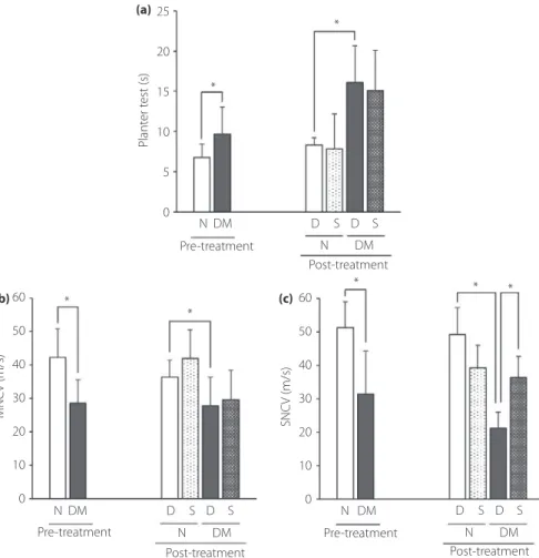
Thermal sensitivities
To evaluate the nociceptive sensitivities, we examined the thermal plantar test. The thermal nociceptive sensitivities became worse in diabetic mice, and SHED-CM did not prevent the
deterioration (Figure 2a). We also used the Von Frey test (data not shown) for evaluating mechanical allodynia. The results tended to become worse in diabetic mice, but did not signifi-cantly change by treatment, the same as the thermal plantar test. 6,000 (a) (d) (h) (e) (f) (g) (b) (c) *** *** 5,000 4,000 3,000 2,000
Total length (µm/cell)
1,000 0 Serum-fr ee DMEM SHED -CM
Exosome >100 kDa 20-100 kDa 6-20 kDa <6 kDa
Figure 1 | Neurite outgrowth of dorsal root ganglion (DRG) neurons. DRG neurons were cultured with (a) Dulbecco's modified Eagle's medium (DMEM), (b) conditioned medium of stem cells from human exfoliated deciduous teeth (SHED-CM), (c) exosome, and SHED-CM of (d)>100 kDa, (e) 20–100 kDa, (f) 6–20 kDa or (g) <6 kDa. Scale bar, 100 μm. (h) Quantitative analyses for neurite outgrowth of DRG. Results are the
means– standard deviation. ***P < 0.001.
Table 1 | Bodyweight and blood glucose in non-diabetic and diabetic mice after treatment
Non-diabetic mice Diabetic mice
DMEM SHED DMEM SHED
n 7 6 10 8
Casual blood glucose (mg/dL) 161– 11 168– 16 615– 276* 607– 289* Bodyweight (g) 27.9– 0.4 27.0– 2.8 19.4– 4.0* 22.7– 3.2* Data are means– standard deviation. *P < 0.05 versus non-diabetic mice. DMEM, treatment with Dulbecco’s modified Eagle’s medium; SHED, treat-ment with conditioned medium of stem cells from human exfoliated deciduous teeth.
Nerve conduction velocities
MNCVs and SNCVs in diabetic mice were significantly delayed compared with those in non-diabetic mice. Administration of SHED-CM significantly prevented the decrease in SNCVs
com-pared with that of DMEM (DMEM, 21.4– 4.7 m/s;
SHED-CM, 36.3– 6.4 m/s; P < 0.05.). Meanwhile, SHED-CM showed no effect on the deterioration of MNCVs (Figure 2b,c). Morphometry of sural nerves
There were no significant differences in the parameters of mor-phometric analyses of myelinatedfibers in sural nerves between non-diabetic and diabetic mice. Treatment with SHED-CM had no effect on the morphometric parameters (Table 2).
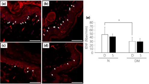
Intraepidermal nervefiber densities
Intraepidermal nerve fiber densities were significantly reduced in diabetic mice, and there was no significant difference between treatment with DMEM and SHED-CM. (Figure 3a–d).
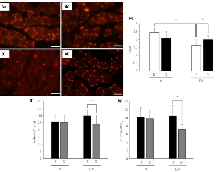
Capillary number-to-musclefiber ratios, and blood flow in hind paw and sciatic nerve
The capillary number-to-muscle fiber ratios in diabetic mice with treatment of DMEM (1.62– 0.36; Figure 4c) were signifi-cantly decreased compared with those in non-diabetic mice (2.44– 1.62; Figure 4a). The treatment of SHED-CM increased the ratios in diabetic mice (1.99– 0.24, P < 0.05 vs diabetic mice with treatment of DMEM; Figure 4d). The hind paw and sciatic nerve blood flow on the side of treatment with SHED-CM increased compared with the opposite side in diabetic mice (Figure 4f,g).
DISCUSSION
In the present study, we showed that SHED-CM promoted neurite outgrowth of cultured DRG neurons. Intramuscular administration of SHED-CM alleviated SNCVs in diabetic mice. Instead of no pathohistological findings in the peripheral nerves, capillary numbers in lower limbs were increased in the
25 20 15 10 5 0 60 50 40 30 20 10 0 60 50 40 30 20 10 0 NDM D S D S N DM Pre-treatment Plant er t est (s) MNC V (m/s) SNC V (m/s) Post-treatment NDM D S D S N DM Pre-treatment Post-treatment N DM D S D S N DM Pre-treatment Post-treatment * * * * * * * (a) (b) (c)
Figure 2 | The results of (a) thermal plantar test, (b) motor nerve conduction velocities (MNCV) and (c) sensory nerve conduction velocities (SNCV) were measured before and after the treatment. Results are the mean– standard deviation. *P < 0.05. D, treatment with Dulbecco's modified Eagle's medium; DM, diabetic mice; N, non-diabetic mice; Post-treatment: 16 weeks after streptozotocin injection; Pre-treatment, after 12 weeks after streptozotocin injection; S, treatment with conditioned medium of stem cells from human exfoliated deciduous teeth.
SHED-CM-injected mice. In addition, SHED-CM increased the viability of HUVECs and blood flow in hind paw and sciatic nerve compared with the opposite side in diabetic mice, indi-cating that the increase in capillary numbers by SHED-CM might be associated with an improvement of the sensory nerve function through increasing the nerve bloodflow. Furthermore, the present study could be reliable and valuable in terms of clinical use for patients with DPN, because the CM was col-lected from human stem cells.
Soluble factors from SHED-CM contributed to the accelera-tion of neurite outgrowth in the culture system of DRG neu-rons from normal mice. It has been reported that a variety of cytokines contained in SHED-CM have a beneficial effect on various diseases26–28. We previously reported that a fraction consisting of molecules of>100 kDa in SHED-CM had protec-tive effects on pancreatic b-cells21, suggesting that SHED-CM might be able to show multiple potent actions for diabetes mel-litus. In the present study, the elongation effect of SHED-CM on the neurite outgrowth was preserved only by the <6 kDa fraction among four fractions with low- to high-molecular weight ranges. It was unlikely that several well-established neu-rotrophic factors, such as FGF2, nerve growth factor and NT3, were active molecules for the neurite outgrowth, because these factors were not contained in the small molecule fraction. In addition, the concentrations of such neurotrophic factors in the fractions of >6 kDa were the levels of pg/mL, meanwhile the previously reported effective doses of the factors for neurite out-growth were the levels of ng/mL29–31. Furthermore, it is inter-esting that <6 kDa fraction also increased the cell viability of HUVECs in the present study (Figure S1e). It is still completely unclear what the candidate factor of <6 kDa is in SHED-CM; therefore, proteomic analysis should be carried out to identify the unknown neuritogenic and angiogenic factors in SHED-CM in the future. Supplementation of purified exosomes did not promote neurite outgrowth, providing no additional signi fi-cance to determine the unidentified neuroregenerative mole-cules. Therefore, our data might predict the existence of undetermined small molecules other than well-known neu-rotrophic factors in SHED-CM for the neurite outgrowth, and further studies to identify the molecules would be required.
We utilized three histopathological approaches to clarify the neuroprotective effects of SHED-CM in DPN. First, the myeli-nated fiber morphometry of sural nerves, which evaluated pathological changes in large- and medium-diameter fibers, showed no significant differences even in the comparison between diabetic and non-diabetic mice. Second, IENFDs that were verified as a standard examination to evaluate small fiber neuropathy in DPN confirmed significant decreases in small fibers in the epidermis of diabetic mice. These findings showed that the duration of diabetes in the current study might be insufficient to induce morphological change in large- and med-ium-diameter fibers, but enough in small-diameter fibers. No alteration of thermal and tactile sensitivities in diabetic mice might associate with the result that IENFDs were not improved
Table 2 | M orphometric d ata o f m yelinated fibers in sural n erves Fas cicle area (l m 2 ) Occupancy rate (%) Fib er area (l m 2 ) Axon area (l m 2 ) Myel in ar ea (l m 2 ) Fiber d iam eter (l m) Axon diam et er (l m) Mye lin thi ck n es s (l m) Axonal circularity Gr at io Non D M-DMEM 21,111.18 – 36 75 .85 50 .44 – 2. 59 24 .9 5 – 1.77 10 .04 – 0.9 3 15.5 0 – 1.3 8 6. 91 – 0.2 8 4.7 2 – 0. 24 1. 09 – 0. 07 0.65 – 0.0 2 0.67 – 0.3 9 Non D M-SHE D 19,589.30 – 17 34 .57 55 .69 – 1. 74 22 .6 2 – 2.65 8.25 – 0.8 9 13.9 5 – 1.8 1 6. 24 – 0.3 7 4.1 9 – 0. 25 1. 02 – 0. 09 0.62 – 0.0 2 0.68 – 0.4 5 DM -DM EM 22 ,74 8. 44 – 18 56 .93 53 .80 – 2. 82 23 .1 8 – 1.17 8.54 – 0.7 1 14.1 3 – 0.6 7 6. 68 – 0.1 5 4.6 4 – 0. 13 1. 02 – 0. 03 0.58 – 0.0 2 0.70 – 0.2 2 D M -SHED 20,8 63.1 1 – 26 93 .40 55 .79 – 2. 95 20 .4 2 – 1.98 7.54 – 0.9 7 12.2 8 – 0.9 9 6. 30 – 0.2 4 4.2 6 – 0. 23 1. 02 – 0. 04 0.61 – 0.0 2 0.68 – 0.3 5 Da ta are the me an – st an da rd de via tio n. DM, d ia be tic m ice; DMEM, treatmen t with Dulbecco ’s m odifie d Eagl e’ s m edium; non DM, n o n -diabet ic m ice ; SHED, treatment with conditioned medium of stem cells from h uman exfol iat ed d ecid u ous te eth .
by SHED-CM. DPN at 12 weeks after STZ administration pro-gressed to an advanced state of hypoalgesia in the present study, and it would be more effective to start the treatment at an earlier point after STZ administration. Third, the capillary-to-musclefiber ratio of soleus muscles was assessed to evaluate the involvement of microcirculation. The series of SHED-CM injections increased the ratio in diabetic mice, and increased the sciatic nerve and hind paw blood flow on the treatment side compared with the opposite side in the very same diabetic mice, suggesting that SHED-CM might ameliorate the microcir-culation of lower limbs, including peripheral nerves, resulting in prevention of further deterioration of sensory function.
SHED secreted several angiogenic factors, including VEGF and FGF2, which play a significant role in promoting angiogen-esis in tissue regeneration32–34. The concentration of VEGF in SHED-CM was relatively higher than those of neurotrophic fac-tors. Therefore, such angiogenic factors in SHED-CM might be associated with the increase in capillary number-to-musclefiber ratios. The concentration of VEGF in SHED-CM was approxi-mately 1,400 pg/mL. In the present study, SHED-CM were injected with 100lL into diabetic mice twice a week over a period of 4 weeks and the total injected amount of VEGF was approximately 1.2 ng, but a much higher amount was adminis-trated for angiogenesis in in vivo studies of previous reports35,36. In addition, SHED-CM improved the cell viability of HUVECs more than the supplementation of 0.5 nmol/L (19.1 ng/mL) VEGF alone. Therefore, not only VEGF, but also various angiogenic factors in SHED-CM might be associated with the increase in capillary number-to-muscle fiber ratios. Meanwhile, it was reported that VEGF induced cancer pain through activation of VEGF receptor expressed in sensory
neurons37. Thus, we should consider the possibility that VEGF might exacerbate diabetic neuropathic pain in future clinical application of SHED-CM in DPN.
Previous studies reported that both MNCVs and SNCVs were improved by transplantation of stem cells23,38,39, but the present study showed that the deterioration of SNCVs, but not MNCVs, was significantly prevented by SHED-CM treatment. MNCVs were not further deteriorated during the period of treatment in both DMEM- and SHED-CM-treated mice in the present study. A decline of MNCVs usually appears subsequently to that of SNCVs in DPN, indicating that deterioration of MNCVs seemed to require longer periods compared with SNCVs due to the dif-ference in nerve fiber size between motor and sensory nerves, and the duration of treatment might be inadequate to evaluate the preventive effect of SHED-CM on MNCVs. Furthermore, in the previous study, transplanted stem cells were from the same species as the recipient animals, whereas in the present study, CM from human stem cells was administered to mice. The dif-ferent animal species of the stem cells for CM and the recipient might be associated with the reduced effects of condition med-ium on improvement of DPN in the present study compared with the previous study. Furthermore, transplanted stem cells were identified at the transplantation site and secreted trophic factors 4 weeks after transplantation9, indicating that the trans-planted stem cells might have more sustained and pronounced effects on DPN compared with the intramuscular administration of CM. Therefore, it might be important to consider the method of administration of SHED-CM to effectively induce neuroregen-eration in DPN. In rat sciatic nerve transection model, silicon conduits containing SHED-CM enhanced sciatic nerve reinner-vation and regeneration20. The continuous intracerebral
80 (a) (b) (c) (d) (e) 70 60 50 40 30 20 10 0 S N * IENF (fiber/mm) DM D D S
Figure 3 | Intraepidermal nerve fiber densities (IENFDs). Non-diabetic mice treated with (a) Dulbecco's modified Eagle's medium and (b) conditioned medium of stem cells from human exfoliated deciduous teeth, and diabetic mice treated with (c) Dulbecco's modified Eagle's medium and (d) conditioned medium of stem cells from human exfoliated deciduous teeth. Scale bar, 50μm. White arrows represent
intraepidermal nerve fibers. (e) Quantitative analyses for IENFDs. Results are means– SD. *P < 0.05. D, treatment with Dulbecco's modified Eagle's medium; DM, diabetic mice; N, non-diabetic mice; S, treatment with conditioned medium of stem cells from human exfoliated deciduous teeth.
administration of SHED-CM significantly improved the neuro-logical outcome in the mice with hypoxic-ischemic brain40, and the continuous intrathecal administration into injured rat spinal cords caused remarkable functional recovery5, implying that the effects of SHED-CM on DPN could be more pronouncedly derived by changing the manner of administration. In addition, a standardized production might be crucial for appropriate uti-lization of CM, because the concentration of VEGF in the pre-sent study was different from that in the previous study20, and was changed according to the passage number (data not shown). More suitable methods for preparation and administration of SHED-CM need to be further elucidated to achieve more remarkable improvement of DPN.
SHED-CM promoted neurite outgrowths of DRG neurons from normal mice, increased capillaries and prevented the deterio-ration of SNCVs, suggesting that SHED-CM might have potential as a new therapeutic strategy for DPN. The use of CM could resolve several problems of cell transplantation: limited survival of transplanted cells, risks of tumor formation or rejection responses. Furthermore, CM is a promising prospect for production as phar-maceuticals for regenerative therapy, because CM could be manu-factured, freeze-dried, packaged and transported more easily. Future studies should be carried out to identify the undetermined small molecules that have favorable effects on nerve regeneration, and to investigate the mechanism of effective actions by SHED-CM for clinical applications in the treatment of DPN.
3 (a) (b) (c) (d) (e) (f) (g) 2.5 2 1.5 1 0.5 0 40 35 30 25 20 15 10 5 0 S O S O O 14 * 12 10 8 6 4 2 0 * N DM S O S N DM (ml/min/100 g) (ml/min/100 g) D S D S N DM * * CNMFR
Figure 4 | Capillary number-to-muscle fiber ratio (CNMFR) and blood flow in hind paw and sciatic nerve. CNMFR of non-diabetic mice treated with (a) Dulbecco's modified Eagle's medium and (b) conditioned medium of stem cells from human exfoliated deciduous teeth, and diabetic mice treated with (c) Dulbecco's modified Eagle's medium and (d) conditioned medium of stem cells from human exfoliated deciduous teeth were measured after the treatment. Scale bar, 50μm. Quantitative analyses for CNMFR (e). Blood flow of (f) hind paw and (g) sciatic nerve were also measured after the treatment. Results are the mean– standard deviation. *P < 0.05. D, treatment with Dulbecco's modified Eagle's medium; DM, diabetic mice; N, non-diabetic mice; O, opposite side of treatment with conditioned medium of stem cells from human exfoliated deciduous teeth; S, treatment with conditioned medium of stem cells from human exfoliated deciduous teeth.
ACKNOWLEDGMENT
This study was supported by a Grant-in-Aid for Scientific Research (17K09851, 16K09742, 15H06720) from the Japanese Ministry of Education, Culture, Sports, Science and Technol-ogy; Grants for Young Researchers from the Japan Associa-tion for Diabetes EducaAssocia-tion and Care; and Grants for Medical Staff from the Japan Association for Diabetes Educa-tion and Care, Manpei Suzuki Diabetes FoundaEduca-tion, Japan Diabetes Foundation and Aichi Medical University Aikeikai. The funders had no role in the study design, data collection and analysis, decision to publish or preparation of the manu-script. The authors thank Norio Ozeki and Rieko Inoue for technical assistance.
DISCLOSURE
The authors declare no conflict of interest.
REFERENCES
1. Boulton AJ, Vinik AI, Arezzo JC, et al. Diabetic neuropathies: a statement by the American Diabetes Association. Diabetes Care 2005; 28: 956–962.
2. Johnson PC, Doll SC, Cromey DW. Pathogenesis of diabetic neuropathy. Ann Neurol 1986; 19: 450–457.
3. Sima AA, Bouchier M, Christensen H. Axonal atrophy in sensory nerves of the diabetic BB-Wistar rat: a possible early correlate of human diabetic neuropathy. Ann Neurol 1983; 13: 264–272.
4. Tesfaye S. Recent advances in the management of diabetic distal symmetrical polyneuropathy. J Diabetes Investig 2011; 2: 33–42.
5. Sakai K, Yamamoto A, Matsubara K, et al. Human dental pulp-derived stem cells promote locomotor recovery after complete transection of the rat spinal cord by multiple neuro-regenerative mechanisms. J Clin Invest 2012; 122: 80– 90.
6. Ruan GP, Han YB, Wang TH, et al. Comparative study among three different methods of bone marrow mesenchymal stem cell transplantation following cerebral infarction in rats. Neurol Res 2013; 35: 212–220.
7. Kolcun JPG, Chang HK, Wang MY. Stem cell transplantation helps alleviate spinal cord injury sequelae in mice.
Neurosurgery 2017; 80: N12–n14.
8. Naruse K, Hamada Y, Nakashima E, et al. Therapeutic neovascularization using cord blood-derived endothelial progenitor cells for diabetic neuropathy. Diabetes 2005; 54: 1823–1828.
9. Shibata T, Naruse K, Kamiya H, et al. Transplantation of bone marrow-derived mesenchymal stem cells improves diabetic polyneuropathy in rats. Diabetes 2008; 57: 3099–3107. 10. Himeno T, Kamiya H, Naruse K, et al. Mesenchymal stem
cell-like cells derived from mouse induced pluripotent stem cells ameliorate diabetic polyneuropathy in mice. Biomed Res Int 2013; 2013: 259187.
11. Hata M, Omi M, Kobayashi Y, et al. Transplantation of cultured dental pulp stem cells into the skeletal muscles ameliorated diabetic polyneuropathy: therapeutic plausibility of freshly isolated and cryopreserved dental pulp stem cells. Stem Cell Res Ther 2015; 6: 162.
12. Zhao L, Jiang X, Shi J, et al. Exosomes derived from bone marrow mesenchymal stem cells overexpressing microRNA-25 protect spinal cords against transient ischemia. J Thorac Cardiovasc Surg 2019; 157: 508–517.
13. Yu B, Zhang X, Li X. Exosomes derived from mesenchymal stem cells. Int J Mol Sci 2014; 15: 4142–4157.
14. Qing L, Chen H, Tang J, et al. Exosomes and their MicroRNA Cargo: new players in peripheral nerve
regeneration. Neurorehabil Neural Repair 2018; 32: 765–776. 15. Cheng C, Kobayashi M, Martinez JA, et al. Evidence for
epigenetic regulation of gene expression and function in chronic experimental diabetic neuropathy. J Neuropathol Exp Neurol 2015; 74: 804–817.
16. Pawitan JA. Prospect of stem cell conditioned medium in regenerative medicine. Biomed Res Int 2014; 2014: 965849. 17. Mizukami H, Yagihashi S. Is stem cell transplantation ready
for prime time in diabetic polyneuropathy? Curr Diab Rep. 2016; 16: 86.
18. Miura M, Gronthos S, Zhao M, et al. SHED: stem cells from human exfoliated deciduous teeth. Proc Natl Acad Sci U S A. 2003; 100: 5807–5812.
19. Huang GT, Gronthos S, Shi S. Mesenchymal stem cells derived from dental tissues vs. those from other sources: their biology and role in regenerative medicine. J Dent Res 2009; 88: 792–806.
20. Sugimura-Wakayama Y, Katagiri W, Osugi M, et al. Peripheral nerve regeneration by secretomes of stem cells from human exfoliated deciduous teeth. Stem Cells Dev 2015; 24: 2687–2699.
21. Izumoto-Akita T, Tsunekawa S, Yamamoto A, et al. Secreted factors from dental pulp stem cells improve glucose intolerance in streptozotocin-induced diabetic mice by increasing pancreatic beta-cell function. BMJ Open Diabetes Res Care 2015; 3: e000128.
22. Tosaki T, Kamiya H, Yasuda Y, et al. Reduced NGF secretion by Schwann cells under the high glucose condition decreases neurite outgrowth of DRG neurons. Exp Neurol 2008; 213: 381–387.
23. Naruse K, Sato J, Funakubo M, et al. Transplantation of bone marrow-derived mononuclear cells improves mechanical hyperalgesia, cold allodynia and nerve function in diabetic neuropathy. PLoS ONE 2011; 6: e27458.
24. Makino E, Nakamura N, Miyabe M, et al. Conditioned media from dental pulp stem cells improved diabetic
polyneuropathy through anti-inflammatory, neuroprotective and angiogenic actions: cell-free regenerative medicine for diabetic polyneuropathy. J Diabetes Investig 2019; 10: 1199– 1208.
25. Himeno T, Kamiya H, Naruse K, et al. Beneficial effects of exendin-4 on experimental polyneuropathy in diabetic mice. Diabetes 2011; 60: 2397–2406.
26. Yamaguchi S, Shibata R, Yamamoto N, et al. Dental pulp-derived stem cell conditioned medium reduces cardiac injury following ischemia-reperfusion. Sci Rep 2015; 5: 16295. 27. Mita T, Furukawa-Hibi Y, Takeuchi H, et al. Conditioned
medium from the stem cells of human dental pulp improves cognitive function in a mouse model of Alzheimer's disease. Behav Brain Res 2015; 293: 189–197. 28. Matsushita Y, Ishigami M, Matsubara K, et al. Multifaceted
therapeutic benefits of factors derived from stem cells from human exfoliated deciduous teeth for acute liver failure in rats. J Tissue Eng Regen Med 2017; 11: 1888–1896.
29. Sierra-Fonseca JA, Najera O, Martinez-Jurado J, et al. Nerve growth factor induces neurite outgrowth of PC12 cells by promoting Gbetagamma-microtubule interaction. BMC Neurosci 2014; 15: 132.
30. Kim MJ, Cotman SL, Halfter W, et al. The heparan sulfate proteoglycan agrin modulates neurite outgrowth mediated by FGF-2. J Neurobiol 2003; 55: 261–277.
31. Colombo F, Racchetti G, Meldolesi J. Neurite outgrowth induced by NGF or L1CAM via activation of the TrkA receptor is sustained also by the exocytosis of
enlargeosomes. Proc Natl Acad Sci U S A 2014; 111: 16943– 16948.
32. Ferrara N, Davis-Smyth T. The Biology of Vascular Endothelial Growth Factor. Endocr Rev 1997; 18: 4–25.
33. Sadat S, Gehmert S, Song YH, et al. The cardioprotective effect of mesenchymal stem cells is mediated by IGF-I and VEGF. Biochem Biophys Res Commun 2007; 363: 674–679. 34. Jeong JO, Kim MO, Kim H, et al. Dual angiogenic and
neurotrophic effects of bone marrow-derived endothelial progenitor cells on diabetic neuropathy. Circulation 2009; 119: 699–708.
35. Adini A, Adini I, Chi ZL, et al. A novel strategy to enhance angiogenesis in vivo using the small VEGF-binding peptide PR1P. Angiogenesis 2017; 20: 399–408.
36. Xin X, Yang S, Ingle G, et al. Hepatocyte growth factor enhances vascular endothelial growth factor-induced angiogenesis in vitro and in vivo. Am J Pathol 2001; 158: 1111–1120.
37. Selvaraj D, Gangadharan V, Michalski CW, et al. A Functional Role for VEGFR1 Expressed in Peripheral Sensory Neurons in Cancer Pain. Cancer Cell 2015; 27: 780–796.
38. Himeno T, Kamiya H, Naruse K, et al. Angioblast derived from ES cells construct blood vessels and ameliorate diabetic polyneuropathy in mice. J Diabetes Res 2015; 2015: 257230.
39. Omi M, Hata M, Nakamura N, et al. Transplantation of dental pulp stem cells improves long-term diabetic polyneuropathy together with improvement of nerve morphometrical evaluation. Stem Cell Res Ther 2017; 8: 279. 40. Yamagata M, Yamamoto A, Kako E, et al. Human dental
pulp-derived stem cells protect against hypoxic-ischemic brain injury in neonatal mice. Stroke 2013; 44: 551–554.
SUPPORTING INFORMATION
Additional supporting information may be found online in the Supporting Information section at the end of the article.
Figure S1 The concentrations of growth factors, (a) nerve growth factor (NGF), (b) brain-derived neurotrophic factor (BDNF), (c) fibroblast growth factor (FGF2) and (d) vascular endothelial growth factor (VEGF), in conditioned medium of stem cells from human exfoliated deciduous teeth (SHED-CM),>6 kDa and <6 kDa of SHED-CM. (e) The cell viability of human umbilical vein endothelial cells (HUVECS).