INTRODUCTION
Transplanted flaps are sometimes subject to con-gestion, especially under conditions such as diabe-tes, arteriosclerosis, radically dissected or irradiated recipient sites and/or insufficient postoperative rest. If flap congestion remains despite surgical reanas-tomosis or unkinking of pedicle vessels, application
of medicinal leeches is indicated ; such therapy has proven useful for management of compromised flaps.
Although there are many reports of leech therapy in cases of flap reconstruction and microsurgical re-plantation (1-4), determining the number of leeches needed and the duration of leech therapy has not been described. Surgeons have relied upon their own experience in making such decisions. The aim of this study was to quantify hemodynamic changes in a congested animal skin flap model during and after leech therapy, to determine the effects of treat-ment according to the number of leeches applied, and to find a good method for determining the
ORIGINAL
Quantitative analysis of hemodynamics of congested
island flaps under leech therapy
Keisuke Kashiwagi, Ichiro Hashimoto, Yoshiro Abe, Kunio Kotsu,
Masahiro Yamano, and Hideki Nakanishi
Department of Plastic and Reconstructive Surgery, the University of Tokushima Graduate School, Tokushima, Japan
Abstract : Background A congested flap is a good indication for leech therapy. However, determining the appropriate number of leeches as well as the appropriate application time in clinical cases is difficult. We analyzed hemodynamics in rabbit island flaps under leech therapy to find a suitable clinical procedure for determining the appropriate number of leeches to be used and the duration of therapy. Methods Island flaps were raised in 35 rabbit ears, and congestion was induced by venous clamping. Treatment involved use of 1 or 3 leeches and was begun 60 minutes after venous occlusion. Flaps were examined for area of surviving tissue, alterations in transcutaneous oxygen and carbon dioxide tension (TcPO2and TcPCO2), and flow volume. Arteriole and venule diameters and flow velocities were examined microscopically. Results Flap survival area in the 3-leech therapy group was significantly larger than the 1-leech therapy group and the control group. With 3-leech therapy, TcPCO2decreased significantly, as did arteriole and venule diameters. After clamp release, TcPCO2and venule diameter continued to decrease in this group, and flow velocity increased significantly. Conclusions Leech therapy may salvage compromised flaps by replacing congested blood with new arterial blood and thus maintaining flap vi-ability. TcPO2and TcPCO2monitoring may help evaluate the therapeutic effect and de-termine the appropriate number of leeches to apply and the duration of therapy. J. Med. Invest. 60 : 213-220, August, 2013
Keywords :leech therapy, congestion, island flap, microcirculation, TcPO2and TcPCO2monitoring
Received for publication February 18, 2013 ; accepted March 12, 2013.
The first two authors should be considered co - first authors. Address correspondence and reprint requests to Ichiro Hashimoto, M.D., Ph.D., Department of Plastic and Recon-structive Surgery, the University of Tokushima Graduate School, Kuramoto cho, Tokushima 770 8503, Japan, and Fax : +81 88 -633 - 7297.
appropriate number of leeches to be used in apy as well as the appropriate duration of such ther-apy. To assess hemodynamics of congested flaps, we measured transcutaneous oxygen/carbon diox-ide tension (TcPO2/TcPCO2), performed laser Dop-pler flowmetry and measured arteriole and venule di-ameters and flow velocities in a rabbit ear chamber model.
MATERIALS AND METHODS
All experimental procedures were approved by the institutional animal care and use committee of the University of Tokushima and were carried out in accordance with committee guidelines.
Congested flap model and leech treatment
Thirty-five Japanese white rabbits (Japan SLC, Inc., Shizuoka, Japan), weighing between 3.0 and 3.5 kg, were used in the study. Animals were main-tained under standard housing conditions and al-lowed water and standard dry rabbit feed ad libitum. Experiments, described below, were performed in animals sedated with pentobarbital administered in-travenously at 30 mg/kg. In each animal, a total-ear pedicled island flap was created by cutting off skin and cartilage completely at the base of the ear, pre-serving only the posterior auricular artery and comi-tant vein (Fig. 1, a). Bleeding at the flap edge was stopped with a bipolar coagulator so as not to affect hemodynamics after congestion. The divided carti-lage was then sutured into its original position (Fig. 1, b) for performance of the experiments. After com-pletion of all experiments, the divided skin was su-tured back into position.
The comitant vein was occluded with a vascu-lar clamp at 0.59 N closing force (TKM-1, Bear Medic, Tokyo, Japan). Sixty minutes after venous
occlusion, medicinal leeches were applied to the ears of animals in the treatment groups described below. The leeches had been purchased from Bio-pharm, Tokyo, Japan, and were maintained dor-mant in a dark cooler (3!!), according to the sup-plier’s instructions. Just before use, the leeches were aroused from dormancy by being moved to a tray kept at room temperature. Leeches that ap-peared weak or small were culled from the original population so that only those weighing 0.29-0.43 g (mean 0.36 g) were applied. Leech feeding on the skin flaps was continued until the leech detached itself from the site (Fig. 2). Passive bleeding at the wound site was allowed to continue ; the wound was covered with gauze soaked in heparinized saline (50 U/mL). The passive bleeding continued at all wound sites throughout the observation period. The clamp was removed 150 minutes after its applica-tion, and venous blood flow resumed. Patency of the vein after removal of the clamp was confirmed mi-croscopically. Measurements were taken for a total of 210 minutes (until 60 minutes after clamp re-moval).
Fig. 1 Rabbit ear flap model. a The island flap is connected only by the posterior auricular artery and vein. b The flap edge is sutured back into its original position. Flap congestion is achieved by occlusion of the comitant vein with a vascular clamp.
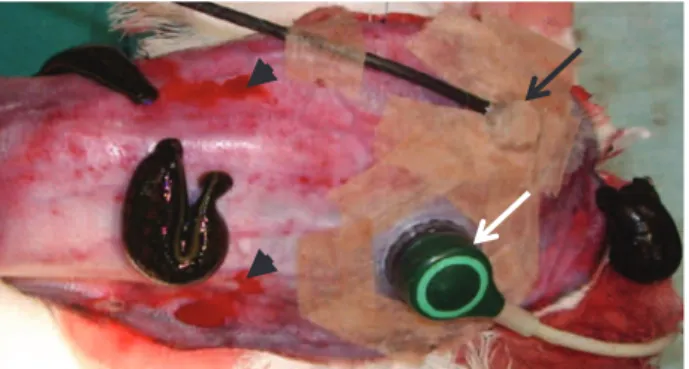
Fig. 2 Leech application and measurement of TcPO2/TcPCO2
and laser Doppler flowmetry. Three leeches are situated on the congested flap. Two sites of passive bleeding from the leech bites are observed (arrowheads). Two probes are attached to the ear-lobe flap (white and black arrows).
Experiment 1
Twenty-one rabbits were divided into 3 groups : a treatment group in which 3 leeches were applied simultaneously to each flap (Group 1-A, n=7), a treatment group in which 1 leech was applied to each flap (Group 1-B, n=7), and a control group in which no leech was applied (Group 1-C, n=7).
TcPO2, TcPCO2and flow volume were measured simultaneously with a TCM3 monitoring system (Radiometer Medical ApS, Copenhagen, Denmark) and an ALF21D laser Doppler flowmeter (Advance, Tokyo, Japan), respectively. Seven days after venous occlusion, flap survival was assessed by comparing the area of surviving flap tissue against the original area of the flap, which had been traced on a trans-parent plastic sheet. Flap survival was calculated as a percentage of the original area.
Experiment 2
Fourteen rabbits were divided into 2 groups : a treatment group in which 3 leeches were applied simultaneously to each flap (Group 2-A, n=7) and a control group in which no leech was applied (Group 2-C, n=7).
A rabbit ear chamber, i.e., a transparent window for microscopic observation of the peripheral mi-crovasculature, was implanted in the distal portion of the rabbit ear. Implantation was performed as pre-viously described (5). A tissue regeneration space (50μm in thickness) was formed in the central hole of the chamber. Five to 7 weeks after implantation, the peripheral microvasculature was examined by intravital microscopy. Arterioles (20-30μm in di-ameter) and venules (45-55μm in diameter) were selected under our previously described intravital microscope-monitor system (5). Diameter and flow velocity of the selected vessels were recorded with a high-speed camera (FASTCAM 1024PCI, Photron, Tokyo, Japan) mounted on the intravital micro-scope, and changes over time were calculated with exclusive camera control software (PFV ver. 2.4.5.1, Photron).
Statistical analysis
Values are expressed as mean!SE. Treatment ef-fects were compared between the 3 Experiment 1 groups by means of the Kruskal-Wallis test and be-tween the 2 Experiment 2 groups by means of the Mann-Whitney U test. All analyses were performed with StatView 5.0 (SAS, Cary, NC, U.S.A.). Signifi-cance was accepted at p!0.05.
RESULTS
Experiment 1 Flap survival
The average size of the original flap area was 85.1!3.0 cm2, 88.6!3.0 cm2and 87.2!1.5 cm2in Groups 1-A (3-leech therapy group), 1-B (1-leech therapy group) and 1-C (control group), respec-tively, with no significant difference between groups (p=0.79).
Flap survival area was 68.6!11.5% (0-95%), 4.3! 4.0% (0-30%) and 5.7!3.3% (0-25%) in Groups 1-A, 1-B and 1-C, respectively, with that in Group 1-A being significant (p!0.01). Total flap necrosis (0% survival) occurred in 14.3% (1/7), 85.7% (6/7) and 57.1% (4/7) of flaps and flap necrosis of more than 90% occurred in 14.3% (1/7), 85.7% (6/7), and 85.7% (6/7) of flaps in Groups 1-A, 1-B and 1-C, respec-tively.
TcPO2and TcPCO2
TcPO2 values decreased immediately with ve-nous occlusion in all 3 experimental groups (Fig. 3). TcPO2increased significantly in Group 1-A starting 30 minutes after leech application until clamp re-lease. After venous clamp removal, no significant difference in TcPO2was observed in this treatment group.
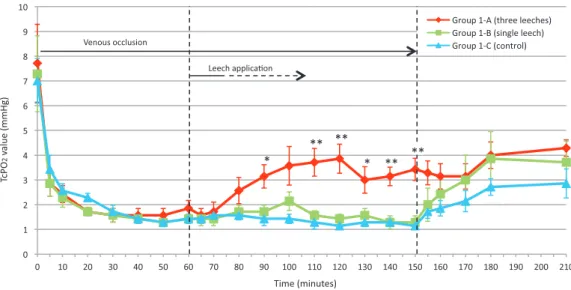
TcPCO2values increased after venous occlusion in all 3 treatment groups (Fig. 4). In Group 1-B, a decrease in TcPCO2was seen 10 minutes after leech application, but the decrease continued only 20 min-utes before leveling off. In Group 1-A, a significant decrease was seen 20 minutes after application of the 3 leeches, and the decrease continued, with some variation, to the last measurement (60 minutes after clamp release), at which point TcPCO2in this group had decreased to the pre-congestion value. TcPCO2values in Groups 1-B and 1-C decreased after release of the venous clamp, but the last-re-corded values were higher than the pre-congestion values.
Flow volume
Flow volume decreased after venous occlusion and increased after clamp removal (Fig. 5) in all 3 groups. No substantial increase in flow volume was observed after leech application during the period of congestion. Flow volume increased after removal of the clamp in Group A-1, but the increase was not significant.
10 9 8 7 6 5 4 3 2 1 0 0 10 20 30 40 50 60 70 80 90 100 110 120 130 140 150 160 170 180 190 200 210 Group 1-A (three leeches) Group 1-B (single leech) Group 1-C (control) 180 160 140 120 100 80 60 40 20 0 0 10 20 30 40 50 60 70 80 90 100 110 120 130 140 150 160 170 180 190 200 210 Group 1-A (three leeches) Group 1-B (single leech) Group 1-C (control) 8 7 6 5 4 3 2 1 0 0 10 20 30 40 50 60 70 80 90 100 110 120 130 140 150 160 170 180 190 200 210 Group 1-A (three leeches) Group 1-B (single leech) Group 1-C (control)
Fig. 5 Flow volume in each group, as monitored by laser Doppler flowmetry (Experiment 1).
Fig. 3 Graph of TcPO2values in each group from venous occlusion to the final measurement (Experiment 1). *p!0.05 ; **p!0.01.
80 70 60 50 40 30 20 10 0 0 10 20 30 40 50 60 70 80 90 100 110 120 130 140 150 160 170 180 190 200 210
Group 2-A (three leeches) Group 2-C (control) Group 2-A (three leeches) Group 2-C (control) 1200 1000 800 600 400 200 0 0 10 20 30 40 50 60 70 80 90 100 110 120 130 140 150 160 170 180 190 200 210
Group 2-A (three leeches) Group 2-C (control)
Experiment 2
Arteriole and venule diameters
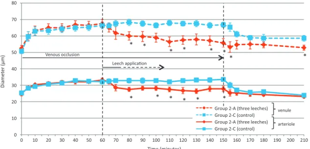
Arteriole and venule diameters increased with ve-nous congestion (Fig. 6). In the treatment group, a significant decrease in the diameters of both the arterioles and venules was seen 20 minutes after leech application until clamp release. In both groups, diameters of the arterioles and venules decreased after clamp removal. Sixty minutes after clamp re-lease, only the venules in the control group had not recovered to their original size (diameter).
Arteriolar and venular flow velocities
Flow velocities in the arterioles and venules de-creased soon after venous occlusion in both groups (Fig. 7 and 8). Flow velocities recovered gradually with treatment during venous occlusion, but the increase was not significant. Flow velocities in the treatment group increased significantly after removal of the clamp and then reached pre-congestion levels by the end of the observation period. Flow veloci-ties in the control group did not reach pre -conges-tion levels.
Fig. 7 Arteriolar flow velocity in each group (Experiment 2). *p!0.05.
600 500 400 300 200 100 0 0 10 20 30 40 50 60 70 80 90 100 110 120 130 140 150 160 170 180 190 200 210 Group 2-A (three leeches) Group 2-C (control)
DISCUSSION
An ischemic random skin flap raised on rat dor-sum (6) and an abdominal skin flap based on the inferior epigastric vessels (7-9) have been reported as flap models useful for testing leech therapy. All are sutured back to their original sites. Our study was characterized by an island ear flap model. Our results showed that survival of the congested flap depends on the number of leeches used. The con-tact area between the flap and the recipient site was very small in our study. This strict condition well dis-played the significant effect of the 3-leech therapy. Improvement in TcPO2 and TcPCO2 values and the diameters of venules and arterioles in the 3-leech treatment group continued even after the leeches detached from the flaps. The decrease in TcPCO2in the 1-leech treatment group, however, was temporary. The difference suggests that suffi-cient leech treatment can improve the microcircu-lation even during the period of passive bleeding.
TcPCO2values in the 3-leech group were main-tained at less than 90 mmHg during leech therapy, and they were restored to the pre-congestion level after removal of the venous clamp ; survival of the flaps in this group was good. However, TcPCO2 val-ues in the other 2 groups, which remained above 90 mmHg, did not return to the pre-congestion levels after removal of the venous clamp, and many flaps in these 2 groups became necrotic. In a clinical series we reported (10), congested flaps with TcPCO2 val-ues of 90 mmHg or more became necrotic, and measurement of TcPCO2was shown to be a good
monitoring system for free flap failure. In the pre-sent study, high TcPCO2values during and after leech therapy corresponded to flap necrosis.
The reported hemodynamic effect of leech ther-apy is limited to a 1.6-cm-diameter area around the bite wound of a single leech (11). That 3 leeches were required in our study to rescue the congested flaps also suggests the limitations of leech therapy. In clinical cases, raised flaps are of various areas and thicknesses ; thus, decisions must be made re-garding the number of leeches to be used and the duration of such therapy. We believe the proper number of medicinal leeches and the proper appli-cation time can be decided by monitoring TcPCO2 in addition to monitoring various factors such as flap color, blood flow by pinprick test, and capillary re-filling.
Expansion of the venules in congested tissue is widely understood because the venule is thought to be a “capacitance vessel”. We observed enlargement of the arterioles also after venous clamping. We be-lieve that venous congestion induced capillary hyper-tension, which in turn caused arterial expansion in the total venous occlusion model. Leech treatment reduced the expansion of both arterioles and ve-nules. However, enlargement of the venules, the in-dicator of flap congestion, remained after clamp re-lease in the control group. As far as we know, this is the first report of visual evaluation of changes in the microvasculature in congested flaps under leech therapy.
A significant increase in blood flow has been reported in congested pig rump flaps under leech Fig. 8 Venular flow velocity in each group (Experiment 2). *p!0.05 ; **p!0.01.
treatment (12). In our study, however, the leech treatment did not increase blood flow volume or flow velocity in the arterioles and venules ; i.e., the leech treatment did not affect blood flow in the microvas-culature during venous congestion in our island flap model. After clamp release, however, improved flow velocity and decreased venule diameter were ob-served in treated flaps but not in untreated flaps, which eventually became necrotic. The no-reflow phenomenon (13) is known as poor or no recovery of microvascular blood flow after restoration of pedi-cle perfusion from severe ischemia. In our study, total loss of the flap occurred only once in the 3-leech group (14.3%), whereas total flap necrosis oc-curred in a combined 71.4% of flaps in the other 2 groups. We surmise that normal blood flow was not restored in most flaps of the 1-leech and control groups but that the 3-leech therapy rescued com-promised flaps from severe circulatory failure.
Because leech therapy did not affect blood flow but did reduce microvessel diameter during conges-tion, we theorize that the treatment reduces blood pressure in the microvasculature, replaces con-gested blood with new arterial blood of lower PCO2 and higher PO2, and thus maintains viability of the flap. Our experimental model mimics clinical situ-ations in which complete obstruction of the venous pedicle and subsequent release of the obstruction occur. Our results suggest that when a surgeon recognizes flap failure before it becomes irrevers-ible, medicinal leeches can be “lifesavers” until surgical re-exploration of the compromised flap is performed. Medicinal leeches have been used for venous-congested free flaps, pedicled flaps, and replants in which surgical re-exploration has been ineffectual (1, 2) or in which venous drainage has been insufficient because of severe damage to the venous pedicle or an improperly designed flap (2-4). The indication for leech therapy can be expanded to immediate use for cases awaiting re-exploration.
Future study
Although hemodynamic effect of leech therapy is limited around the bite wound of a leech, relation-ship between specific placement of the leeches and effect of this therapy was not evaluated in our study. It follows, then, that necrosis was not studied in re-lation to this factor. The effect of leech therapy in inducing passive bleeding was confirmed, however, an interesting question remains to be explored.
CONCLUSIONS
We investigated the hemodynamic effects of leech therapy in congested rabbit ear island flaps. The bloodletting effects of the leeches contributed to salvaging the compromised flaps by replacing con-gested blood with new arterial blood and maintain-ing viability of the flaps. The leech treatment pre-vented the no-reflow phenomenon in the congested island flaps. Tracking TcPO2and TcPCO2may be useful in monitoring the therapeutic effect of leech application and determining the appropriate number of leeches to be applied as well as the appropriate duration of the leech therapy.
DISCLOSURES
Drs. Kashiwagi, Hashimoto, Abe, Kotsu, Yamano, and Nakanishi have no conflicts of interest or finan-cial ties to disclose.
REFERENCES
1. Flurry M, Natoli NB, Mesa JM, Moyer KE : Tunneling of a leech into a free flap breast reconstruction. J Plast Reconstr Aesthet Surg 64 : 1687-1688, 2011
2. Batchelor AGG, Davison P, Sully L : The sal-vage of congested skin flaps by the applica-tion of leeches. Br J Plast Surg 37 : 358-360, 1984
3. Dabb RW, Malone JM, Leverett LC : The use of medicinal leeches in the salvage of flaps with venous congestion. Ann Plast Surg 29 : 250-256, 1992
4. Walton RL, Beahm EK, Brown RE, Upton J, Reinke K, Fudem G, Banis J, Davidson J, Dabb R, Kalimuthu R, Kitzmiller WJ, Gottlieb LJ, Buncke HJ : Microsurgical replantation of the lip : A multi-institutional experience. Plast Re-constr Surg 102 : 358-368, 1998
5. Hashimoto I, Nakanishi H, Shono Y, Yamano M, Toda M : The features of thrombus in a mi-crovessel injury model and the antithrombotic efficacy of heparin, urokinase, and prostaglandin E1. Plast Reconstr Surg 111 : 2307-2314, 2003 6. Schlaudraff KU, Bezzola T, Montandon D,
Pepper MS, Pittet B : Mixed arterio-venous in-sufficiency in random skin flaps in the rat : Is the application of medicinal leeches beneficial?
J Surg Res 150 : 85-91, 2008
7. Cottler PS, Skalak TC : Development of a clini-cally useful mechanical leech device that pro-motes flap survival in an animal model of ve-nous-congested skin flaps. Ann Plast Surg 47 : 138-147, 2001
8. Lozano DD, Stephenson LL, Zamboni WA : Ef-fect of hyperbaric oxygen and medicinal leech-ing on survival of axial skin flaps subjected to total venous occlusion. Plast Reconstr Surg 104 : 1029-1032, 1999
9. Lee C, Mehran RJ, Lessard ML, Kerrigan CL : Leeches : Controlled trial in venous compro-mised rat epigastric flaps. Br J Plast Surg 45 : 235-238, 1992
10. Hashimoto I, Nakanishi H, Takiwaki H, Takase MT, Yamano M, Sedo H : Flap monitoring by
transcutaneous PO2and PCO2: Importance of transcutaneous PCO2in determining follow-up treatment for compromised free flaps. J Reconstr Microsurg 23 : 269-274, 2007
11. Conforti ML, Connor NP, Heisey DM, Hartig GK : Evaluation of performance characteristics of the medicinal leech (Hirudo medicinalis) for the treatment of venous congestion. Plast Reconstr Surg 109 : 228-235, 2002
12. Hayden RE, Phillips JG, McLear PW : Leeches ; Objective monitoring of altered perfusion in congested flaps. Arch Otolaryngol Head Neck Surg 114 : 1395-1399, 1988
13. May JW Jr., Chait LA, O’Brien BM, Hurley JV : The no-flow phenomenon in experimental free flaps. Plast Reconstr Surg 61 : 256-267, 1978